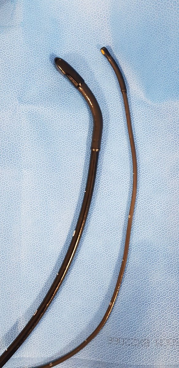
On the left is a conventional adult transoesophageal echo probe
On the right is a neonatal TOE (TEE) probe
Occasionally, just occasionally, the latter is your friend (for an adult cardiologist/physiologist)...
A brief 🧵 on safe performance of TOE
#ECHOFIRST #cardiotwitter
On the right is a neonatal TOE (TEE) probe
Occasionally, just occasionally, the latter is your friend (for an adult cardiologist/physiologist)...
A brief 🧵 on safe performance of TOE
#ECHOFIRST #cardiotwitter
TOE/TEE is actually one of the safer procedures we do, complications are less likely than with:
- Coronary angiography
- Pacemaker insertion
- Ablations
- PCI
- Valve interventions
And that's natural as it's an investigation, not a treatment/therapeutic procedure
- Coronary angiography
- Pacemaker insertion
- Ablations
- PCI
- Valve interventions
And that's natural as it's an investigation, not a treatment/therapeutic procedure
Pharyngeal or oesophageal perforation/rupture is the most feared complication of TOE/TEE
It's incredibly rare - but does happen
TOE technique varies & how it is performed is crucial to minimizing the risk of major complication
It's incredibly rare - but does happen
TOE technique varies & how it is performed is crucial to minimizing the risk of major complication
This classic study from Werner Daniel & colleagues is now > 30yrs old!
>10000 patients
No reports of oesophageal rupture
But...
>10000 patients
No reports of oesophageal rupture
But...


Note the incidence of not being able to insert the probe was 201/10419 - 1.9%
Higher than what we'd see today, but the point is it is NOT zero
And this message is crucial - do NOT approach TOE thinking "I'm going to get this probe down no matter what..."
No! That's trouble!
Higher than what we'd see today, but the point is it is NOT zero
And this message is crucial - do NOT approach TOE thinking "I'm going to get this probe down no matter what..."
No! That's trouble!

In this series of intra-op TOE (different to elective TOE under sedation), the risk of oesophageal perforation was 0.01% - 1 in 10000 cases


Calm voice
Reassurance
Clear explanation
Quiet room
Adequate throat spray
Gentle sedation (too much is as problematic as too little)
Now, if you can get over the tongue & to the back of the throat, you know you're in the right place
Ask pt to swallow & advance as they do...
Reassurance
Clear explanation
Quiet room
Adequate throat spray
Gentle sedation (too much is as problematic as too little)
Now, if you can get over the tongue & to the back of the throat, you know you're in the right place
Ask pt to swallow & advance as they do...
If you feel resistance at this point... STOP!
Try again
Resistance twice?
Try something different
Maybe sit them up more
Talk to pt, explain what you need them to do
Try different neck position
Try again
*Gentle* pressure is ok if you're in the right place
Still won't go?
Try again
Resistance twice?
Try something different
Maybe sit them up more
Talk to pt, explain what you need them to do
Try different neck position
Try again
*Gentle* pressure is ok if you're in the right place
Still won't go?
STOP!
Ask for help
If help can't help
This is when I've found a smaller probe (paediatric or even neonatal) very helpful...if you have one
It can't do 3D
But, more often than not, it's sufficient to answer your Q
- Is there a vegetation?
- Is there LAA thrombus?
Ask for help
If help can't help
This is when I've found a smaller probe (paediatric or even neonatal) very helpful...if you have one
It can't do 3D
But, more often than not, it's sufficient to answer your Q
- Is there a vegetation?
- Is there LAA thrombus?
The image quality is different of course as the frequency is different, but images still diagnostic in most cases
Here's an example
Adult probe just wouldn't go
Neonatal probe went down ok
Clinical Q - why is there AR? (poor TTE views)
Look what we found!
Here's an example
Adult probe just wouldn't go
Neonatal probe went down ok
Clinical Q - why is there AR? (poor TTE views)
Look what we found!
Severe LVSD also!
BAV with cusp prolapse and eccentric AR
Mechanism? ✅
Severity? ✅
3D of AV? ❌
But that's ok!
Mechanism? ✅
Severity? ✅
3D of AV? ❌
But that's ok!
I've had to use the paediatric/neonatal probe about 5 times in the past 10 years
Like I said, just occasionally, it's very helpful!
End/
Like I said, just occasionally, it's very helpful!
End/
• • •
Missing some Tweet in this thread? You can try to
force a refresh