Lea Alhilali, MD Profile picture
Jun 30, 2023 12 tweets 5 min read Read on X
1/Controversy in radiology can get tense! The Mt Fuji sign for tension pnemocephalus is under scrutiny. When should you call it?

A #tweetorial about imaging this important #neurosurgery complication
#medtwitter #FOAMed #FOAMrad #medstudent #radres #meded #neurotwitter
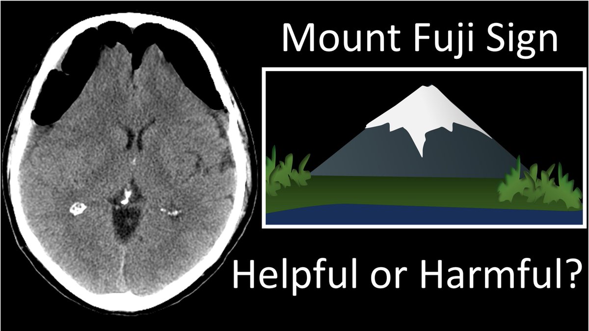
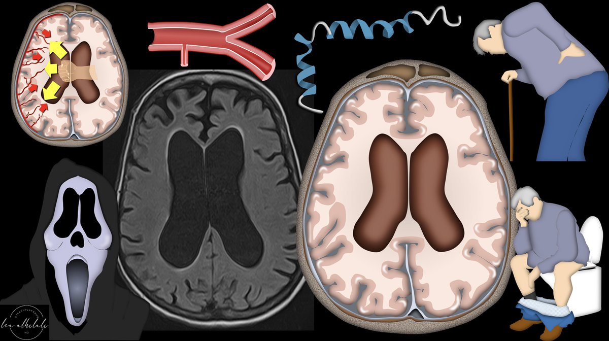
2/Some believe that peaked, mountain like appearance of the frontal lobes is a critical sign of a life-threatening complication & should be called & reported. Others believe it is too non-specific, commonly seen when there isn’t tension & should be retired. Who’s right?
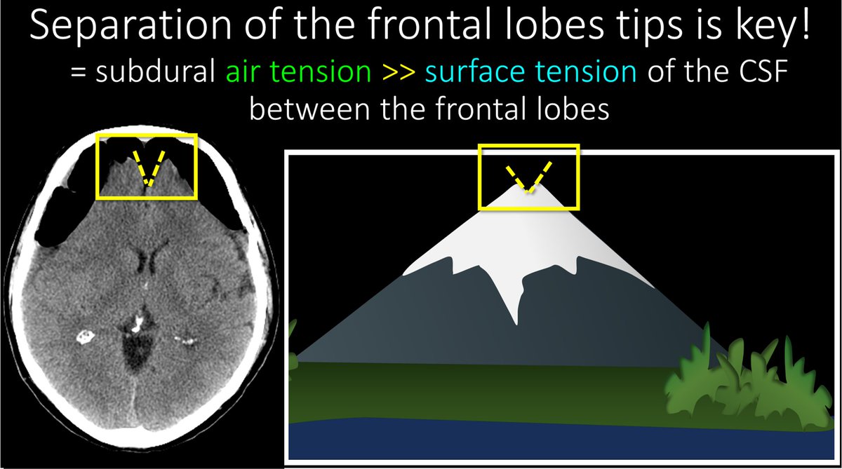
3/First, let’s clarify about what the Mt Fuji sign actually is. Most are familiar with the fact that large collections of pneumocephalus can compress the frontal lobes—making them look like the slopes of a mountain. But this isn’t actually enough to call Mt Fuji.
4/You also need to see frontal lobe separation. This means the subdural air tension is greater than CSF surface tension between the frontal lobes--one of the highest liquid surface tensions—so you know pressure is high. This little V is why it looks like Mt Fuji, not any mountain
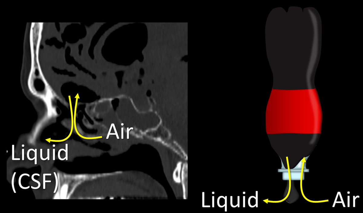
5/Why do we get tension pneumocephalus? 3 main ways. (1) Upside down coke bottle effect w/a CSF leak. As liquid drips out w/a CSF leak, nature abhors a vacuum, so air rushes in to replace it. If outside pressure is higher than CSF pressure, more air will come in & create tension
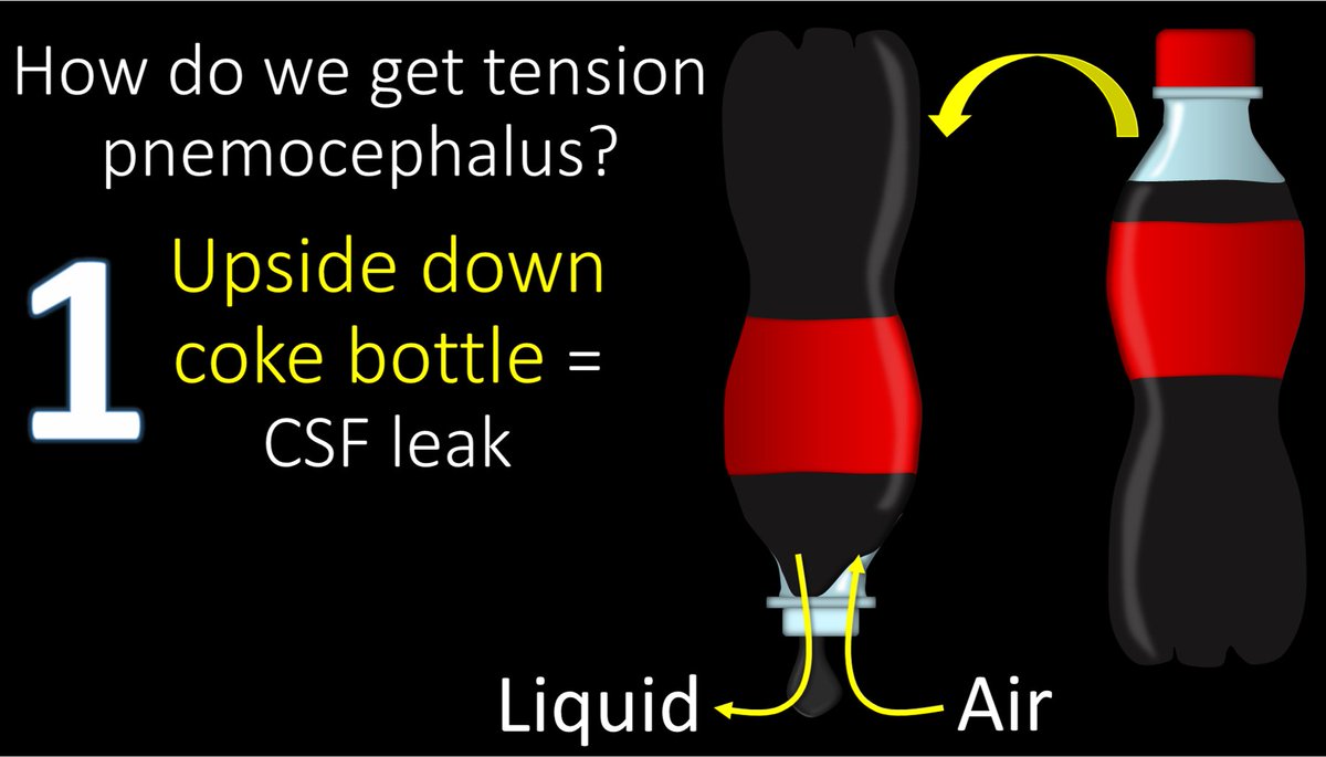
6/Here’s a skullbase CSF leak creating pneumocephalus. As CSF leaks out, air replaces it. If air pressure is higher than intracranial pressure, more air will come in. The worst tension pneumocephalus I ever saw was a pt w/an unknown sphenoid sinus skullbase leak they put on CPAP!
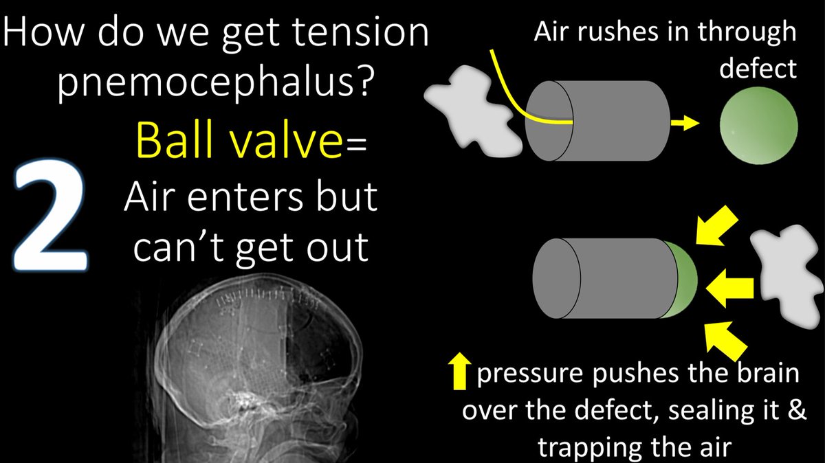
7/Next mechanism is the ball valve mechanism. Air gets in through a defect (from trauma, surgery, etc). Increased pressure eventually pushes down on the brain, causing the brain to close the defect so the air can’t escape. This is the same mechanism seen w/tension pneumothorax.
8/Final mechanism is use of nitrous oxide in neuroanesthesia. If the pt has a subdural collection (usually w/air in the operative setting), nitrous oxide enters the subdural 34 times faster than it diffuses out as nitrogen into the blood stream—creating increased pressure/tension
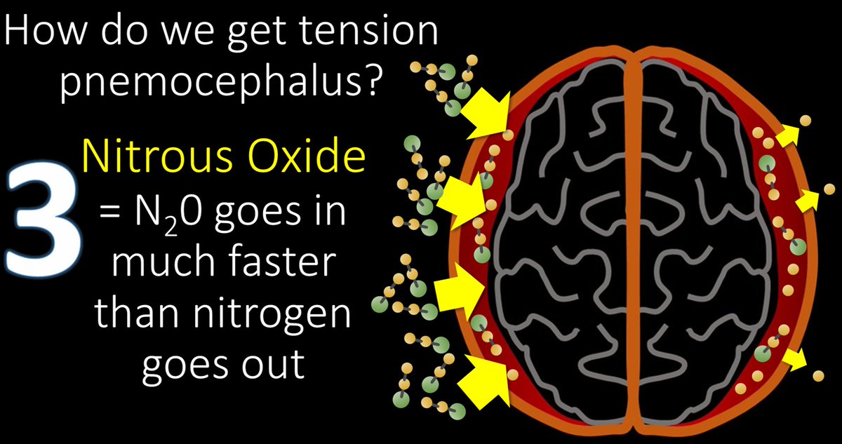
9/This is why nitrous oxide is no longer commonly used in neuroanesthesia. It is also why tension pneumocephalus is less common in the post-operative setting than it was in the past.
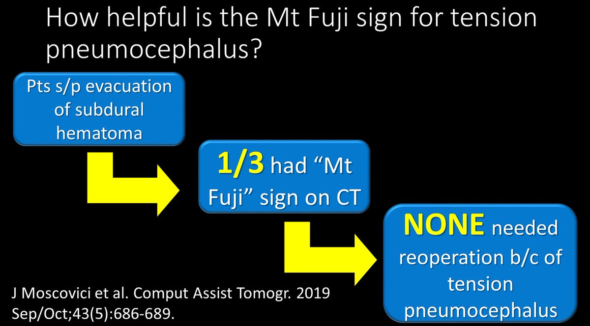
10/So how helpful is the Mt Fuji sign for determining tension pneumocephalus post-operatively, when pneumocephalus is common? Well it turns out, the sign can be seen in 1/3 pts without a neurosurgical emergency. So it is not very specific and can cause overcalling
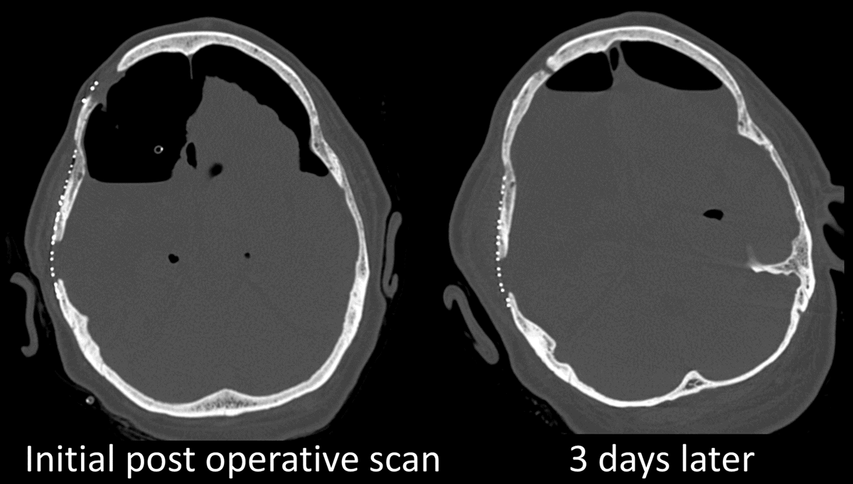
11/More importantly is how the pt is doing clinically. Significant pneumocephalus can be seen post op—but if the pt is not declining, even large amounts can be managed by putting the pt on 100% O2, just like a pneumothorax, w/good results.
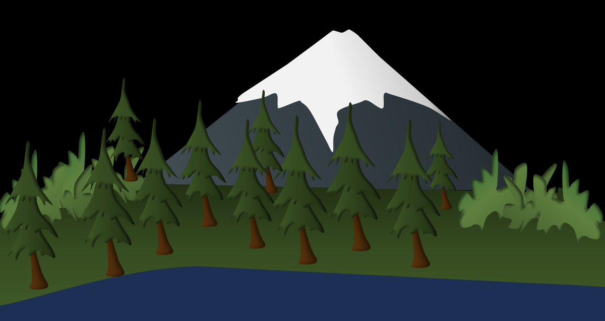
12/So remember it isn’t just one sign—it is the whole picture of how the pt is doing clinically. Don’t lose sight of the forest for the mountain!

• • •

Missing some Tweet in this thread? You can try to force a refresh
 

Keep Current with Lea Alhilali, MD

Lea Alhilali, MD Profile picture

Stay in touch and get notified when new unrolls are available from this author!

Read all threads

This Thread may be Removed Anytime!

PDF

Twitter may remove this content at anytime! Save it as PDF for later use!

Try unrolling a thread yourself!

how to unroll video
  1. Follow @ThreadReaderApp to mention us!

  2. From a Twitter thread mention us with a keyword "unroll"
@threadreaderapp unroll

Practice here first or read more on our help page!

More from @teachplaygrub

Feb 24
1/”That’s a ninja turtle looking at me!” I exclaimed. My fellow rolled his eyes at me, “Why do I feel I’m going to see this a thread on this soon…”

He was right! A thread about one of my favorite imaging findings & pathology behind it Image
2/Now the ninja turtle isn’t an actual sign—yet!

But I am hoping to make it go viral as one. To understand what this ninja turtle is, you have to know the anatomy.

I have always thought the medulla looks like a 3 leaf clover in this region.

The most medial bump of the clover is the medullary pyramid (motor fibers).

Next to it is the inferior olivary nucleus (ION), & finally, the last largest leaf is the inferior cerebellar peduncle.

Now you can see that the ninja turtle eyes correspond to the ION.Image
3/But why are IONs large & bright in our ninja turtle?

This is hypertrophic olivary degeneration.

It is how ION degenerates when input to it is disrupted. Input to ION comes from a circuit called the triangle of Guillain & Mollaret—which sounds like a fine French wine label! Image
Read 9 tweets
Feb 21
1/They say form follows function!

Brain MRI anatomy is best understood in terms of both form & function.

Here’s a thread to help you to remember important functional brain anatomy! Image
2/Let’s start at the top.

At the vertex is the superior frontal gyrus.

This is easy to remember, bc it’s at the top—and being at the top is superior. It’s like the superior king at the top of the vertex. Image
3/It is also easy to recognize on imaging.

It looks like a big thumb pointing straight up out of the brain

I always look for that thumbs up when I am looking for the superior frontal gyrus (SFG) Image
Read 11 tweets
Feb 14
1/”I LOVE spinal cord syndromes!” is a phrase that has NEVER, EVER been said by anyone.

Do you become paralyzed when you see cord signal abnormality?

Never fear—here is a thread on all the incomplete spinal cord syndromes to get you moving again! Image
2/Spinal cord anatomy can be complex.

On imaging, we can see the ant & post nerve roots. We can also see the gray & white matter

Hidden w/in the white matter, however, are numerous efferent & afferent tracts—enough to make your head spin. Image
3/Lucky for you, for the incomplete cord syndromes, all you need to know is gray matter & 3 main tracts

Anterolaterally, spinothalamic tract (pain & temp)

Posteriorly, dorsal columns (vibration, proprioception, & light touch), & next to it, corticospinal tracts—providing motor Image
Read 20 tweets
Feb 9
1/To be or not 2b?? That is the question!

Do you have questions about how to remember cervical lymph node anatomy & levels?

Here’s a SUPERBOWL thread to show you how! Image
2/Google cervical lymph node anatomy & you always get this anatomic picture w/the head flung back like a model posing.

But unless you live in LA, your patients don’t look like this & understanding anatomy from this image is difficult Image
3/First, you need to know how lymph node drainage works in the neck

Nodes drain like rivers—smaller streams drain into larger rivers

In the neck, there are outer circle nodes (peripheral) & inner circle nodes—both drain into the large river of the deep cervical nodes Image
Read 17 tweets
Jan 31
1/Can’t remember what to look for on scans for memory loss?

New Alzheimer’s treatments are changing these scans!

Read on for the latest @theAJNR SCANtastic on imaging in AD:

ajnr.org/content/early/Image
2/Current hypothesis in Alzheimer’s disease (AD) is that accumulation of amyloid beta (AB) protein result sin inflammation & neuronal death

Thus, new treatments in AD are focused on anti-AB antibodies that remove this protein in AD patients. Image
3/However, AB protein is also deposited in vessel walls in AD pts—just like in cerebral amyloid
angiopathy

So removing it from vessel walls increases vascular permeability, leading to edema & hemorrhage

Think of it like how a baby gets mad when you take its candy away! Image
Read 15 tweets
Jan 31
1/Can’t remember what to look for on scans for memory loss?

New Alzheimer’s treatments are changing the way we look at these scans!

Read on to get up to date w/the latest @theAJNR SCANtastic on imaging for Alzheimer’s Disease:

ajnr.org/content/early/…Image
@TheAJNR 2/Current hypothesis in Alzheimer’s disease (AD) is that accumulation of amyloid beta (AB) protein result sin inflammation & neuronal death

Thus, new treatments in AD are focused on anti-AB antibodies that remove this protein in AD patients. Image
@TheAJNR 3/However, AB protein is also deposited in vessel walls in AD pts—just like in cerebral amyloid angiopathy

So removing it from vessel walls increases vascular permeability, leading to edema & hemorrhage

Think of it like how a baby gets mad when you take its candy away! Image
Read 15 tweets

Did Thread Reader help you today?

Support us! We are indie developers!


This site is made by just two indie developers on a laptop doing marketing, support and development! Read more about the story.

Become a Premium Member ($3/month or $30/year) and get exclusive features!

Become Premium

Don't want to be a Premium member but still want to support us?

Make a small donation by buying us coffee ($5) or help with server cost ($10)

Donate via Paypal

Or Donate anonymously using crypto!

Ethereum

0xfe58350B80634f60Fa6Dc149a72b4DFbc17D341E copy

Bitcoin

3ATGMxNzCUFzxpMCHL5sWSt4DVtS8UqXpi copy

Thank you for your support!

Follow Us!

:(