
1/Correlate clinically!
It’s harder than you think in the THALAMUS—where its size is small & but the clinical symptoms are large.
Here’s a #tweetorial to help you remember the main thalamic syndromes & their locations!
#meded #medtwitter #neurotwitter #stroke #radres #FOAMed
It’s harder than you think in the THALAMUS—where its size is small & but the clinical symptoms are large.
Here’s a #tweetorial to help you remember the main thalamic syndromes & their locations!
#meded #medtwitter #neurotwitter #stroke #radres #FOAMed
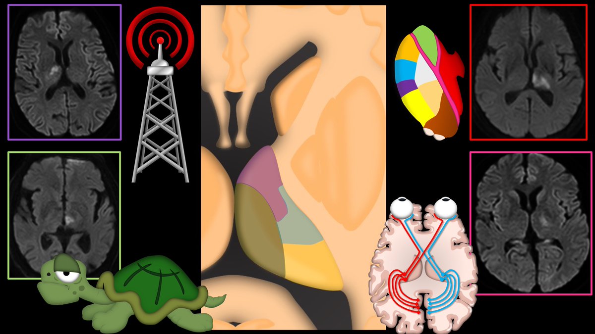
2/Thalamus is a dense network of nuclei & tracts connected to almost everything in the brain. So almost any symptom can be correlated to it.
So saying “thalamus” as the answer when asked where a lesion is located is always reasonable—even w/o knowing what the symptoms are!
So saying “thalamus” as the answer when asked where a lesion is located is always reasonable—even w/o knowing what the symptoms are!

3/Think of the thalamus like the internet service provider or ISP for the brain. Like an ISP, everywhere is connected through it.
And like an ISP, things go bad when it goes down.
But just like an ISP, the problems created depend on where in the network the outage is located
And like an ISP, things go bad when it goes down.
But just like an ISP, the problems created depend on where in the network the outage is located

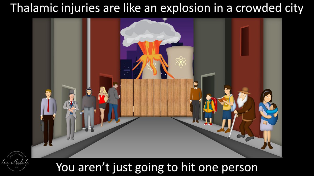
4/Different outages cause different symptoms. Classic symptoms are associated w/specific thalamic locations.
But bc the thalamus is so tightly packed, like a crowded city, every tiny variations in location can change symptoms by affecting different, neighboring nuclei & tracts
But bc the thalamus is so tightly packed, like a crowded city, every tiny variations in location can change symptoms by affecting different, neighboring nuclei & tracts
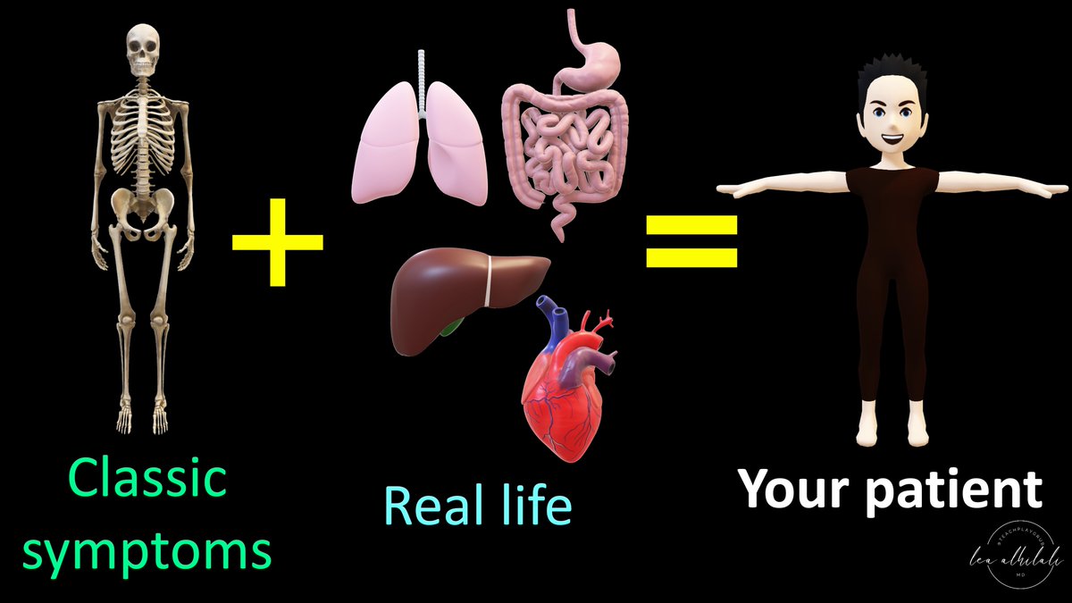
5/Think of the classic thalamic syndromes like a skeleton—they are your starting point. They can help get you a basic gestalt of where a lesion might be.
But real life is never classic & the variations from the classic thalamic syndromes give you your patient’s presentation.
But real life is never classic & the variations from the classic thalamic syndromes give you your patient’s presentation.
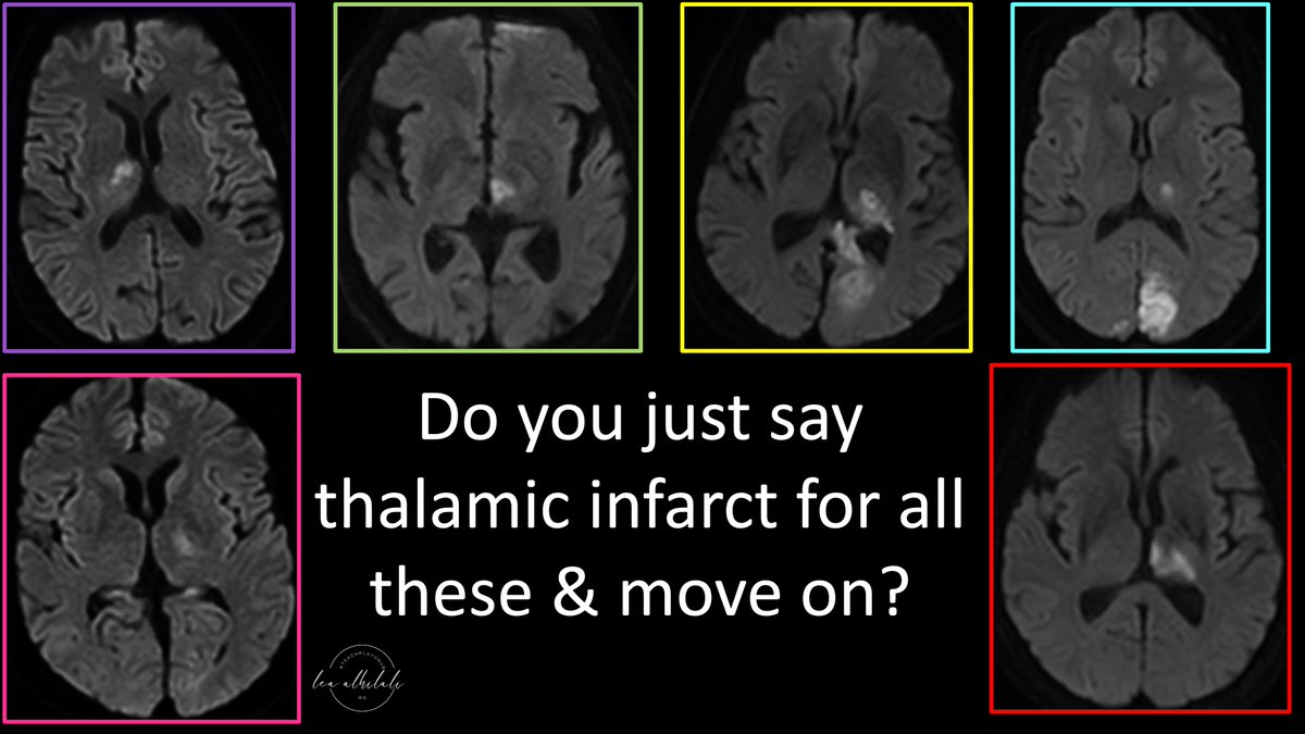
6/So to all the radiologists out there, do you just say “thalamic infarct” for these & move on?
While they’re all in the thalamus, they’re in very different locations w/different symptoms
You can actually tell your clinician WHERE exactly they are & what the SYMPTOMS might be
While they’re all in the thalamus, they’re in very different locations w/different symptoms
You can actually tell your clinician WHERE exactly they are & what the SYMPTOMS might be
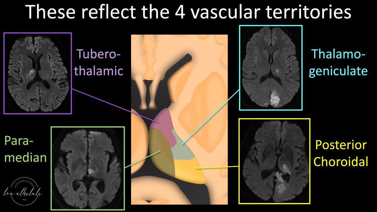
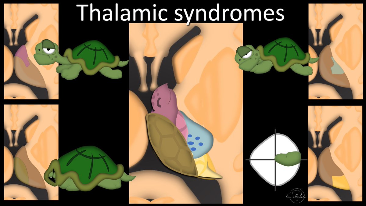
7/These infarcts reflect the four main thalamic vascular territories:
Tuberothalamic anteriorly, paramedian medially, thalamogeniculate laterally, & posterior choroidal posteriorly.
Each has a different syndrome associated w/them. So how do you remember these territories?
Tuberothalamic anteriorly, paramedian medially, thalamogeniculate laterally, & posterior choroidal posteriorly.
Each has a different syndrome associated w/them. So how do you remember these territories?
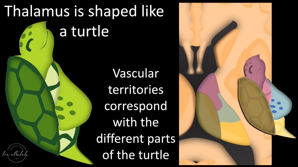
8/Thalamus looks like a turtle. In fact, thalamus means turtle in Greek. Just kidding—but it sounds true, doesn’t it?
The turtle head, arm, shell, & tail each correspond w/one of the vascular territories—& the turtle anatomy can help you remember the associated syndromes too!
The turtle head, arm, shell, & tail each correspond w/one of the vascular territories—& the turtle anatomy can help you remember the associated syndromes too!
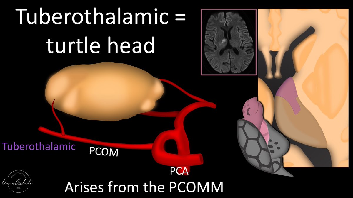
9/Let’s start w/the turtle head. This is the territory of the tuberothalamic artery, which is a branch of the posterior communicating artery (PCOMM).
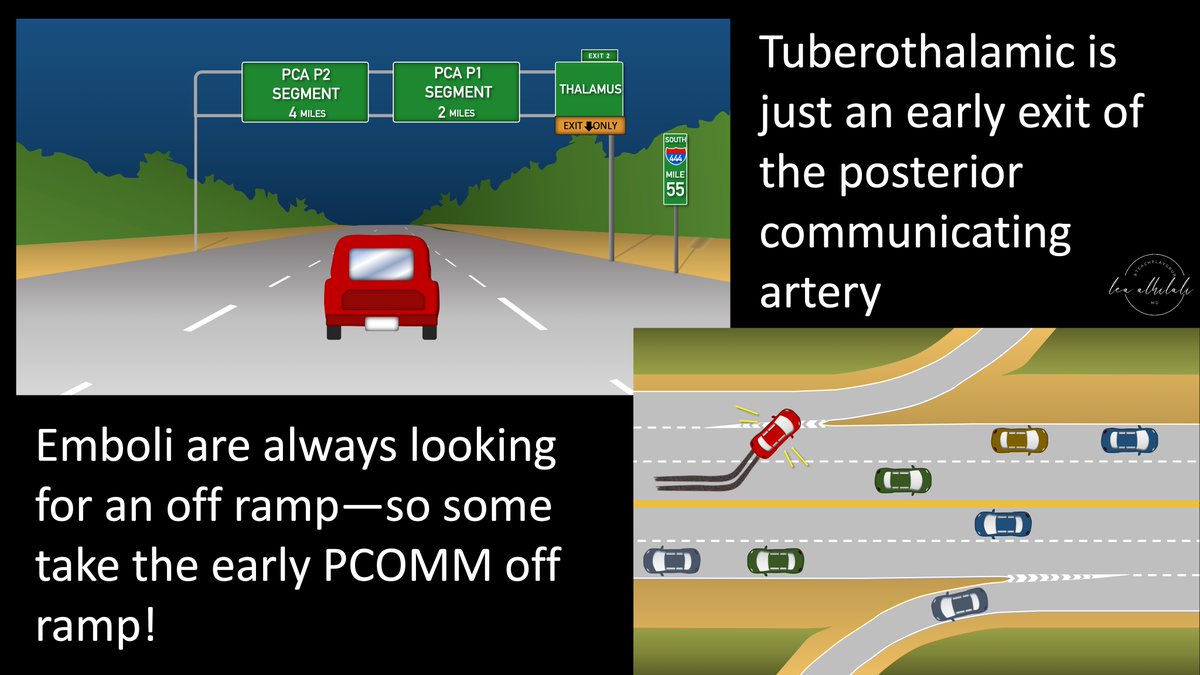
10/Tuberothalamic territory infarcts tend to be cardioembolic.
You can remember this bc the PCOMM is basically a highway from the ICA to the PCA & emboli always exit as soon as there is an opportunity.
Tuberothalamic is just an early exit off of the PCOMM, so emboli exit here
You can remember this bc the PCOMM is basically a highway from the ICA to the PCA & emboli always exit as soon as there is an opportunity.
Tuberothalamic is just an early exit off of the PCOMM, so emboli exit here
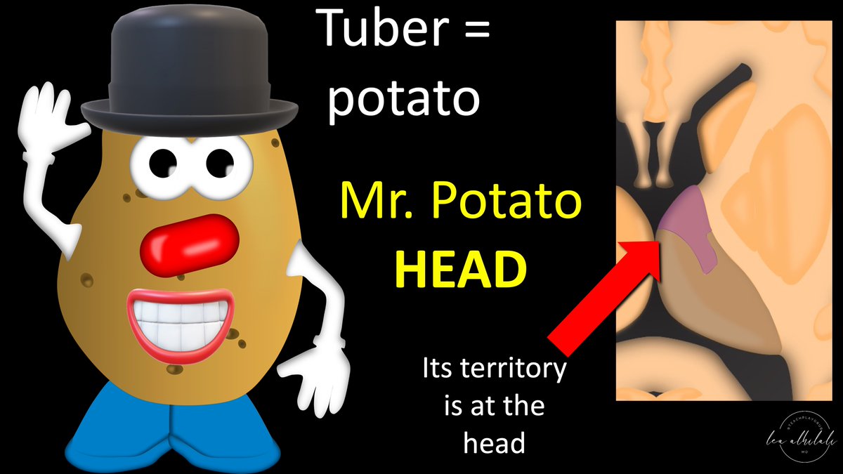
11/You can remember that the tuberothalamic artery supplies the anterior aspect of the thalamus bc tuber is a potato & I always think of Mr. Potato HEAD.
So the tuberothalamic supplies the HEAD of the thalamic turtle.
So the tuberothalamic supplies the HEAD of the thalamic turtle.
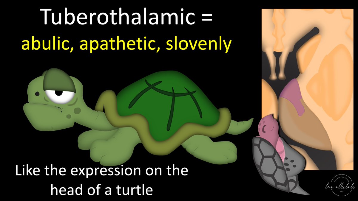
12/The turtle head can also help you remember the associated syndrome. Tuberothalamic infarcts result in neuropsychological syndrome w/an abulic, apathetic, & slovenly patient.
Just think of the expression on a turtle’s face—that is like the tuberothalamic syndrome!
Just think of the expression on a turtle’s face—that is like the tuberothalamic syndrome!
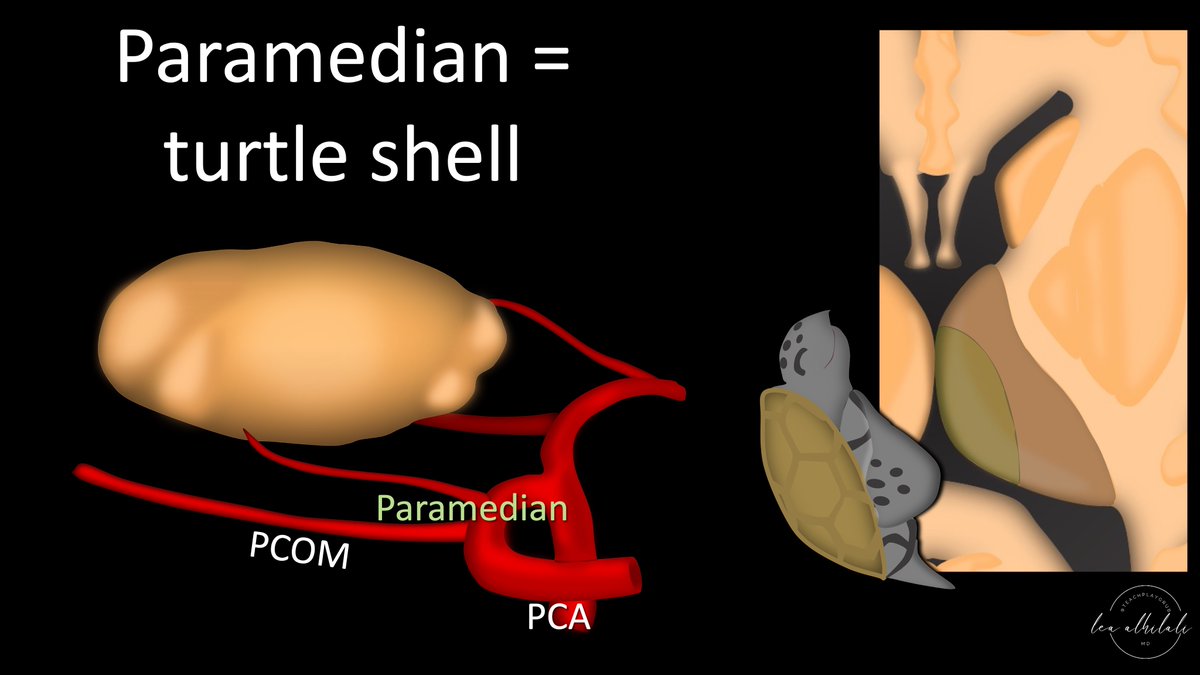
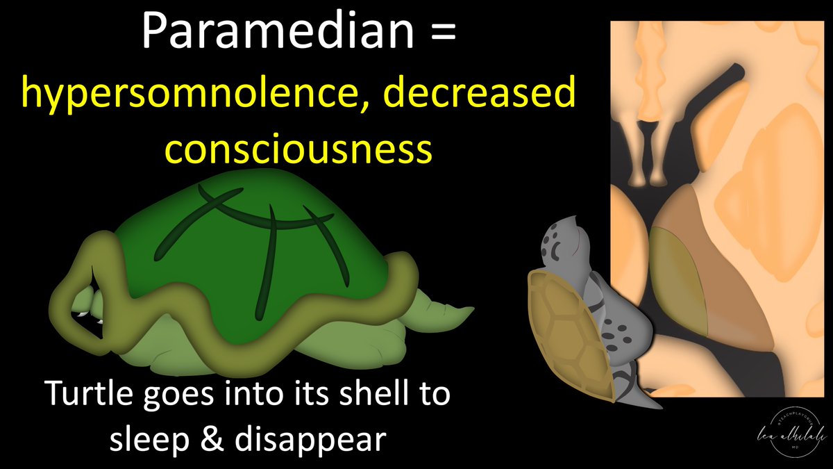
13/Next is the turtle shell. This is the paramedian artery territory. Paramedian artery is a branch of the P1 segment. Bc it’s a small perforator, it is susceptible to small vessel disease & large artery plaques impinging on their origin.
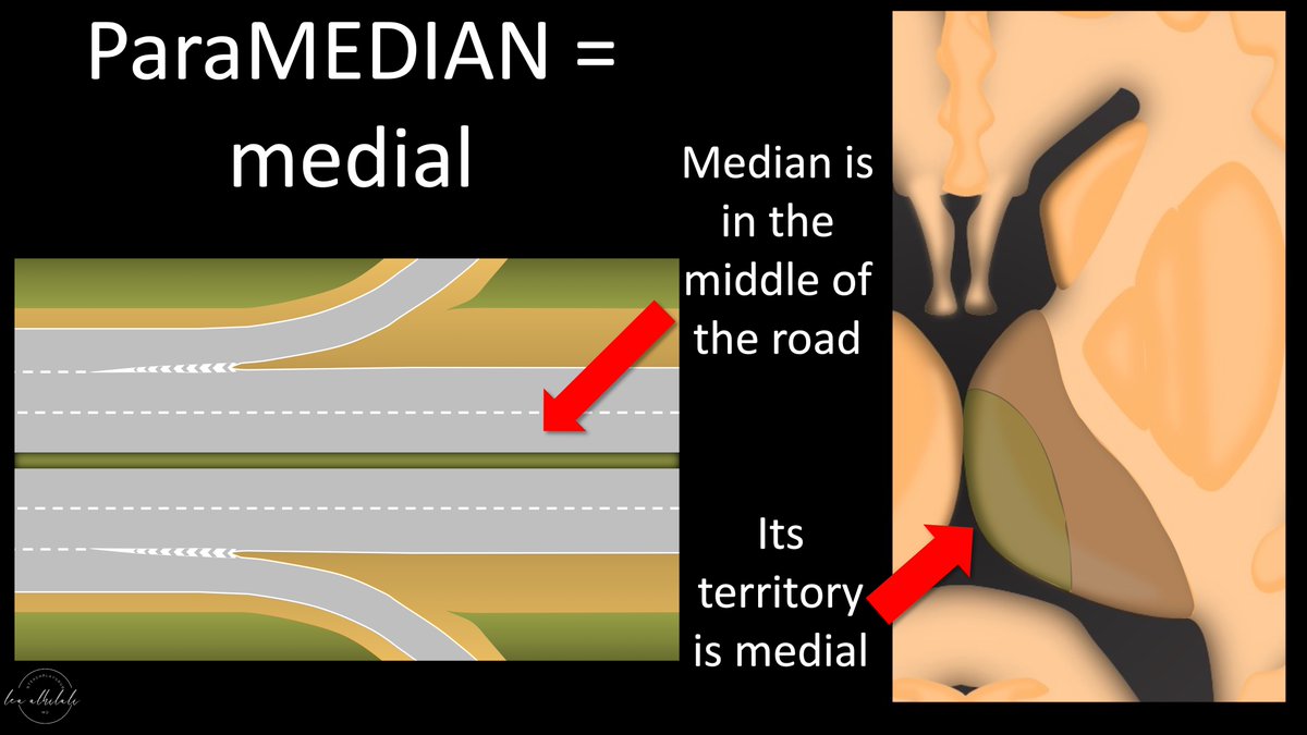
14/You can remember the paramedian artery supplies the medial aspect of the thalamus from its name: ParaMEDIAN.
Medians are the middle of the road, so this artery supplies the medial aspect of the thalamus
Medians are the middle of the road, so this artery supplies the medial aspect of the thalamus
15/The turtle shell can help you remember the syndrome. Paramedian infarcts result in hypersomulence & decreased consciousness
The turtle shell is where the turtle goes to sleep. So infarcts in the thalamus turtle shell—means the patient has withdrawn into their shell as well
The turtle shell is where the turtle goes to sleep. So infarcts in the thalamus turtle shell—means the patient has withdrawn into their shell as well
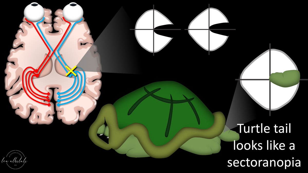
16/Next is the turtle tail. This is the posterior choroidal artery territory. This also arises from the P1 segment to supply the lateral geniculate nucleus & surrounding structures. Like the paramedian artery, it is also susceptible to small vessel disease.
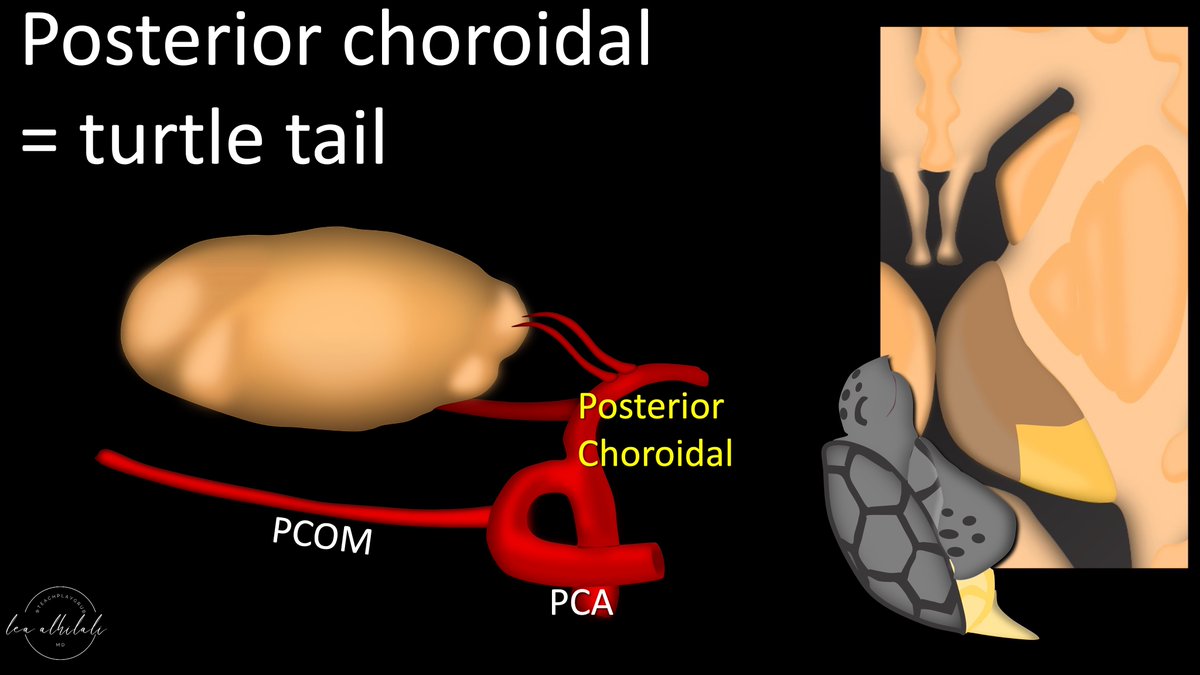
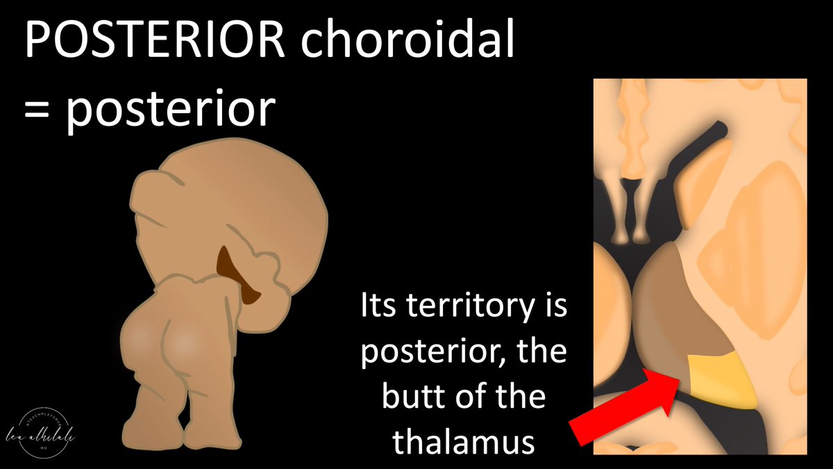
17/You can remember that the posterior choroidal artery supplies the posterior aspect of the thalamus from its name—POSTERIOR choroidal.
So the posterior choroidal supplies the posterior thalamus—or the butt/tail of the turtle.
So the posterior choroidal supplies the posterior thalamus—or the butt/tail of the turtle.
18/Posterior choroidal artery actually only supplies a part of the lateral geniculate nucleus. So a posterior choroidal infarct doesn’t take out the full lateral geniculate nucleus & give you a quadrantanopia. Instead, it gives you only a part of a quadrant, a sector.
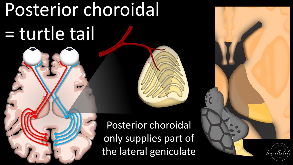
19/So posterior choroidal arteries give you a sectoranopia.
This sector defect actually looks like a little turtle tail!
So you can remember than an infarct of the turtle tail gives you a visual field defect that looks like a turtle tail!
This sector defect actually looks like a little turtle tail!
So you can remember than an infarct of the turtle tail gives you a visual field defect that looks like a turtle tail!
20/Last is the turtle arm. This is the territory of the thalamogeniculate artery. It arises later, from the P2 segment of the PCA. Like the paramedian & posterior choroidal arteries, it is also susceptible to small vessel disease.
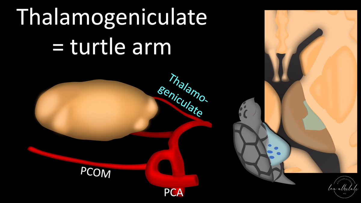
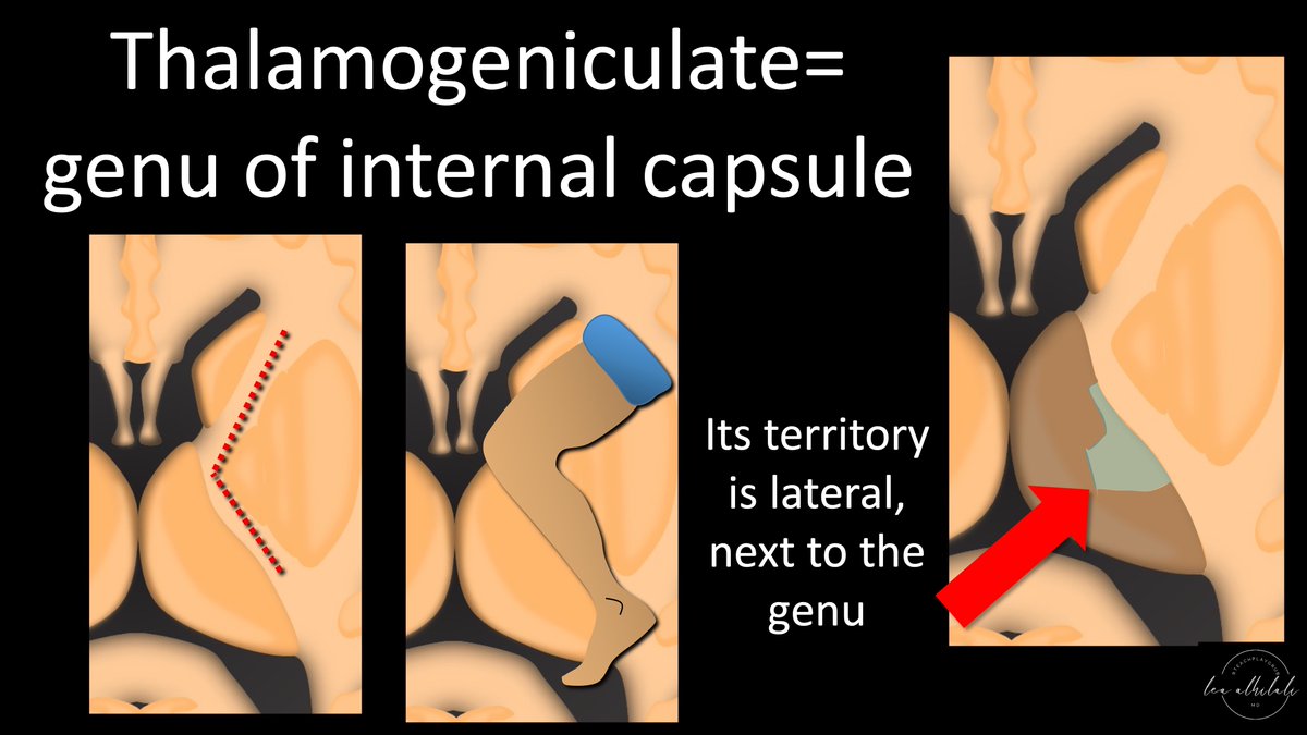
21/ You can remember that the thalamogeniculate artery supplies the lateral thalamus bc the “geniculate” part of the name refers to the genu of the internal capsule (IC)
Genu means knee & this is where the IC turns like a knee. So it supplies the lateral thalamus next to the IC
Genu means knee & this is where the IC turns like a knee. So it supplies the lateral thalamus next to the IC
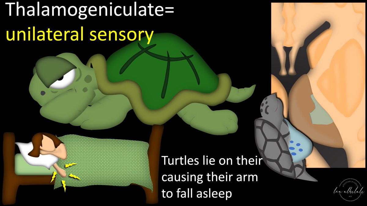
22/These infarcts give a hemisensory defect.
Remember this bc if you look at the thalamic turtle, it is basically laying on its arm. What happens when you lay on your arm too much? It falls asleep—giving you a hemisensory defect.
Turtle arm infarcts = hemisensory defects
Remember this bc if you look at the thalamic turtle, it is basically laying on its arm. What happens when you lay on your arm too much? It falls asleep—giving you a hemisensory defect.
Turtle arm infarcts = hemisensory defects
23/So next time you see a thalamic infarct, ask yourself—what part of the thalamic turtle has it taken out? Head, shell, arm, or tail? That will give you an outline of what symptoms to expect.
And guess what—you’ve just correlated clinically yourself!
And guess what—you’ve just correlated clinically yourself!
• • •
Missing some Tweet in this thread? You can try to
force a refresh


