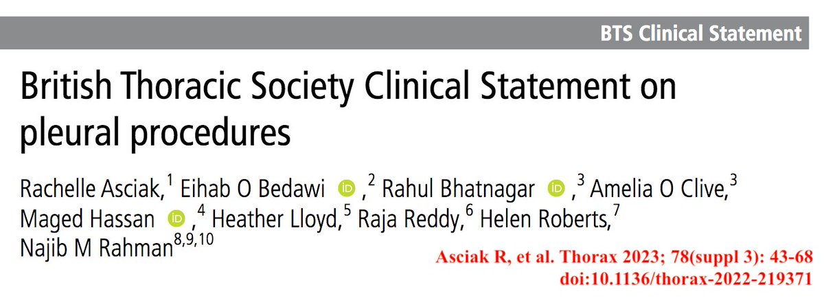
British Thoracic Society recently published a must-read Statement on pleural procedures. It is 26 pages long & is accompanied by 13 online supplementary appendices 

These are the highlights (I skip paragraphs on pleural biopsy & malignant effusions that are not among an intensivist's daily routine)
Summary of clinical practice points:
Before carrying out a pleural procedure, a review of indications & contraindications should be performed:


Summary of clinical practice points:
Before carrying out a pleural procedure, a review of indications & contraindications should be performed:


Pleural aspiration (PA) (diagnostic & therapeutic):
Thoracentesis should be performed ABOVE A RIB
Thoracic ultrasound MUST be used for aspiration
SMALL bore needles are preferred
For therapeutic aspiration >60 mL, a catheter should be used rather than a needle alone
Thoracentesis should be performed ABOVE A RIB
Thoracic ultrasound MUST be used for aspiration
SMALL bore needles are preferred
For therapeutic aspiration >60 mL, a catheter should be used rather than a needle alone
Use of Veress needle may reduce risk of damaging underlying structures
Therapeutic PA should be performed SLOWLY using manual syringe aspiration or gravity drainage. Vacuum bottles or wall suction should NOT be used.
In general, a max of 1.5 L should be drained in 1 attempt
Therapeutic PA should be performed SLOWLY using manual syringe aspiration or gravity drainage. Vacuum bottles or wall suction should NOT be used.
In general, a max of 1.5 L should be drained in 1 attempt
Routine use of manometry DOES NOT HELP to reduce the risk associated with large volume PA
The procedure should be stopped if symptoms of chest tightness, pain, persistent cough or worsening breathlessness develop
The procedure should be stopped if symptoms of chest tightness, pain, persistent cough or worsening breathlessness develop
Intercostal drain insertion:
Small-bore drains (< 14 Fr) are suitable for most indications including draining empyema
Larger drains should be considered in unstable trauma & pneumothorax complicating mechanical ventilation
Consider a drain > 14 Fr if pleurodesis is intended
Small-bore drains (< 14 Fr) are suitable for most indications including draining empyema
Larger drains should be considered in unstable trauma & pneumothorax complicating mechanical ventilation
Consider a drain > 14 Fr if pleurodesis is intended
BEFORE drain insertion, aspiration of air or fluid w the needle applying the anaesthetic is necessary, & failure to do so should prompt further assessment
All chest drains should be fixed with a holding suture to prevent fall out
All chest drains should be fixed with a holding suture to prevent fall out
A chest drain inserted for managing pleural effusion should be clamped promptly in patients with repetitive coughing or chest pain to avoid re-expansion pulmonary which is a potentially fatal complication
A follow-up chest xray should be conducted within a few hours of insertion
A follow-up chest xray should be conducted within a few hours of insertion
For pleural fluid, the volume to be drained over specific time periods should be specified in the procedure report & in handover (eg, 500 mL/hr)
In cases of non-functioning intercostal drain where another drain is required, the old track must be avoided when inserting a new one
In cases of non-functioning intercostal drain where another drain is required, the old track must be avoided when inserting a new one
How to set up a chest drain bottle & underwater seal drain:
Aseptic non-touch technique should be used when changing a chest drain bottle/underwater seal drain or drain tubing
The drain bottle must be kept below the insertion site & the drain must be kept upright at all times
Aseptic non-touch technique should be used when changing a chest drain bottle/underwater seal drain or drain tubing
The drain bottle must be kept below the insertion site & the drain must be kept upright at all times
The drain must have adequate water in the system to cover the end of the tube
For patients with pneumothorax and suspected/confirmed COVID-19, a viral filter should be considered to minimise the risk of droplet exposure via the chest drain circuit
For patients with pneumothorax and suspected/confirmed COVID-19, a viral filter should be considered to minimise the risk of droplet exposure via the chest drain circuit
Drains should be checked daily for wound infection, fluid drainage volumes & presence of resp swinging and/or bubbling
CLAMPING a bubbling chest tube should be AVOIDED unless under specialist's supervision & in specific circumstances only
CLAMPING a bubbling chest tube should be AVOIDED unless under specialist's supervision & in specific circumstances only
Drainage of a large pleural effusion should be controlled to prevent the potential complication of re-expansion pulm edema
Suction & digital chest drain devices:
SUCTION should be AVOIDED soon after drain insertion to minimise the risk of re-expansion pulm edema
Routine use of thoracic suction should be avoided given a lack of data demonstrating clinical benefit
SUCTION should be AVOIDED soon after drain insertion to minimise the risk of re-expansion pulm edema
Routine use of thoracic suction should be avoided given a lack of data demonstrating clinical benefit
If suction is used, low-pressure, high-volume thoracic suction should be used
Patients receiving suction should have a viral filter or a digital device should be used to minimise the risk of aerosol generation
Patients receiving suction should have a viral filter or a digital device should be used to minimise the risk of aerosol generation
Other points:
Pleural procedures should be undertaken in normal working hours. They should only be undertaken out of hours in an emergency
No large studies accurately define bleeding risk associated w pleural procedures in pts on antiPLT/anticog meds, or those w coagulopathy
Pleural procedures should be undertaken in normal working hours. They should only be undertaken out of hours in an emergency
No large studies accurately define bleeding risk associated w pleural procedures in pts on antiPLT/anticog meds, or those w coagulopathy
Several small studies have found no increased bleeding risk of thoracentesis or small-bore chest drain insertion in patients on clopidogrel, or with an uncorrected bleeding risk
Assessment of a non-functioning chest drain:
Cessation of fluid swinging in the tubing is usually a
manifestation of drain blockage which can be resolved with simple saline flushing. The full length of the drain/tubing should be inspected to rule out any kinking
Cessation of fluid swinging in the tubing is usually a
manifestation of drain blockage which can be resolved with simple saline flushing. The full length of the drain/tubing should be inspected to rule out any kinking

Management of problematic sc emphysema:
Surgical (subcutaneous) emphysema following
chest drain insertion is common & often of no clinical consequence. However, sometimes substantial amounts of air can accumulate. Risk factors: drain blockage & poor drain placement or fixation
Surgical (subcutaneous) emphysema following
chest drain insertion is common & often of no clinical consequence. However, sometimes substantial amounts of air can accumulate. Risk factors: drain blockage & poor drain placement or fixation

Kudos to the authors for this comprehensive document!
#FOAMed #FOAMcc #MedTwitter #MedEd #MedStudentTwitter
#FOAMed #FOAMcc #MedTwitter #MedEd #MedStudentTwitter
• • •
Missing some Tweet in this thread? You can try to
force a refresh

 Read on Twitter


