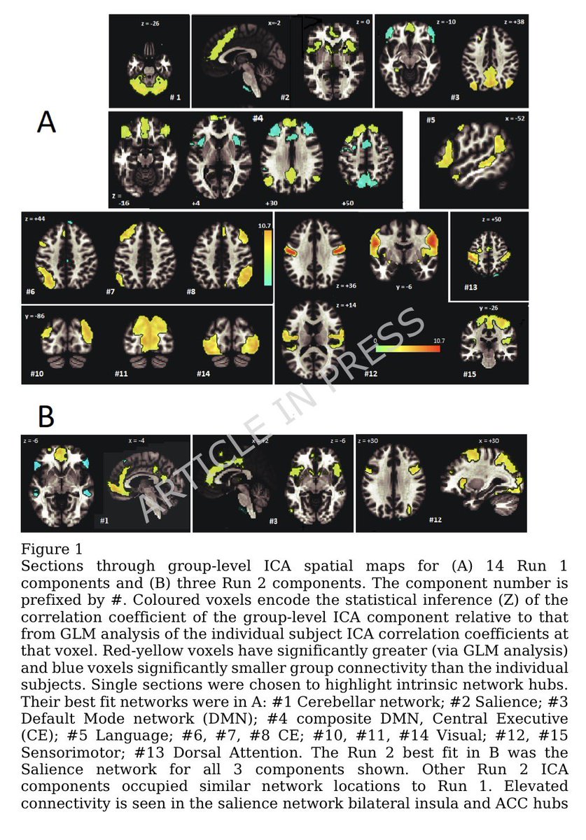
A NEW discovery, akin to a black-swan event-—unexpected but making perfect sense once revealed, would offer an approach to fixing broken immune responses in #LongCovid and some autoimmune diseases! 1/
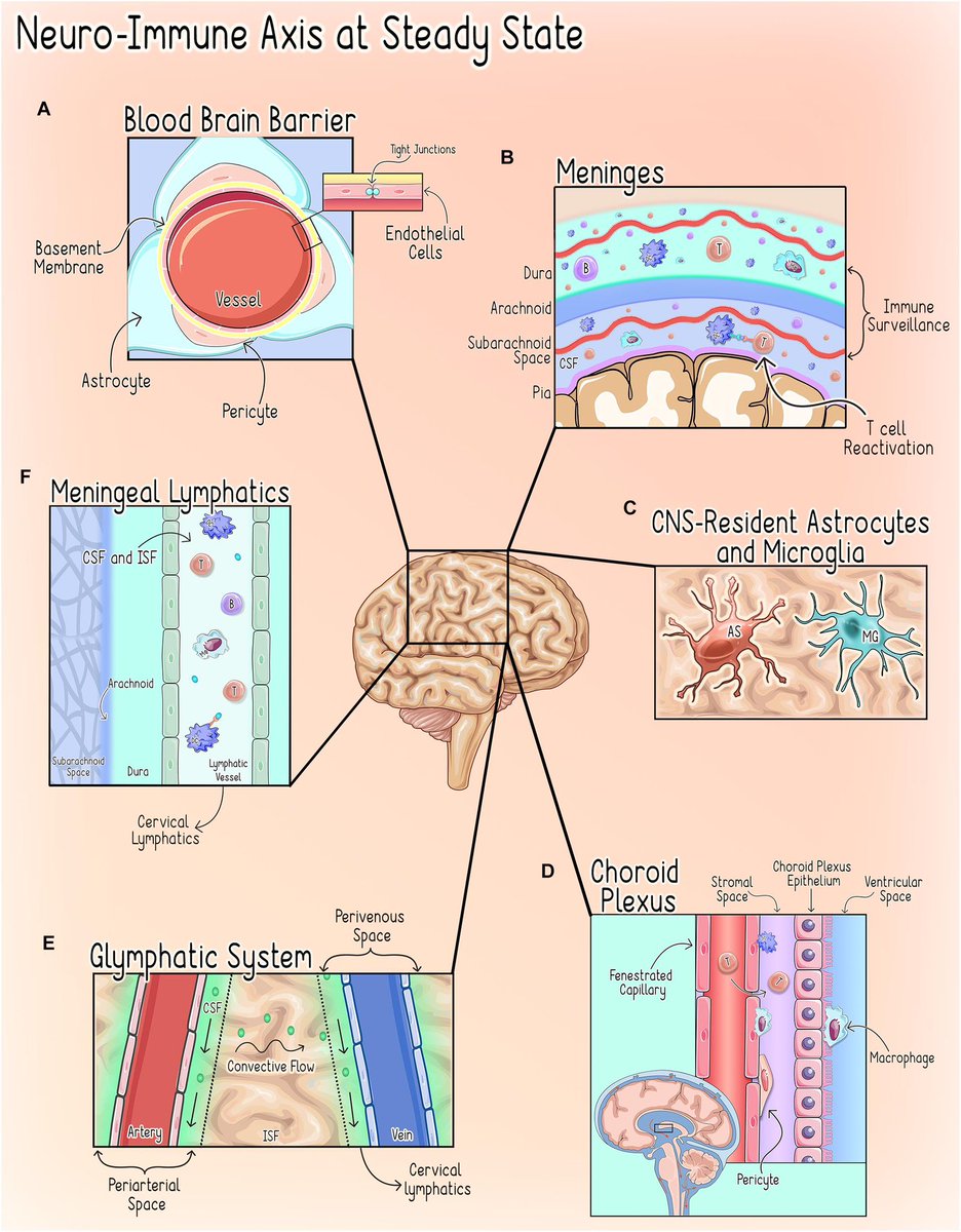
Scientists have long known that brain plays a part in the immune system — but how it does so has been a mystery. Now, scientists have identified cells in brainstem that sense immune cues from the periphery of body & act as master regulators of body’s inflammatory response 2/
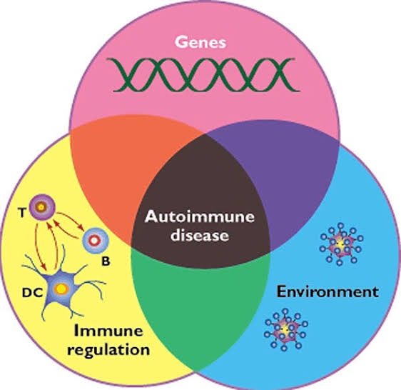
The results suggest that the brain maintains a delicate balance between the molecular signals that promote inflammation and those that dampen it —a finding that could lead to treatments for autoimmune diseases and other conditions caused by an excessive immune response. 3/
Scientists have known that the brainstem has many functions, such as controlling basic processes such as breathing. However, it shows that there is whole layer of biology that we haven’t even anticipated! 4/
After sensing an intruder, the immune system unleashes a flood of immune cells that promote inflammation. This response must be controlled w/ exquisite precision: if it’s too weak, the body is at risk of becoming infected; if it’s too strong, it can damage body’s own tissues 5/
Previous work has shown that the vagus nerve, a large network of nerve fibres that links the body with the brain, influences immune responses. However, the specific brain neurons that are activated by immune stimuli remained elusive. 6/
To investigate how the brain controls the body’s immune response, researchers monitored the activity of brain cells after injecting the abdomen of mice with bacterial compounds that trigger inflammation. 7/
They used single-cell RNA sequencing, combined with functional imaging, to identify the circuit components of this neuro-immune axis. 8/
The researchers identified neurons in the brainstem that switched on in response to the immune triggers. Activating these neurons with a drug reduced the levels of inflammatory molecules in the mice’s blood. 9/
Silencing the neurons led to an uncontrolled immune response, with the number of inflammatory molecules increasing by 300% compared with the levels observed in mice with functional brainstem neurons. 10/
These nerve cells act as a rheostat in the brain that ensures that an inflammatory response is maintained within the appropriate levels. 11/
Further experiments showed two discrete groups of neurons in vagus nerve: one that responds to pro-inflammatory immune molecules & another that responds to anti-inflammatory molecules. These neurons relay their signals to the brain, allowing it to monitor the immune response 12/
In mice with conditions characterized by an excessive immune response, artificially activating the vagal neurons that carry anti-inflammatory signals diminished inflammation. 13/
Finding ways to control this newly discovered body–brain network would offer an approach to fixing broken immune responses in various conditions such as autoimmune diseases and even long COVID. 14/
There’s evidence that therapies targeting the vagus nerve can treat diseases such as multiple sclerosisand rheumatoid arthritis, suggesting that targeting the specific vagal neurons that carry immune signals might work in people. 15/15
➡️ Compared with healthy controls,
✔ Long COVID patients had blunted morning cortisol peaks
✔ Higher evening cortisol
✔ Loss of normal circadian pattern
Blood cortisol alone failed to detect these changes. 2/
Key insight:
➡️ Salivary cortisol profiling may be a more sensitive marker of stress-system dysfunction in LongCOVID than standard blood tests.
➡️ HPA axis disruption could underlie:
• Fatigue
• Brain fog
• Sleep disturbance
• Dysautonomia. 3/
➡️ New review highlights that persistent cognitive symptoms in COVID survivors are strongly linked to pro-inflammatory cytokines and blood–brain barrier (BBB) dysfunction.
➡️ Key culprits include IL-6, TNF-α, IL-1β, IL-8, IL-13 and MCP-1 — many remain elevated months after infection.
🔥 COVID-19 is not just a respiratory disease.
➡️ Evidence suggests cognitive impairment can occur due to:
Post-COVID fatigue isn’t just subjective.
Using advanced MRI, researchers found real changes in brain blood flow and oxygen metabolism in people with Post-COVID-19 Syndrome (PCS) after mild infection.
➡️ Key finding:
PCS patients showed increased oxygen metabolism in the hippocampus (memory hub) but reduced metabolism in the anterior cingulate cortex (ACC) — despite no visible brain atrophy. 1/
Why this matters:
➡️ Higher hippocampal metabolism was linked to better cognitive performance, suggesting a compensatory response to maintain thinking and memory in PCS. 2/
In contrast, lower anterior cingulate cortex (ACC) metabolism correlated with:
Why do some people feel exhausted long after COVID-19?
➡️ New brain-imaging research shows that even after mild COVID, people with persistent fatigue can have subtle but real changes in brain structure.
➡️ These changes are not large or widespread, but tend to appear in connected brain networks, especially areas involved in attention, decision-making, and sensory processing. 1/
Importantly, the brain regions affected overlap with areas that naturally express TMPRSS2, a protein that helps SARS-CoV-2 enter cells — suggesting certain brain circuits may be more vulnerable to the virus. 2/
The study also links these changes to brain chemical systems involved in mood, energy, and cognition (serotonin, acetylcholine, glutamate, and cannabinoids). 3/
COVID-19 doesn’t just affect the lungs — it can disrupt how cells produce energy. New research shows that COVID-19 alters the genetic “switches” that control mitochondria, the structures that power our cells. 1/
By comparing people who died from severe COVID-19, those who recovered, and healthy individuals, researchers found lasting changes in how mitochondrial genes are regulated. These changes were most prominent in genes involved in energy production and metabolism. 2/
Importantly, people with COVID-19 showed abnormally high levels of proteins that control mitochondrial structure and stress responses, suggesting long-term damage to the cell’s energy system. 3/
#LongCOVID (LC) shares striking symptom overlap with hypermobility spectrum disorders (HSD/hEDS): fatigue, brain fog, dysautonomia, pain—especially in women.
➡️ A new case series explores whether some “intractable” LC may reflect undiagnosed hypermobility disorders.
➡️ Five women with persistent LC symptoms were evaluated at an hEDS/HSD clinic.
All met Beighton score criteria for hypermobility.
➡️ 4 diagnosed with hEDS, 1 with HSD
➡️ 3 had dysautonomia
None had prior hypermobility diagnoses. 1/
All patients carried MTHFR polymorphisms (C677T or A1298C)—recently linked to hEDS/HSD.
➡️ Several also showed features of mast cell activation, suggesting immune dysregulation may unmask latent connective tissue disorders after SARS-CoV-2 infection.
➡️ Targeted management (physical therapy, methylfolate/B12, mast cell stabilization, pain interventions) led to clinical improvement in all cases.
🔑 Takeaway: Consider hEDS/HSD in women with refractory Long COVID, especially with multisystem pain and dysautonomia. 2/
This case series suggests that some patients with severe, persistent #LongCOVID—especially women—may have previously undiagnosed hypermobility disorders (hEDS/HSD).
➡️ Five women with refractory LongCOVID symptoms were found to meet criteria for hypermobility, often with dysautonomia, mast cell–related features, and MTHFR polymorphisms.
➡️ Targeted management led to clinical improvement, highlighting the need to consider hEDS/HSD in patients with intractable Long COVID symptoms. 3/