Emmanuel Profile picture
May 24, 2024 โ€ข 10 tweets โ€ข 4 min read โ€ข Read on X
๐—™๐—ฟ๐—ผ๐—บ ๐—ฏ๐—ถ๐—ฟ๐—ฑ๐˜€ ๐˜๐—ผ ๐—บ๐—ฎ๐—บ๐—บ๐—ฎ๐—น๐˜€: ๐˜€๐—ฝ๐—ถ๐—น๐—น๐—ผ๐˜ƒ๐—ฒ๐—ฟ ๐—ผ๐—ณ ๐—ต๐—ถ๐—ด๐—ต๐—น๐˜† ๐—ฝ๐—ฎ๐˜๐—ต๐—ผ๐—ด๐—ฒ๐—ป๐—ถ๐—ฐ ๐—ฎ๐˜ƒ๐—ถ๐—ฎ๐—ป ๐—ถ๐—ป๐—ณ๐—น๐˜‚๐—ฒ๐—ป๐˜‡๐—ฎ ๐—›5๐—ก1,
led to efficient intra- and interspecies transmission

H/t @DuckSwabber
biorxiv.org/content/10.110โ€ฆ
Image
2) Highly pathogenic avian influenza H5N1 virus was detected infecting dairy cattle herds in several states in the U.S. in early 2024, representing the first report of H5N1 in a livestock species. Image
3) Affected cattle displayed clinical signs like decreased appetite, respiratory distress, diarrhea, decreased milk production, and abnormal milk. Virus was detected in milk, nasal swabs, blood, and tissues of infected cows. Image
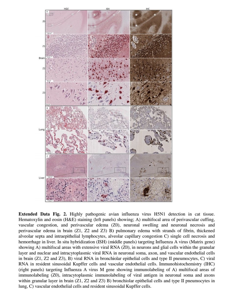
4) Extensive virus replication was observed in the mammary glands of infected cows, resulting in high viral loads and shedding in milk. Histological analysis and virus staining showed tropism for milk-secreting epithelial cells. Image
5) Virus transmission between cows was determined to be efficient based on epidemiological and genomic analysis. Virus was also transmitted to wild birds, domestic cats, and a raccoon on infected farms. Image
6) Genomic analysis identified a reassortment event that produced a new genotype (B3.13) with enhanced ability to infect cows. Mutations accumulated after transmission to cows. Image
7) Between-farm transmission occurred through livestock movement, contact with infected wild birds, and fomites/personnel movement between locations. Image
8) This represents the first sustained transmission of H5N1 in a livestock species. Efficient shedding and transmission in cows is a concern that could allow the virus to further adapt to mammals. Image
9) So in summary, this paper describes the spillover and transmission of H5N1 virus in dairy cattle herds in multiple U.S. states, highlighting its ability to cross species barriers into a new host.

Thanks for reading ๐Ÿ™ Image
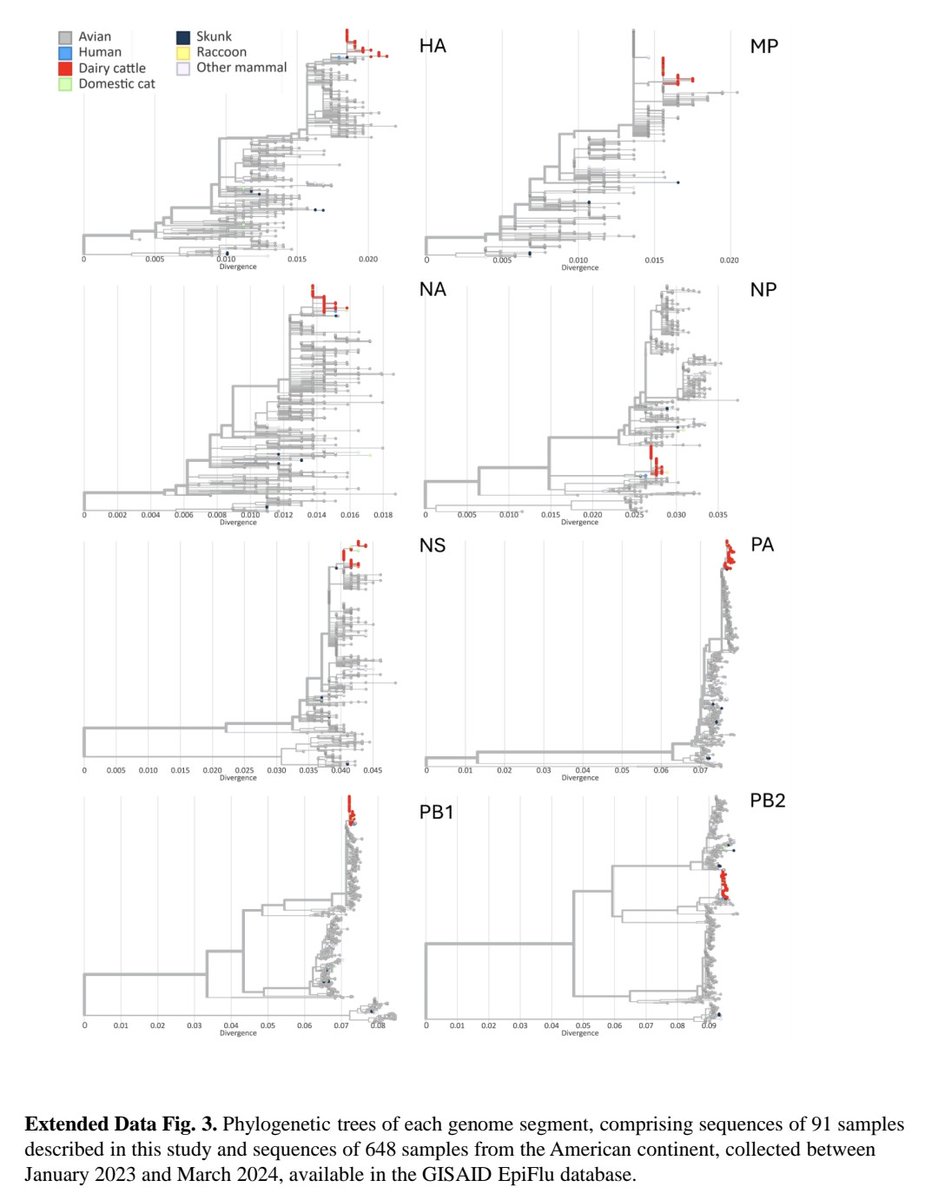
10) Other extended data Image

โ€ข โ€ข โ€ข

Missing some Tweet in this thread? You can try to force a refresh
ใ€€

Keep Current with Emmanuel

Emmanuel Profile picture

Stay in touch and get notified when new unrolls are available from this author!

Read all threads

This Thread may be Removed Anytime!

PDF

Twitter may remove this content at anytime! Save it as PDF for later use!

Try unrolling a thread yourself!

how to unroll video
  1. Follow @ThreadReaderApp to mention us!

  2. From a Twitter thread mention us with a keyword "unroll"
@threadreaderapp unroll

Practice here first or read more on our help page!

More from @ejustin46

Apr 17
The incredible world of T-CELLS and why are they exhausted fighting the SARS-COV-2 virus ?

(An explanation๐Ÿงต for kids based on an astonish study Thanks to @DavidJoffe64)
sciencedirect.com/science/articlโ€ฆImage
2) T-cells are like the superheroes of our immune system - they work hard to protect us from germs and viruses.

When a virus like SARS-COV-2 gets into our body, the T cells spring into action. There are different types of T cells, each with a special job to do. Image
3) - Some T cells are like the fighters, killing off the virus-infected cells.
- Other T cells are the coordinators, telling the other immune cells what to do.
- There are also T cells that act like the police, making sure the immune response doesn't go overboard.
Read 11 tweets
Apr 16
"COVID-19 CRUSADERS:
The TRIO of NASAL IMMUNE CELLS That BATTLE the VIRUS"

A masterpiece study in Nature ๐Ÿ’ฏ๐Ÿ’ฅ
nature.com/articles/s4159โ€ฆImage
2) The most important immune components in the nasal tissue for fighting COVID-19 are:

โ–ถ๏ธ Monocytes and neutrophils
- These immune cells moved into the area under the nose lining during active COVID-19 infection and helped protect the underlying lymphoid tissue. Image
3)โ–ถ๏ธ Macrophages
- As people recovered from COVID-19, macrophages started to help repair the nasal tissue.
โ–ถ๏ธ Germinal centers and B cells
- The specialized germinal center structures in the nose contained active B cells that were producing antibodies against the COVID-19 virus Image
Read 6 tweets
Apr 16
How SARS-COV-2 spreads (Re) and why there are differences between countries ?

An explanation for kids based on the astonishing new study of @firefoxx66 @richardneher and colleagues
pubmed.ncbi.nlm.nih.gov/40233303/Image
2) With a really fast-spreading virus going around, like the one that causes COVID-19, when someone gets sick, they can pass the virus to other people. But some sick people end up spreading it to a lot more people than others. Image
2) The scientists in this study wanted to figure out how the virus is spreading and how much it's spreading from person to person. To do this, they looked at the genetic code, or the "DNA", of the virus.

Whenever the virus infects someone new, it can change a little bit.
Read 7 tweets
Apr 12
STRUCTURAL BRAIN CHANGE with LONG COVID ๐Ÿ˜ฐ

Brain scans of Long COVID patients showed reduced volumes in critical brainstem regions like superior and middle cerebellar peduncles, along with decreased connectivity.
These structural and functional changes were linked to motor ... Image
2) ... sensory, and autonomic dysfunction in patients. Researchers propose a "Broken Bridge Syndrome" where COVID-19 disrupts the vital connection between the brainstem and cerebellum, contributing to the diverse neurological symptoms of Long COVID. Image
3) Figure 4 clearly shows the structural dissolution and volume reduction of the 4th ventricle in the superior cerebellar peduncle in one and the same COVID-19 patient. Image
Read 4 tweets
Apr 12
SURPRISING T CELLS !

This study looked at the immune response to COVID-19 vaccines and natural infection. It found that both vaccines and prior infection can generate strong T cell responses that recognize different parts of the virus.
biorxiv.org/content/10.110โ€ฆImage
2) These T cell responses are important for fighting off the virus, even as it mutates.

The study first looked at people who got the mRNA COVID-19 vaccines. It found that the vaccines quickly induced T cells that could produce multiple immune molecules like interferon-gamma ... Image
3) ...TNF-alpha, and IL-2. This shows the vaccines are generating a robust and versatile T cell response.

The study then looked at how well the T cell response from vaccines and prior infection could recognize different variants of the virus. Image
Read 7 tweets
Apr 11
CANCER+ COVID-19 !
The Increased Risk of Viral Co-Infections (Herpesviruses (HHV) and Measles Virus (MeV)) in Cancer Patients with COVID-19
jenci.springeropen.com/articles/10.11โ€ฆImage
2) Herpes viruses, like the one that causes cold sores, were 3 times more common in cancer patients with COVID-19 compared to others. Nearly 30% of cancer patients with COVID-19 had the herpes virus reactivate, while it didn't reactivate at all in non-cancer patients with COVID Image
3) The Epstein-Barr virus, which can cause mononucleosis, also reactivated in 20% of cancer patients, but only 5% of non-cancer patients.

Measles antibodies were found in 78% of cancer patients with COVID-19, much higher than the 17.5% in non-cancer COVID-19 patients. Image
Read 5 tweets

Did Thread Reader help you today?

Support us! We are indie developers!


This site is made by just two indie developers on a laptop doing marketing, support and development! Read more about the story.

Become a Premium Member ($3/month or $30/year) and get exclusive features!

Become Premium

Don't want to be a Premium member but still want to support us?

Make a small donation by buying us coffee ($5) or help with server cost ($10)

Donate via Paypal

Or Donate anonymously using crypto!

Ethereum

0xfe58350B80634f60Fa6Dc149a72b4DFbc17D341E copy

Bitcoin

3ATGMxNzCUFzxpMCHL5sWSt4DVtS8UqXpi copy

Thank you for your support!

Follow Us!

:(