Lea Alhilali, MD Profile picture
Aug 2 7 tweets 3 min read Read on X
1/Wish that your knowledge of autoimmune encephalitis was automatic?

Do you feel in limbo when it comes to the causes of limbic encephalitis?

Do you know the patterns of autoimmune encephalitis?

Here’s a thread with some hints to help you figure it all out! Image
2/Two pearls:
(1) Most common pattern is limbic encephalitis
(2) Small cell can cause any autoimmune pattern.

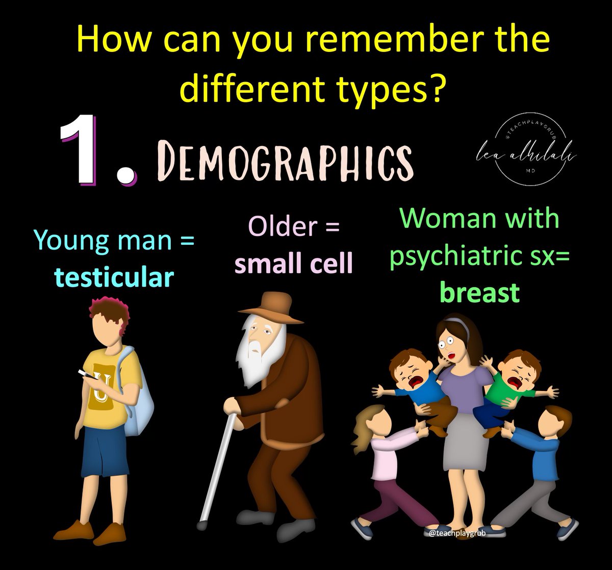
You can also remember the causes by the demographic:
🔸Young man: testicular
🔸Older: Small cell
🔸Woman with psychiatric symptoms: breast Image
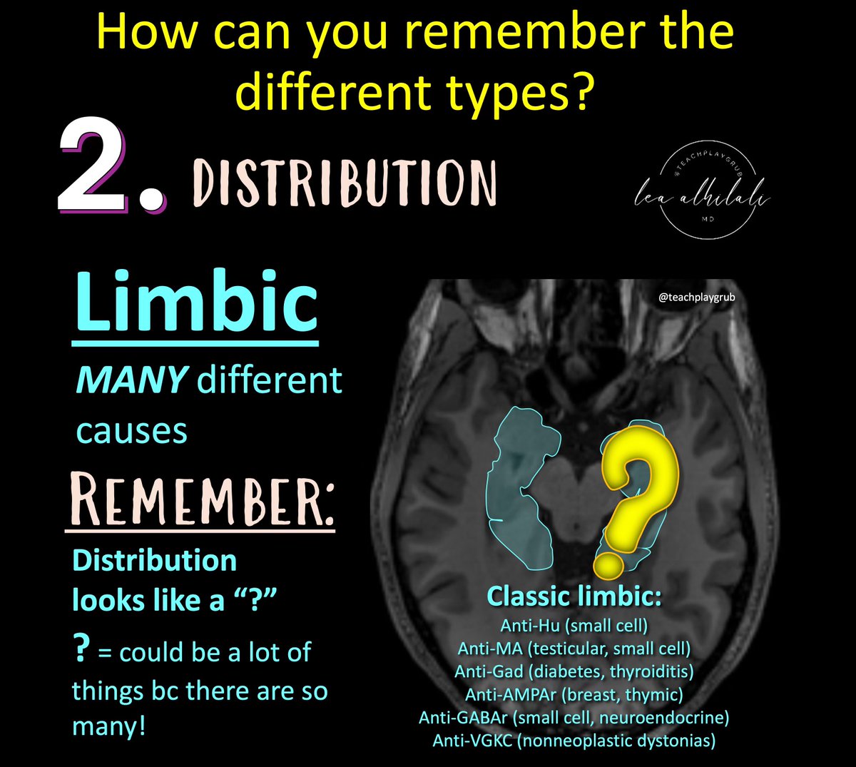
3/Limbic encephalitis is the most common pattern

But it has many, many different causes

Remember--limbic involvement is shaped like a question mark!

So for limbic encephalitis, the cause remains a question bc the differential is so broad

Must question & clinically correlate! Image
4/Some other patterns to remember:

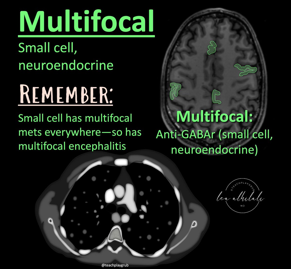
Multifocal = small cell and neuroendocrine

Remember:
🔸Small cell gives you mets everywhere = small encephalitis everywhere
🔸Neuroendocrine can arise from different places all over your body = encephalitis different places all over your brain Image
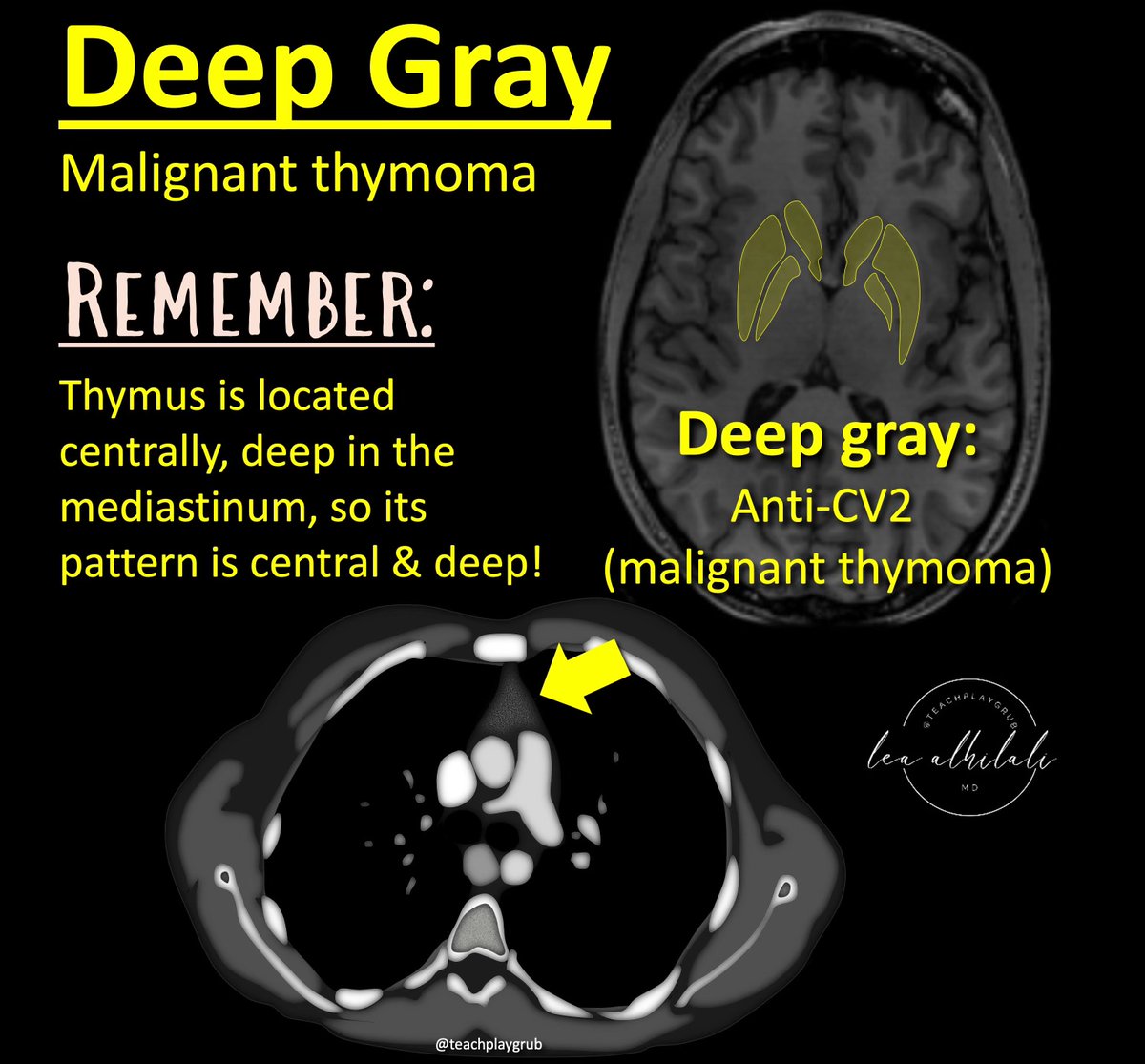
5/Central/Deep gray = malignant thymoma

Remember:
Thymus is located central and deep in your chest, so its pattern is central and deep! Image
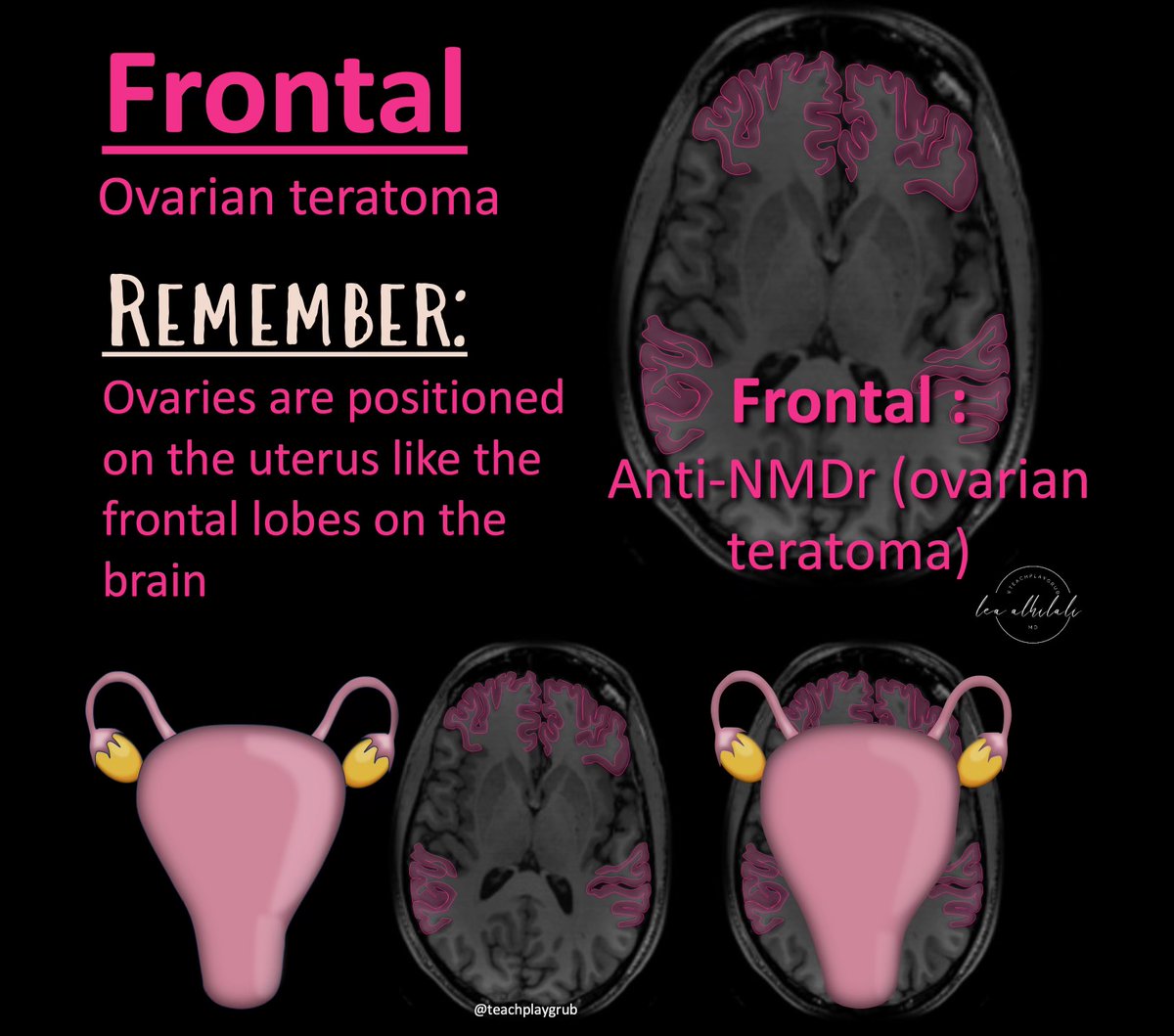
6/Frontal = ovarian teratoma

Remember:
Ovaries are situated on the uterus right where the frontal lobes are situated on the brain! Image
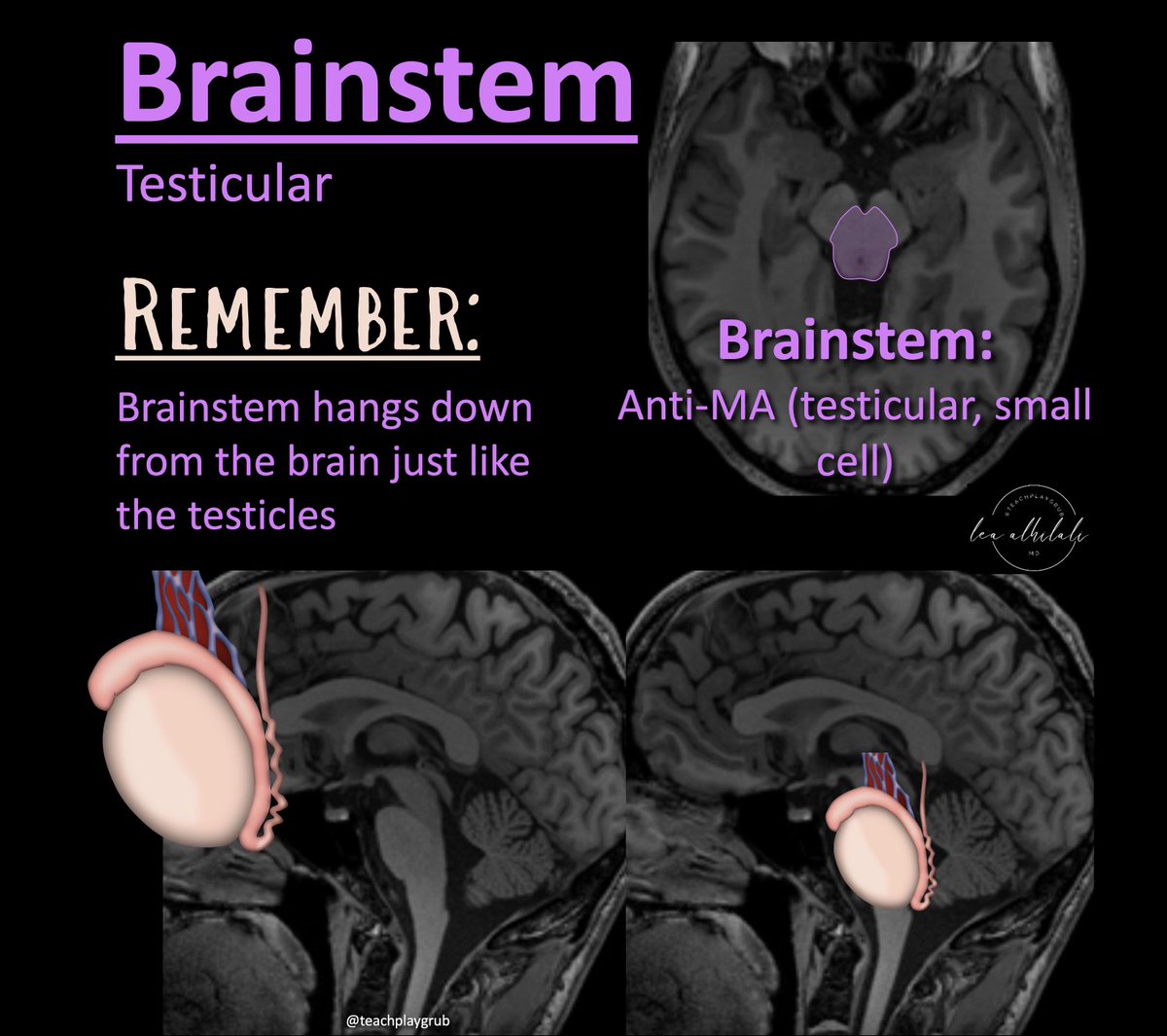
7/Brainstem = testicular

Remember:
Brainstem hangs down from the cerebrum like the testicles

Hopefully, these hints will help you, so that the next time you see autoimmune encephalitis, you can be on autopilot! Image

• • •

Missing some Tweet in this thread? You can try to force a refresh
 

Keep Current with Lea Alhilali, MD

Lea Alhilali, MD Profile picture

Stay in touch and get notified when new unrolls are available from this author!

Read all threads

This Thread may be Removed Anytime!

PDF

Twitter may remove this content at anytime! Save it as PDF for later use!

Try unrolling a thread yourself!

how to unroll video
  1. Follow @ThreadReaderApp to mention us!

  2. From a Twitter thread mention us with a keyword "unroll"
@threadreaderapp unroll

Practice here first or read more on our help page!

More from @teachplaygrub

Jul 23
1/To call it or not to call it? That is the question!

Do you feel a bit wacky & wobbly when it comes to calling normal pressure hydrocephalus on imaging?

You don’t want to overcall it, but you don’t want to miss it either!

Let me help you out w/a thread about imaging in NPH! Image
2/First, you must understand the pathophysiology of “idiopathic” or iNPH.

It was first described in 1965—but, of the original six in the 1965 cohort, 4 were found to have underlying causes for hydrocephalus.

This begs the question—when do you stop looking & call it idiopathic? Image
3/Thus, some don’t believe true idiopathic NPH exists.

After all, it’s a syndrome defined essentially only by response to a treatment w/o ever a placebo-controlled trial.

However, most believe iNPH does exist--but underlying etiology is controversial. Several theories exist Image
Read 19 tweets
Jun 27
1/Blast from the past!

Sometimes to be next gen, you gotta to go old school!

Cutting edge pituitary imaging must be MRI, right?

Or can we go back to the future w/CT?

Here’s the latest in pituitary imaging in this month’s @theAJNR SCANtastic!

ajnr.org/content/45/6/7…
Image
2/Pituitary imaging is actually very difficult.

First challenge is the small size of the gland & even smaller adenomas, requiring high resolution.

And the difference between adenomas & the gland is subtle—both enhance, but adenomas enhance SLIGHTLY less Image
3/The difference in enhancement is transient & ends quickly

So pituitary imaging must be done dynamically to catch this small window of difference

So we have to do very high resolution imaging very quickly—the worst of both worlds! Image
Read 12 tweets
Jun 21
1/”I LOVE spinal cord syndromes!” is a phrase that has NEVER, EVER been said by anyone.

Do you become paralyzed when you see cord signal abnormality?

Never fear—here is a thread on all the incomplete spinal cord syndromes to get you moving again! Image
2/Spinal cord anatomy can be complex.

On imaging, we can see the ant & post nerve roots.

We can also see the gray & white matter.

Hidden w/in the white matter, however, are numerous efferent & afferent tracts—enough to make your head spin. Image
3/Lucky for you, for the incomplete cord syndromes, all you need to know is gray matter & 3 main tracts

Anterolaterally, spinothalamic tract (pain & temp). Posteriorly, dorsal columns (vibration, proprioception, & light touch), & next to it, corticospinal tracts—providing motor Image
Read 20 tweets
Jun 19
1/”Tell me where it hurts.”

How back pain radiates can tell you where the lesion is—if you know where to look!

Remembering lumbar radicular pain distributions can be back breaking work--but here's a thread to help you! Image
2/Let’s start with L1.

L1 radiates to the groin.

I remember that b/c the number 1 is, well, um…phallic.

So the phallic number 1 radiates to the groin. Image
3/Let’s skip to L3 for a second.

I remember L3 is to the knee—easy, it rhymes! Image
Read 8 tweets
Jun 10
1/Do you know all the aspects of, well, ASPECTS?

Many know the anterior circulation stroke system—but posterior circulation (pc) ASPECTS is often left behind

25% of infarcts are posterior circulation

Do you know pc-ASPECTS?!

Here’s a thread to help you remember pc-ASPECTS Image
2/Many know anterior circulation ASPECTS.

It uses a 10-point scoring system to semi-quantitation the amount of the MCA territory infarcted on non-contrast head CT

If you need a review: here’s my thread on ASPECTS:
Image
3/But it’s only useful for the anterior circulation.

Posterior circulation accounts for ~25% of infarcts

Even w/recanalization, many of these pts do poorly bc of the extent of already infarcted tissue

So there’s a need to quantitate the amount of infarcted tissue in these pts Image
Read 8 tweets
May 29
1/Waving the white flag when it comes to white matter anatomy?

Turns out white matter anatomy isn’t black & white!

This months @theAJNR SCANtastic is the white knight you need to rescue you!

Here’s the white matter anatomy you NEED to know!

ajnr.org/content/45/5/5…
Image
2/Gray matter or cortical functional anatomy is well known.

Everyone knows the motor & sensory strips. Most know Broca’s & Wernicke’s

But most forget that white matter has similar functional topography & just bc it’s white matter doesn’t mean it doesn’t have function! Image
3/But too often we don’t refer to this white matter functional anatomy.

Instead we use general terms like “corona radiata”

But that’s the equivalent of using the word “body.”

Just like the body has many different systems in it, so does the corona radiata! Image
Read 12 tweets

Did Thread Reader help you today?

Support us! We are indie developers!


This site is made by just two indie developers on a laptop doing marketing, support and development! Read more about the story.

Become a Premium Member ($3/month or $30/year) and get exclusive features!

Become Premium

Don't want to be a Premium member but still want to support us?

Make a small donation by buying us coffee ($5) or help with server cost ($10)

Donate via Paypal

Or Donate anonymously using crypto!

Ethereum

0xfe58350B80634f60Fa6Dc149a72b4DFbc17D341E copy

Bitcoin

3ATGMxNzCUFzxpMCHL5sWSt4DVtS8UqXpi copy

Thank you for your support!

Follow Us!

:(