This preprint (from April 5) is FASCINATING. Someone requested I have a look at it, and I'm glad they did, because it made SO MANY separate things suddenly make sense.

Here's a look at how a specific type of MICROCLOTS may be associated with LONG COVID pathology...

1/many Preprint posted April 5, 2024: "Increased fibrinaloid microclot counts in platelet-poor plasma are associated with Long COVID"  Abstract: "...The pathophysiology underlying Long COVID remains unclear but appears to involve multiple mechanisms including persistent inflammation, coagulopathy, autoimmunity, and organ damage. Studies suggest that microclots, also known as fibrinaloids, play a role in Long COVID. In this context, we developed a method to quantify microclots and investigated the relationship between microclot counts and Long COVID. We show that as a cohort, platele...
This thread will have three things:
- Takeaways of this study
- Breakdown of the method they developed
- How these findings connect with other known patterns

This is a study that looks at some rugged little blood clots that come courtesy of the SARS-CoV-2 spike protein.

2/ "Studies suggest that microclots, also known as fibrinaloids, play a role in Long COVID by blocking capillaries, limiting oxygen exchange, and potentially causing microvascular pulmonary thrombosis and multiple organ failure. Amyloid-containing deposits, resembling microclots, have also been observed in increased numbers in muscle tissue biopsies from people with Long COVID compared to samples from controls, and these numbers increase after exercise, an important observation as many people with Long COVID experience post-exertional symptom exacerbation after activity. However, the depo...
Some important background info:

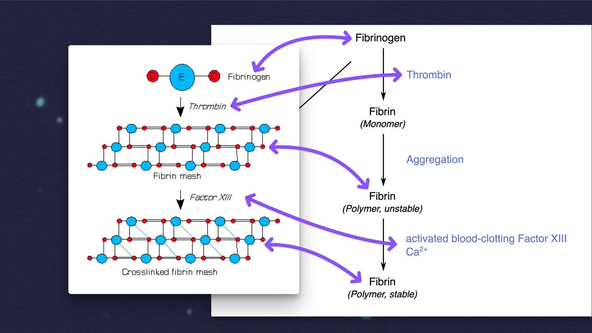
- FIBRINOGEN is a protein that just kinda hangs out in the blood, waiting.

- FIBRIN is a fibrous protein that forms a mesh to hold platelets together to form a blood clot.

- THROMBIN is the enzyme that converts FIBRINOGEN into FIBRIN.

3/ Fibrinogen + Thrombin = Fibrin (monomer)  The fibrin monomers aggregate to form a mesh.  Fibrin mesh + clotting factor = cross-linked mesh that's much more stable, and which contracts tightly.  Image source: https://commons.wikimedia.org/wiki/File:Stabilisation_de_la_fibrine_par_le_factor_XIII.png
If you add thrombin to blood plasma "from people with various chronic inflammatory and neurodegenerative diseases," anomalous microclots can form that are "more resistant to breakdown than normal clots."

These can also be induced JUST by adding the spike protein to plasma.

4/ "Fibrinaloid microclots can be induced by adding thrombin in vitro to plasma samples from people with various chronic inflammatory and neurodegenerative diseases such as Alzheimer’s, Parkinson’s, type 2 diabetes and rheumatoid arthritis. Following considerable work using electron microscopy (that uncovered ‘dense matted deposits’), it was demonstrated that tiny amounts (1 molecule per 100,000,000 fibrinogen molecules) of bacterial lipopolysaccharide causes blood to clot into an anomalous ‘amyloid’ type form... These ‘microclots’ are significantly more resistant to breakdown than normal...
These microclots may impair oxygen exchange in various ways, which would lead to a cascade of issues, and it's a plausible explanation for why PEM occurs. If the virus directly enters pulmonary vasculature through the mouth, these spike protein clots could be a huge problem.

5/ "Studies suggest that microclots, also known as fibrinaloids, play a role in Long COVID by blocking capillaries, limiting oxygen exchange, and potentially causing microvascular pulmonary thrombosis and multiple organ failure. Amyloid-containing deposits, resembling microclots, have also been observed in increased numbers in muscle tissue biopsies from people with Long COVID compared to samples from controls, and these numbers increase after exercise,...  An alternative hypothesis based on the radiological evidence of lung changes in acute COVID suggests that the oral cavity may provide...
In this study, they developed a new method to measure the microclot counts for individuals, then demonstrated "that as a cohort, samples from people with Long COVID have a higher mean microclot count compared to samples from control groups."

6/ "The primary goal of this study was to investigate the relationship between the microclot count present in PPP, and the occurrence of Long COVID. To determine microclot counts for individuals, we have developed a robust, medium-throughput assay using automated image analysis. In addition, relationships between microclots counts and sex, age, body mass index (BMI), time since COVID-19 infection, and symptoms of Long COVID were investigated. We demonstrate that as a cohort, samples from people with Long COVID have a higher mean microclot count compared to samples from control groups. We ...
What are the takeaways?

- Microclot counts are raised in the LC cohort compared to controls
- SARS-CoV-2 infection raises microclot counts, which "take several months to return to control levels."
- Microclots may be a biomarker to screen, or even a treatment target.

7/ "... Significant differences were seen between the COVID controls and Long COVID individuals (** = p < 0.01), between the uninfected controls and Long COVID individuals (**** = p < 0.0001) and between both groups of controls and individuals who had tested positive for COVID in the previous three weeks (**** = p < 0.0001, *** = p < 0.001).  ... We discovered that microclot counts were raised in Long COVID samples compared to controls and that recent COVID-19 cases also had raised counts. However, we also observed that not all people with Long COVID have microclot counts above levels obs...
What is the new method they devloped? It's a way to screen for microclots, and it specifically addresses a few potential issues with past studies.

In particular, the type of slidse typically used often have specks of reflective material about the size of the microclots!

8/ "Fibrinaloid microclots are known to consist of amyloid-like material and, hence, can bind to the amyloid-specific dye ThT. This dye intercalates with the Beta-sheet structure of amyloid material with a resultant shift in excitation emission and quantum yield. Existing assays have used a blood smear approach .... During the development of our method, we identified several processing issues that affected the observed microclot counts with this method. Firstly, we noticed that auto-fluorescent material was visible on untreated glass slides, comparable in size to previously reported micro...
Critically, they also found that leaving the blood tubes sitting around for an hour before separating out the components affected the microclot count, as did repeated freeze-thaw cycles.

So, they designed a method that was quick and had as little handling as possible.

9/ "We also identified processing issues relating to the handling and storage of the blood samples. We established that when whole blood tubes were left standing for more than an hour before centrifugation, the resulting PPP showed reduced or no microclot count. After observing this effect, to ensure the accuracy of our analysis, we only used PPP samples that were processed within 30 minutes of collection. Additionally, repeated freeze-thaw cycles of the PPP following processing also resulted in alterations in microclot counts. As such, PPP was stored in aliquots, and analysis was carried...
In this method, they're able to keep the blood samples in little wells, rather than smearing them on a slide. An imaging module focuses at different depths to capture a multi-layered image, and the microclots are then automatically identified and counted.

10/ "To optimise the efficiency of our process and minimise manual handling, we used 15-well µ-Slides that come pre-sealed and were handled in a dust-free flow cabinet. We also used low protein binding plasticware at all stages. Each PPP sample was imaged directly in the well of the slide, allowing us to obtain 3D images within approximately 10 minutes per well. To minimise variability, we took triplicate technical repeats and analysis parameters were set to automatically identify the boundaries of the well to remove edge effects (Figure 1). Figure 1A shows the captured image, whereas Figu...
Importantly, they did repeat data collection and found that they got consistent results up to six hours after initial data collection, which means it can be done with an autosampler.

This method seems robust both for research and, potentially, diagnostics.

11/ "...obtain 3D images within approximately 10 minutes per well. To minimise variability, we took triplicate technical repeats and analysis parameters were set to automatically identify the boundaries of the well to remove edge effects (Figure 1). Figure 1A shows the captured image, whereas Figure 1B shows the processed image and associated microclot counts, automatically identified by the software. Control experiments and repeat data collection on the same slide gave consistent results up to six hours after sample handling, allowing the use of an autosampler for medium throughput. The r...
What was found, specifically?

LC group has significantly higher clot counts compared to both uninfected controls and infected-but-no-LC controls.

Interestingly, there was ALSO significant difference in clot counts between the "recent COVID" group and other control groups.

12/ "Microclots in Long COVID samples vs control samples  Having developed a robust assay for the quantification of microclots in PPP samples, we determined the distribution of microclot counts in each of the four groups. Figure 2 shows the average microclot count for each individual. Within the groups, mean microclot counts can be determined for each of the cohorts with the Uninfected Control (mean 13.6 ÷ S.D. 7.4), COVID Control (20.7 ÷ 10.1), Long COVID (40.1 ÷ 28.1) and Recent COVID (50.5 ÷ 20.4) groups. Significantly higher mean microclot counts were observed in the Long COVID group c...
It's very notable that "only one [control] had a microclot count >50 while approximately half the LC group had counts above this level."

Also very interesting: there was a statistically significant difference in LC vs. control for microclot counts for women, but not men.

13/ "Strikingly, within the data is that only one outlier in the control groups had a microclot count >50 while approximately half the Long COVID group had counts above this level. However, there is overlap between the groups; although microclots are detectable in all samples from people with Long COVID, in about half of the samples the counts are similar to the counts observed in samples from controls.  Pooling the data from the two control groups, Figure 3 shows that female Long COVID participants had a significantly higher mean microclot count than female controls p < 0.0001 whereas no ...
The difference in immune response seen between men and women may pattern with what has been observed elsewhere (including a stronger innate immune response in men, but a more robust adaptive response in women).

14/ "Our study also found that females had more microclots, which is consistent with the fact that females are more affected by Long COVID. Most Long COVID patients experience severe fatigue, with the female sex group as an independent risk factor in alignment with our symptom data Female COVID-19 patients exhibit more robust T cell activation, in contrast, male patients have higher levels of innate immune cytokines and a greater presence of non-classical monocytes, potentially accounting for the sex differences in disease outcomes. Males, but not females, who have recovered from COVID-19 ...
Very notably, the "Recent COVID samples have microclot counts comparable to" around the *top 25% of the LC group!*

"Together, these data indicate that exposure to SARS-CoV-2 initially increases the microclot count..., but these microclots are cleared over time."

15/ "Recent COVID samples have microclot counts comparable to the upper quartile of the Long COVID group, indicating that recent exposure to the virus leads to higher microclot counts (Figure 2). Within the control group where we have the dates of a confirmed infection from either lateral flow of PCR a significant time dependent decrease in microclot count was observed (Figure 4, r = -0.628, p < 0.0001). By ~450 days post-infection, all samples were within the same range as the COVID control group (20.7 + 10.1). This indicates that the control group have a decrease in microclot counts to b...
IMO, this finding is a really strong explanation as to why there seems to be an increased risk of cardiac incidents in the 3-6 months following a SARS-CoV-2 infection: The presence of the spike protein may be creating microclots throughout the cardiovascular system!

16/
"Recent COVID samples have microclot counts comparable to the upper quartile of the Long COVID group, indicating that recent exposure to the virus leads to higher microclot counts (Figure 2). Within the control group where we have the dates of a confirmed infection from either lateral flow of PCR a significant time dependent decrease in microclot count was observed (Figure 4, r = -0.628, p < 0.0001). By ~450 days post-infection, all samples were within the same range as the COVID control group (20.7 + 10.1). This indicates that the control group have a decrease in microclot counts to b...
Figure 4 - Microclot count as a function of time since infection within the COVID control and recent COVID groups combined. Participants reported the date that they first experienced symptoms during their most recent COVID infection. A Pearson rank correlation showed a significant inverse relationship between microclot count and date of previous COVID infection (r = -0.628 and p < 0.0001).
How did the microclots relate to symptoms? Their questionnaire was basic, but they generally found the symptoms you'd expect being prominent in the LC group: fatigue, post-exertional malaise, difficulties concentrating, etc.

However, high microclots didn't GUARANTEE LC!

17/
"The self-reported symptom scores indicate that over 93% of the group experienced "Feeling tired or having low energy", 84% experienced symptoms triggered by physical, mental, or emotional effort, 93% experienced symptoms that occur one or two days after physical, mental, or emotional effort, and 93% experienced difficulty concentrating. These symptoms were experienced at a higher rate than other symptoms (figure 5). These symptoms are typical of post-exertional symptom exacerbation (PESE) and persistent fatigue, which are strongly associated with Long COVID. Using this sympt...
Figure 5 – Relationship of microclot counts to symptoms. Long COVID participants rated how much they had been affected by a range of symptoms, and were then split by group based on their response from “not bothered” green, “bothered a little” blue to “bothered a lot” orange. Individuals’ microclot counts were then plotted against these groups. Figures are ordered based on the percentage of participants reporting to be ‘bothered a lot’ by a symptom. Comparative analyses across study groups were conducted employing the Kruskal-Wallis tests to ascertain statistical differences, utilising PRISM...
So, do microclots cause LC? I mean, they probably don't *help*.

One possible explanation is that LC may reflect a persistent dysregulated state (e.g. a "pro-coagulable state"), and external intervention would be required to shift things back to normal.

18/ "It is not fully understood how microclotting could relate to the development of Long COVID symptoms. One theory is that the balance of 'clotting vs anticlotting' (i.e. fibrin formation vs fibrinolysis) is shifted towards clotting by SARS-COV2 infection. The longitudinal data presented here suggests that this 'balance' can be restored within 6 months in those that recover. However, a subgroup of people may have persistent microclots due to ongoing inflammation e.g. due to viral persistence or abnormal immune response, possibly combined with ineffective fibrinolysis, leading to a pro-co...
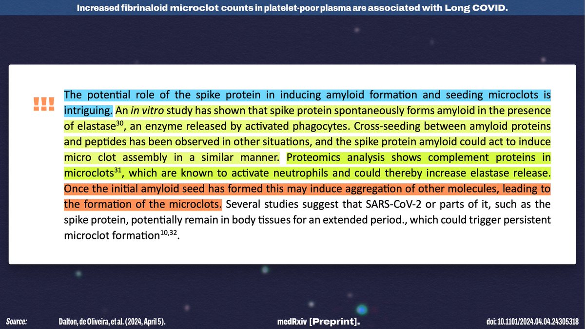
I think the "potential role of the spike protein in inducing amyloid formation and seeding microclots" is a FASCINATING possibility—especially given that there seems to be the possibility of a runaway process of consistent clotting.

(It may also explain vCJD!)

19/ "The potential role of the spike protein in inducing amyloid formation and seeding microclots is intriguing. An in vitro study has shown that spike protein spontaneously forms amyloid in the presence of elastase, an enzyme released by activated phagocytes. Cross-seeding between amyloid proteins and peptides has been observed in other situations, and the spike protein amyloid could act to induce micro clot assembly in a similar manner. Proteomics analysis shows complement proteins in microclots, which are known to activate neutrophils and could thereby increase elastase release.  Once t...
The authors also note that, since this is a new line of research, we have no idea what role microclots may play in other conditions—but they're beginning by investigating whether pwME have similar microclot profiles to pwLC!

I'm also very curious how EBV impacts clotting!

20/ "We did not investigate samples from people with other acute viral infections, and it is possible that microclots may be raised in response to other infections. This has implications for other post-viral conditions such as myalgic encephalomyelitis (ME), often associated with infection by Epstein-Barr virus, and we are currently investigating whether people with ME have similar microclot profiles to people with Long COVID."
Of course, this isn't the only way that the SARS-CoV-2 spike protein has been shown to cause cardiac damage: It may also lead to fusion of uninfected cells!

21/
Overall, the implications are significant. At the very least, I think this is a plausible explanation for why there are often cardiac issues *in the time period shortly after infection,* even if it doesn't explain LC!

Source: (h/t @JPeaceJPeace)

22/22medrxiv.org/content/10.110…

• • •

Missing some Tweet in this thread? You can try to force a refresh
 

Keep Current with Nick #RespiratorsFilterPathogens😷 Anderegg

Nick #RespiratorsFilterPathogens😷 Anderegg Profile picture

Stay in touch and get notified when new unrolls are available from this author!

Read all threads

This Thread may be Removed Anytime!

PDF

Twitter may remove this content at anytime! Save it as PDF for later use!

Try unrolling a thread yourself!

how to unroll video
  1. Follow @ThreadReaderApp to mention us!

  2. From a Twitter thread mention us with a keyword "unroll"
@threadreaderapp unroll

Practice here first or read more on our help page!

More from @NickAnderegg

Oct 9
NEW STUDY! This exploratory study identifies a SPECIFIC PHENOTYPE OF LONG COVID that appears related to NEUROMUSCULAR DISTURBANCE rather than lung damage—and they've termed it Complex Ventilatory Dysfunction!

Breakdown of the paper (thread written for a general audience!)...

1/ Published Oct 7, 2024: "A new phenotype of patients with post-COVID-19 condition is characterised by a pattern of complex ventilatory dysfunction, neuromuscular disturbance and fatigue symptoms"  Abstract:  Background Patients with post-COVID-19 condition frequently suffer from chronic dyspnoea. The causes and mechanism for dyspnoea in these patients without evidence of structural lung disease are unclear.  ...  Results ... A pattern of reduced forced vital capacity (FVC), but normal total lung capacity (TLC), termed complex ventilatory dysfunction ... was observed and occurred mo...
Broadly speaking, there are two groups of acute covid outcomes involving dyspnea (shortness of breath) as a long-term symptom:

- Severe cases that may have physical lung damage
- "Mild" cases that now have ME/CFS-like features, but who have no evidence of lung damage!

2/ "Current evidence suggests that cellular damage, a robust innate immune response with inflammatory cytokine production and a procoagulant state induced by SARS-CoV-2 infection are factors potentially contributing to post-COVID-19 sequelae such as dyspnoea, fatigue, and cognitive and mental disturbances... Dyspnoea has been well characterised as a major clinical symptom of post-COVID condition after severe and critical COVID-19 and is correlated with impaired lung function in terms of pulmonary restriction, and with reduced diffusion capacity as a possible consequence of pulmonary remod...
In this study, they explored this distinction further and identified a distinct subset of patients with a pattern of breathing abnormality that they have termed complex ventilatory dysfunction (CVD).

So how did they arrive at this conclusion? Let's dig in!

3/16 "We hypothesise that patients suffering from post-COVID-19 condition who have fatigue and exertional intolerance also have a reduction in respiratory muscle strength, causing a dysfunctional breathing pattern which is distinct from typical pulmonary sequelae after COVID-19 such as obstruction, restriction or impaired diffusion capacity. Based on clinical observations, we describe a new breathing abnormality termed complex ventilatory dysfunction (CVD), defined as total lung capacity (TLC) - forced vital capacity (FVC) >10% predicted value and absence of restriction (TLC ≥ lower limit o...
Read 16 tweets
Sep 22
NEW STUDY! It VERY thoroughly supports the hypothesis that SARS-CoV-2 emerged as a zoonotic spillover event in the Huanan Seafood Wholesale Market—using multiple methods!

Breakdown of the paper (written for a general audience!)...

1/many (but it's worth it, I promise!) Published Sep 19, 2024 in Cell: "Genetic tracing of market wildlife and viruses at the epicenter of the COVID-19 pandemic"  Highlights: - Common ancestor of SARS-CoV-2 linked to Huanan market matches the global common ancestor - Wildlife mitochondrial DNA identified in samples from stalls positive for SARS-CoV-2  Abstract:  "... We demonstrate that market-linked severe acute respiratory syndrome coronavirus 2 (SARS-CoV-2) genetic diversity is consistent with market emergence and find increased SARS-CoV-2 positivity near and within a wildlife stall. We identify wildlife DNA in...
This paper reanalyzes the same data from the April 2023 paper in Nature that cast doubt on the Huanan Market hypothesis (pictured).

In the new paper published in Cell this week, another group conducted far more detailed (and statistically sound) analyses!

2/
Original paper that analyzed this same data: "Surveillance ofSARS-CoV-2 at the Huanan Seafood Market"  "Severe acute respiratory syndrome coronavirus 2 (SARS-CoV-2), the causative agent of coronavirus disease 2019, emerged in December 2019. Its origins remain uncertain. It has been reported that a number of the early human cases of coronavirus disease 2019 had a history of contact with the Huanan Seafood Market...."
"...It should be noted that the selection of shops for sampling was biased because shops selling wildlife as well as shops linked to early cases were prioritized for sampling. The origin of the virus cannot be determined from the analyses available so far. Although gene barcode analysis of animal species in the study suggested that Myotis, Nyctereutes and Melogale-species that have been recognized as potential host species of sarbecoviruses-were present at the market, these barcodes were mostly detected within the SARS-CoV-2 RT-qPCR-negative samples from the environment. It remains pos...
This new paper starts by reviewing the evidence supporting the Huanan Market hypothesis, and some of the details are FASCINATING!

To begin with, of the 174 COVID cases identified with an onset of December 2019, 32% had a link to the Huanan Market.

In a city of 12 million.

3/ "INTRODUCTION Many of the earliest known cases of COVID-19 worked at or visited the Huanan Seafood Wholesale Market ("Huanan market") in the city of Wuhan, a link first made by clinicians at different hospitals throughout the city. Retrospective review of early COVID-19 cases identified 174 patients with onset in December 2019, 32% of whom had an ascertained link to this location, within a city of over 12 million."
Read 24 tweets
Sep 10
Want to see 13 academic cry-bullies throw a hilarious, peer-reviewed tantrum?

The real gold is in the 943-word "Competing Interests" section!

I also discovered that ONE OF THE AUTHORS WROTE HIS OWN WIKIPEDIA PAGE 🤣🤣

Thread...

1/19
Zero-covid advocacy during the COVID-19 pandemic: a case study of views on Twitter/ X  by Kasper P. Kepp, Kevin Bardosh, Tijl De Bie, Louise Emilsson, Justin Greaves, Tea Lallukka, Taulant Muka, J. Christian Rangel, Niclas Sandström, Michaéla C. Schippers, Jonas Schmidt-Chanasit & Tracy Vaillancourt
"The advocacy, although timely and informative, often appealed to emotions and values using anecdotes and strong criticism of authorities and other scientists."  So what's the problem? The rest of this sentence is just tone policing and/or paternalism.   "Risks were emphasized about children’s vulnerability, Long COVID, variant severity, and Mpox, and via comparisons with human immunodeficiency viruses (HIV)."  Why is this being framed as a bad thing if the advocacy is timely and informative?  "Far-reaching policies and promotion of remedies were advocated without s...
Kasper P. Kepp "has been engaged in the pandemic debate in Danish media and social media, where he has been critical of the studied zero-covid groups"

It's wildly unethical to conduct a study *specifically* targeting entities you've personally had conflict with.

2/ Ethics declarations  Competing interests  The authors do research in public health, epidemiology, biochemistry, virology, biostatistics, policy, politics, education and student experience, pediatrics, mathematical modeling, data science, and psychology relevant to the claims made by the studied advocacy in the paper but with no direct association to the studied advocacy.  Kasper P. Kepp has unpaid research affiliations with METRICS, Stanford, and Epistudia, Bern, has published or submitted a dozen papers on COVID-19-related research (SARS-CoV-2 mutation evolution, public health, and epidemi...
"Kevin Bardosh is Director of Collateral Global, a UK-based research and education charity that is focused on understanding the impact of COVID policies around the world"

Let's have a look at the latest news from Collateral Global! Hmmm maybe not a neutral source either?

3/

Kevin Bardosh is Director of Collateral Global, a UK-based research and education charity that is focused on understanding the impact of COVID policies around the world, and has been active in the pandemic debate on social media and in the popular press.
News:  - Record levels of speech and language problems among youngsters linked to lockdown - Zuckerberg censorship revelation tip of "widespread and chilling" silencing of Covid science - New Covid school closures condemned by scientists  Latest podcasts, all staring Kevin Bardosh:  - What happened in California? Missing science and murky emergency laws during Covid - Covid Models: What can Philosophy teach us? - Pandemic Panic: Civil Liberties in Canada during Covid
10 WAYS THE COVID RESPONSE HARMED SOCIETY:  - Episode 10 Governance - 10 Ways the Covid Response Harmed Society - Episode 9 Environment and Ecosystems - 10 Ways the Covid Response Harmed Society - Episode 8 Community - 10 Ways the Covid Response Harmed Society  IN THE PRESS  - We still need to reckon with the folly of lockdown - Did the Covid inquiry just admit lockdown was a mistake? - The lesson they're determined to ignore: lockdown was a disaster, writes infectious diseases expert Kevin Bardosh
Read 25 tweets
Sep 9
NEW PREPRINT! Another study about ABNORMAL BLOOD CLOTTING related to SARS-CoV-2, but unlike the others I've covered, this isn't related to the spike protein.

Turns out that Mpro, a viral protease [pro-tee-ace], can START the cascade.

Thread (written for everybody!)...

1/many bioRxiv preprint posted Sept 5, 2024: "The Main protease (Mpro) from SARS-CoV-2 triggers plasma clotting in vitro by activating coagulation factors VII and FXII."  "Here we show that the SARS-CoV-2 main protease (Mpro) can play a direct role in the activation of the coagulation cascade. Adding Mpro to human plasma from healthy donors increased clotting probability by 2.5-fold. The results of enzymatic assays and degradomics analysis indicate that Mpro triggers plasma clotting by proteolytically activating coagulation factors zymogens VII and XII at their physiological activat...
Here's the takeaway: The *Main protease* (Mpro) of SARS-CoV-2—an enzyme that cuts up viral polyproteins—can also cleave a few host coagulation factors in a way that ACTIVATES them and BEGINS the blood clot cascade.

So that's uh... that's not ideal.

2/ "In conclusion, in this work we provided several pieces of experimental evidence showing that Mpro can induce plasma clotting by proteolytically activating FVII and FXII, which in turn can initiate the extrinsic and intrinsic pathways of blood coagulation, with a final pro-coagulant effect. This non-canonical mechanism highlights a possible novel function of Mpro in vivo that, in addition to the 'cytokine and bradykinin storm' mechanism, can contribute to the pathogenicity of SARS-CoV-2 in COVID-19."  ---  Activates blood clotting factors that are at the beginning of the coagulati...
Now, let's look at how they figured it out!

Mpro plays an important role in the self-replication process of the virus, but we also ALREADY know that Mpro (and the other protease of SARS-CoV-2) can also modify the cellular machinery of its host cell to evade defenses.

3/ "...R1AB assumes the crucial role of generating the non-structural proteins forming the replicase-transcriptase complex, essential for the RNA-synthesizing machinery. During viral maturation, two key proteases encoded in the R1AB gene, i.e. the main protease Mpro (also known as 3CL protease nsp5) and the papain like PLpro nsp protease, cleave the replicase polyprotein R1AB promoting the assembly of the replicase-transcriptase complex that encodes for the four structural proteins, i.e. the envelope (E), membrane (M), spike (S) and nucleocansid (N) proteins. ... Mpro and PLpro are pleiot...
Read 23 tweets
Aug 30
Whenever I summarize a research paper about the SARS-CoV-2 spike protein, people always ask if the S proteins from the vaccines will do the same thing. It's a fair question!

mRNA vaccines are MUCH less likely to cause spike-related problems than an infection! Here's why...

1/
First, the spike protein used in the mRNA vaccines isn't the same as the spike protein on the actual virus! The US-approved mRNA vaccines (and Novavax) use a stabilized version of the protein that DOES NOT cause many of the issues that the wild SARS-2-S protein does!

2/ Image
But let's ignore the difference in spike design. What is the difference in QUANTITY?

We can do some fairly simple back-of-the-envelope calculations, using numbers pulled from the scientific literature!

3/
Read 11 tweets
Aug 26
NEW STUDY! This one confirms A LOT of other results.

Turns out, SARS-CoV-2 infection can cause lasting EPIGENETIC MODIFICATIONS, which effectively results in ACCELERATED BIOLOGICAL AGING.

Let's dive in! (Breakdown thread written for a general audience...)

1/many Published Aug 20, 2024 in "Clinical Epigenetics": "Epigenetic patterns, accelerated biological aging, and enhanced epigenetic drift detected 6 months following COVID-19 infection: insights from a genome-wide DNA methylation study"  ---  "Our study provides valuable insights into the epigenetic consequences of COVID-19. Results suggest possible associations with accelerated aging, epigenetic drift, and the disruption of critical biological pathways linked to insulin resistance, immune response, and vascular health. Understanding these epigenetic changes could be cruc...
This one is dense, so I'll start with the takeaways, then some background, then the details.

The authors conclude that SARS-CoV-2 exposure may have "associations with aging, [stochastic epigenetic mutation] accumulation, and dysregulation in critical pathways."

2/ "In conclusion, these results provide comprehensive insights into the epigenetic consequences of SARSCoV-2 exposure after 6 months, emphasizing potential associations with aging, SEM accumulation, and dysregulation in critical pathways linked to insulin resistance, immune response, and vascular function."
Between the post-COVID group and the healthy controls, they found differences in epigenetic markers for genes related to a few different pathways, including:

- Hypoxia and vascular maintenance
- Insulin resistance
- Cell death signalling
- T-cell signaling and activation

3/


"Moreover, to assess epigenetic drift at the gene level (Gene-EML), we employed a sequence kernel association test (SKAT). Designed initially for rare variant studies, this method has recently found applications in other areas like copy number variations (CNVs) and epigenetic modifications. It has been widely used in numerous studies aiming to identify genetic associations with diseases such as Alzheimer's disease, schizophrenia, and autism spectrum disorder, amyotrophic lateral sclerosis. This analysis has identified a list of genes that exhibit significantly different epigenetic drif...
"The "VEGF signaling pathway" and "Hypoxia response via HIF activation" pathways have been associated with COVID-19 due to their involvement in vascular dysfunction and inflammation observed during disease progression. Vascular endothelial growth factor (VEGF) plays a crucial role in angiogenesis and regulates various activities such as vascular permeability, cell migration, proliferation, and survival. Hypoxia, or low oxygen conditions, can activate the hypoxia-inducible factor (HIF), a key regulator in the response to hypoxia. The concurrent activation of HIF and ...
"Moreover, pathways like "Insulin Resistance" and the "Insulin/IGF pathway-protein kinase B signaling cascade" could become relevant due to COVID-19's association with various metabolic alterations, such as impacts on insulin sensitivity and glucose metabolism. Studies have shown that COVID-19 patients, even those with mild cases, may experience increased insulin resistance, which can persist long after the acute phase of the infection.  Another attractive deregulated pathway is represented by the "Apoptosis signaling pathway"; this pathway plays a central...
"The "T-cell receptor signaling pathway" and "T-cell activation" pathways are highly suggestive concerning COVID-19. These pathways are relevant because the SARS-CoV-2 virus can directly impact the immune response of T-cells. During COVID-19 infection, there has been significant observed impact on T-cell populations and their activation, as T-cells play a crucial role in the immune response against the virus: Longitudinal studies show that immune abnormalities may persist after a severe COVID-19 progression, with sustained activation of myeloid cells, the presence o...
Read 27 tweets

Did Thread Reader help you today?

Support us! We are indie developers!


This site is made by just two indie developers on a laptop doing marketing, support and development! Read more about the story.

Become a Premium Member ($3/month or $30/year) and get exclusive features!

Become Premium

Don't want to be a Premium member but still want to support us?

Make a small donation by buying us coffee ($5) or help with server cost ($10)

Donate via Paypal

Or Donate anonymously using crypto!

Ethereum

0xfe58350B80634f60Fa6Dc149a72b4DFbc17D341E copy

Bitcoin

3ATGMxNzCUFzxpMCHL5sWSt4DVtS8UqXpi copy

Thank you for your support!

Follow Us!

:(