NEW PREPRINT! This one seems potentially very, VERY SIGNIFICANT!

This study found "significant reductions in measures of mitochondrial content and impaired muscle energetics" in LC fatigue—AND a corresponding biomarker!

Summary thread (for a general audience!)...

1/many Preprint posted on Aug 20, 2024 to medRxiv: "Soluble IL-2R impairs muscle cell mitochondrial respiration in fatigued individuals with post-acute sequelae of COVID-19."  Abstract: "...This study aimed to investigate the underlying mechanisms of PASC-related fatigue by examining skeletal muscle function and circulating factors in affected individuals.  We conducted a cross-sectional case-control study of patients with fatigue-associated PASC who had experienced mild to moderate COVID-19 without hospitalization. Skeletal muscle biopsies revealed reduced mitochondrial respiration ...
So as you probably know (or at least, should know), prolonged fatigue is one of the most common and significant long-term consequences of COVID, even after mild cases.

Studies have pointed to the T cell response having an impact on mitochondrial function.

2/ "Prolonged fatigue stands out as one of the most prevalent and debilitating complaints in patients with PASC. This fatigue is often accompanied by unexplained muscle and joint pain, suggesting a potential link to underlying musculoskeletal pathology. Recent studies have associated PASC fatigue with structural and functional mitochondrial abnormalities in skeletal muscle, pointing towards a possible mechanistic explanation for the persistent symptoms. Notably, PASC patients exhibit elevated T cell levels and dysregulation of both humoral and cellular immune responses. This immune dysreg...
They sought to examine if there are specific factors that stand out in relation to mitochondria. They found two major things:

- Mitochondrial function is impaired.
- The "T cell-derived soluble IL-2 receptor alpha subunit" (sIL2R) may play a role in this impairment.

3/ "The observed deficits in mitochondrial respiration and muscle performance in PASC patients, coupled with prolonged immunological changes, led us to hypothesize that a humoral response to SARS-CoV-2 is the primary cause of skeletal muscle pathology. Specifically, we suggest that certain circulating factors, most likely T-cell derived, are elevated in PASC and induce mitochondrial abnormalities. To test this hypothesis, we conducted an analysis of plasma immune markers of PASC participants to identify candidates that may directly or indirectly impair muscle cell mitochondrial respiratory ...
So, what did they find specifically?

This study looked at those with "mild to moderate" COVID, meaning they had at least one symptom but didn't require oxygen or hospitalization. For some of the experiments, they also included pre-pandemic biopsies as controls!

4/ "Inclusion criteria for PASC participants were defined as follows: individuals who reported a positive RTPCR test or at home antigen test for SARS-COV-2, reported “mild to moderate illness” and experienced post-COVID fatigue at least 4 weeks after positive COVID test were eligible. Mild to moderate illness was defined as having at least one sign or symptom of COVID but not requiring supplemental oxygen or hospitalization. ... Subjects were recruited to participate in a comprehensive assessment of muscle and physical function, and muscle biopsy and blood collection at least 4 weeks afte...
As is commonly found in these types of studies, many of the participants in the Long COVID group actually had fairly high levels of physical activity before they developed Long COVID.

For the majority of the people in that group, COVID forced them to reduce their activity.

5/ "RESULTS  PASC participants report reduced physical activity, but fatigue is not correlated with compromised muscle mass and contractility.  Answers to physical activity prior to CoV-2 infection and current symptom questions revealed that 18% reported high activity levels (4 to 6 days of intense exercise), 36% reported intermediate activity levels (3 to 5 days of exercise) and 45% reported low activity levels (occasional walks, yoga, or activities of daily living, etc.) prior to infection (Table 1). Seventy-three percent of participants (n = 8) reported reduced activity since initial i...
They also conducted some basic physical and mental exertion tasks, and they found no major difference between the LC group and the healthy controls. That is, "there was no significant difference in the muscle's ability to produce force."

However, endurance took a hit.

6/
"We assessed healthy and PASC participants for their ability to perform timed-up and go (TUG) tests both with (Fig 1A) and without (Fig 1B) simultaneous cognitive testing and found no significant differences between the two groups (n = 7 healthy, 11 PASC). We also assessed knee extensor force and found that there was no significant difference in the muscle’s ability to produce force (Fig 1C, n = 7 healthy, 11 PASC). Subjects with PASC achieved on average 9.8% lower of their expected distance for 6-minute minute walk test based on age, sex and height (p = 0.0894) when compared to healthy co...
Figure 1. Muscle function testing and ultrasonography show no significant differences between PASC and healthy controls.  A and B. Timed up and go (TUG) tests both with and without simultaneous cognitive testing in healthy (n=7) and PASC (n=11).  C. Maximum force generated in knee extensors was not different between healthy (n=7) and PASC (n=11).  D. Percent of expected minute walk testing (6 MWT) was not statistically different but was trending (p=0.0894, n=7 healthy, 11 PASC).
Likewise, they found no significant differences in the thickness of the muscles, and no significant differences in muscle fiber size between the Long COVID group and the healthy controls. There was also no difference in fiber type composition.

So what's going on?

7/ "There was no difference in rectus femoris (Fig 1E) or vastus lateralis (VL) complex (Fig 1F) thickness when assessed via sonography (n = 7 healthy, 10 PASC). Immunohistochemical analysis of VL biopsies supported the ultrasound findings, with no significant differences in muscle fiber size (Supplemental Fig 1A). We also found no significant differences in fiber type composition (Supplemental Fig 1B). Although fiber type is coupled to oxidative capacity in healthy individuals, in some disease states these two variables are disconnected."
The muscle fibers seem to be morphologically healthy, so why aren't they working as intended?

The examination of the muscle fibers suggest that there might be some difference in mitochondrial activity within the cells. Mitochondria are the powerhouse of the cell!

8/ "We sought to gain insight into muscle mitochondrial activity using common histological methods: succinate dehydrogenase (SDH) histochemistry, which is a surrogate indicator of SDH enzyme activity. The intensity of SDH histochemistry was quantified as a percent of all fibers showing dark, intermediate, or light coloration. In the PASC group (n = 9), there was a significantly (p < 0.05) higher percentage of intermediate intensity fibers (Supplemental Fig 1C) than in the healthy group (n = 7), suggesting lower mitochondrial activity. Type I fibers trended (p < 0.1) towards lower levels of dar...
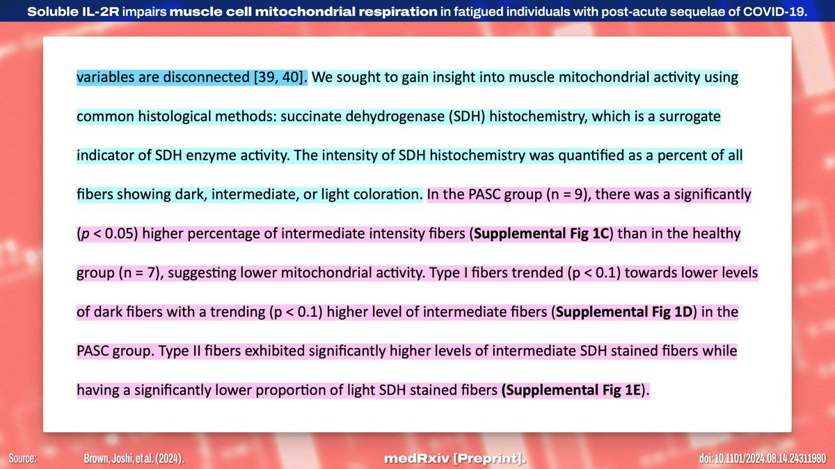
When they compared mitochondrial respiration between healthy controls and the LC group, they found a lower *quantity* of active mitochondria in the LC group, but the energy capacity of the present mitochondria seemed mostly unchanged.

9/


"PASC participants have compromised skeletal muscle mitochondrial function.  Several recent studies have linked PASC fatigue to reduced mitochondrial respiratory capacity in skeletal muscle [12, 41]. The observation that SDH activity differed in type II fibers prompted us to explore potential differences in mitochondrial respiration between healthy individuals and those with PASC. To investigate this, we used high resolution respirometry (HRR) to measure mitochondrial oxygen flux (JO2). State 3 respiration for complex I and combined complex I+II were significantly (p < 0.05) lower in PASC m...
Figure 2. Mitochondrial respiration, enzyme activity, and OXPHOS protein abundance between healthy and PASC participants. A - C. Respiration is lower for complex I, state 3 + glutamate, and complex I+ll, but unchanged in complex Il, state 3 (n= 7 healthy, 11 PASC). D. Electron transport activity is not different between groups (n=10 healthy, 7 PASC).
"In the electron transport chain (ETC), the electrons go through a chain of proteins that increases its reduction potential and causes a release in energy. Most of this energy is dissipated as heat or utilized to pump hydrogen ions (H+) from the mitochondrial matrix to the intermembrane space and create a proton gradient.  ...  Complex I, ... During this process, 4 hydrogen ions pass from the mitochondrial matrix to the intermembrane space, contributing to the electrochemical gradient.  Complex II, ... Complex II, also known as succinate dehydrogenase, accepts electrons from succinate ...
Diagram showing the components of the electron transport chain for mitochondria.  Source: https://www.ncbi.nlm.nih.gov/books/NBK553192/
On further examination, they found a similar pattern to the previous one.

These findings suggest "mainly mitochondrial content and not activity was lower in [LC] compared to healthy muscle." Electron microscopy also suggests the mitochondria are defective in some way.

10/
"Citrate synthase activity and cytochrome C oxidase activity were further measured in tissue homogenates (Fig 2E-F). We found that citrate synthase (CS, a proxy for mitochondrial content) was significantly lower, but cytochrome C oxidase (CCO, a proxy for complex IV activity) did not reach statistical significance (p = 0.0848, n = 7 healthy, 11 PASC). The ratio of CCO to CS was not significantly different between groups (Fig 2G), suggesting that mainly mitochondrial content and not activity was lower in PASC compared to healthy muscle. Electron microscopy demonstrated several alterations i...
Supplemental Figure 2. Increased autophagosomes localized near mitochondria. Pannels A and B show representative micrographs of a PASC participant's skeletal muscle sarcomere and mitochondrial features at 2um and 600nm. Compared to control muscle (C) PASC displayed fewer mitochondrial pairs at Z-discs and increased autophagosome (black arrows) localization near mitochondria (white arrows).
They then asked if the reduced mitochondrial content is a result of deficient production of new mitochondria. The results indicate "that mitochondrial biogenesis was not impaired."

So like... what the fuck is happening here? To figure out the mechanism...

11/ "To determine if mitochondrial complex protein levels were stoichiometric, we performed western blot analysis (Fig 2H) of 4 key subunits of complex I (NDUFB8, Fig 2I), II (SDHB, Fig 2J), III (UQCRC2, Fig 2K), and V (ATP5A, Fig 2L). Quantification of those blots indicated complex II protein was significantly lower, and complex III approached significance (p = 0.0544, n = 7 healthy, 11 PASC). To determine if changes in mitochondrial content and function from SARs-CoV-2 infection were due to altered mitochondrial biogenesis, we measured synthesis rates of mitochondrial protein. However, ther...
They examined the potential factors circulating in the blood. They identified sIL2R as "the most highly abundant circulating inflammatory marker, with the largest fold difference between healthy" and LC groups.

sIL2R is a receptor that comes from T cells!

12/ "Circulating sIL2R is significantly elevated in PASC participants and can directly impair mitochondrial function in muscle cells in vitro.  Prolonged inflammatory response and T cell dysfunction have been linked to PASC fatigue, and many inflammatory markers are reported to be elevated systemically with PASC. To assess circulating markers in our participants, we quantified 65 circulating factors (Human Immune Monitoring Panel) in the serum of PASC and healthy participants (Fig 3, n = 8 healthy, 6 PASC). We found sIL2R was the most highly abundant circulating inflammatory marker, with the l...
They treated some muscle cell tissue cultures with either sIL2R or the regular IL-2 cytokine.

They found the *baseline* oxygen consumption rate was unchanged, but the *maximum* respiration rate was limited by sIL2R specifically! This suggests a previously unknown pathway!

13/
"To assess if sIL2R has an inhibitory effect on mitochondrial respiration, we treated C2 C 12 myotubes with human recombinant sIL2R or IL-2 for 24 hours and used a Seahorse Extracellular Flux Analyzer to assess oxygen consumption rate (OCR) (Fig 4A, n = 4). We found that basal respiration (Fig 4B) was unchanged, but maximal respiration (Fig 4C) was significantly reduced after treatment with sIL2R when compared to vehicle (PBS). Administration of IL-2 had no significant effect on OCR when compared to vehicle (Supplemental Fig 4), suggesting that the mechanism of action for sIL2R is distinct...
Figure 4. C2C12 myotubes treated with sIL2R exhibit reduced mitochondrial respiration and complex III protein content.  IL-2R application to myotubes. A. Oxygen consumption is reduced in myotubes after sIL2R administration but is not affected by IL-2 administration (n=4). B. Basal respiration was not different between PBS and sIL2R treatment. C. Maximal respiration was lower in sIL2R cells. D. Cytochrome C oxidase activity was significantly different between PBS (n=3) and sIL2R (n=4).
What does this all mean? Despite no apparent differences in their muscles, those with LC fatigue "exhibited significant impairment of mitochondrial respiration across multiple components of the respiratory chain." sIL2R "emerged as the most significantly elevated factor."

14/ "Discussion  In this study we investigated skeletal muscle and systemic factors potentially responsible for persistent fatigue in PASC participants. Despite reporting chronic fatigue and achieving 10% less expected 6-minute walking distance compared to controls, PASC participants showed no apparent differences in muscle size or strength. However, they exhibited significant impairment of mitochondrial respiration across multiple components of the respiratory chain. To elucidate the mechanism behind this skeletal muscle mitochondrial respiratory deficiency, we assessed circulating factors -...
Critically, elevated sIL2R levels are already:

1. Associated with muscle wasting.
2. A known biomarker that is "predictive of respiratory failure and ICU outcomes" in COVID!

Thus, the authors "posit that PASC fatigue is partially attributable to elevated systemic sIL2R."

15/ "Among the elevated immune markers in our PASC cohort, sIL2R (CD25) showed the highest expression and greatest difference from healthy controls. Elevated sIL2R levels have been correlated to muscle wasting like sarcopenia and cachexia and, in COVID, sIL2R was predictive of respiratory failure and ICU outcomes. Our findings extend these observations, proposing a mechanism by which sIL2R might contribute to worsened outcomes and persistent fatigue after recovery from acute COVID. Our in vitro data suggest that IL-2 alone does not significantly affect muscle cell mitochondrial respiration, wh...
The specifics of the effect sIL2R has are still unclear—although the authors have some hypotheses—but future research will need to explore this line of questioning further to pick apart the mechanism.

16/ "It would be valuable to address the mechanistic aspects of soluble interleukin-2 receptor (sIL2R) action on mitochondria in muscle tissue. While the exact pathway remains speculative, exploring potential mechanisms could provide a framework for future investigations. One possibility is that sIL2R interacts with membrane-bound receptors on muscle cells, initiating a signaling cascade that ultimately affects mitochondrial function. One such possibility is the β1-integrin, which may interact with extracellular IL2R to mediate intracellular signaling that regulates myoblast proliferation a...
The study has a few limitations, but they're largely the type of limitations that would mean there's only a significant result if there's a large effect size to be measured! Of course, more research is needed to fully understand what's happening here.

17/ "Some limitations exist for this study. The availability of samples with known negative COVID status is limited, but we made every effort to optimize our research within these constraints. We included both pre- and post-pandemic controls samples, which resulted in higher than optimal levels of variability in age and BMI. Variability can act as a potential confounding factor, and therefore the significance of our results, despite high variability, suggests a strong biological effect. It is currently unclear if the alterations in OXPHOS are due to total number of mitochondria or due to decr...
Overall, these findings suggest "that mitochondria may function adequately under resting conditions but fail to meet increased energy demands during exertion," and that this "could explain post-exertional malaise" in LC despite otherwise normal muscle function.

18/ "Our experiments revealed that sIL2R administration in myotubes did not affect basal respiration, but had decreased maximal respiration. In addition, mitochondrial content was decreased in muscles from patients with PACS compared to controls. There findings could explain post-exertional malaise in PASC patients despite seemingly normal baseline muscle function. It suggests that mitochondria may function adequately under resting conditions but fail to meet increased energy demands during exertion. The link between post-viral fatigue and mitochondrial abnormalities in muscle has been recog...
This feels like a critical finding, and I agree with the authors' conclusion that their study "provides new insights into the physiological basis of" fatigue in Long COVID, opening "avenues for potential therapeutic interventions."

Source:

19/19 medrxiv.org/content/10.110…
"In conclusion, our study provides new insights into the physiological basis of PASC fatigue, highlighting the role of sIL2R in mitochondrial dysfunction. These findings open avenues for potential therapeutic interventions and underscore the need for comprehensive post-viral care strategies."
Huh. Latent herpesvirus reactivation leading to increased sIL2R… well that’s really the whole thing end-to-end isn’t it?

20/19
This quote is fascinating, because I’ve always thought the evidence lines up fairly well with post-infection chronic fatigue being the body’s rest-forcing illness response basically getting “stuck on.”

I wonder if sIL2R is a regulator for that process?

21/19

• • •

Missing some Tweet in this thread? You can try to force a refresh
 

Keep Current with Nick #RespiratorsFilterPathogens😷 Anderegg

Nick #RespiratorsFilterPathogens😷 Anderegg Profile picture

Stay in touch and get notified when new unrolls are available from this author!

Read all threads

This Thread may be Removed Anytime!

PDF

Twitter may remove this content at anytime! Save it as PDF for later use!

Try unrolling a thread yourself!

how to unroll video
  1. Follow @ThreadReaderApp to mention us!

  2. From a Twitter thread mention us with a keyword "unroll"
@threadreaderapp unroll

Practice here first or read more on our help page!

More from @NickAnderegg

Aug 18
This preprint (from April 5) is FASCINATING. Someone requested I have a look at it, and I'm glad they did, because it made SO MANY separate things suddenly make sense.

Here's a look at how a specific type of MICROCLOTS may be associated with LONG COVID pathology...

1/many Preprint posted April 5, 2024: "Increased fibrinaloid microclot counts in platelet-poor plasma are associated with Long COVID"  Abstract: "...The pathophysiology underlying Long COVID remains unclear but appears to involve multiple mechanisms including persistent inflammation, coagulopathy, autoimmunity, and organ damage. Studies suggest that microclots, also known as fibrinaloids, play a role in Long COVID. In this context, we developed a method to quantify microclots and investigated the relationship between microclot counts and Long COVID. We show that as a cohort, platele...
This thread will have three things:
- Takeaways of this study
- Breakdown of the method they developed
- How these findings connect with other known patterns

This is a study that looks at some rugged little blood clots that come courtesy of the SARS-CoV-2 spike protein.

2/ "Studies suggest that microclots, also known as fibrinaloids, play a role in Long COVID by blocking capillaries, limiting oxygen exchange, and potentially causing microvascular pulmonary thrombosis and multiple organ failure. Amyloid-containing deposits, resembling microclots, have also been observed in increased numbers in muscle tissue biopsies from people with Long COVID compared to samples from controls, and these numbers increase after exercise, an important observation as many people with Long COVID experience post-exertional symptom exacerbation after activity. However, the depo...
Some important background info:

- FIBRINOGEN is a protein that just kinda hangs out in the blood, waiting.

- FIBRIN is a fibrous protein that forms a mesh to hold platelets together to form a blood clot.

- THROMBIN is the enzyme that converts FIBRINOGEN into FIBRIN.

3/ Fibrinogen + Thrombin = Fibrin (monomer)  The fibrin monomers aggregate to form a mesh.  Fibrin mesh + clotting factor = cross-linked mesh that's much more stable, and which contracts tightly.  Image source: https://commons.wikimedia.org/wiki/File:Stabilisation_de_la_fibrine_par_le_factor_XIII.png
Read 22 tweets
Aug 15
My thoughts on this new finding? AAAAAAAAAAAAAAAAAAAAAAAAAAAAAA

"The consumption of BW [bottled water] is associated with heightened risk for certain health conditions," such as:
- hypertension (+5% increased risk)
- diabetes (+9%)
- GI ulcers (+21%)
- kidney stones (+17%)

1/10 Published Aug 15, 2024 in IJERPH: "Consumption of Bottled Water and Chronic Diseases: A Nationwide Cross-Sectional Study"  Abstract: "...On average, a liter of bottled water includes about 240,000 tiny pieces of plastic. ... Utilizing data from the Italian National Institute of Statistics’ “Aspects of Daily Life” survey (N = 45,597), we employed logistic regression to explore the correlation between BW consumption and the prevalence of various chronic diseases,... Adjustments were made for covariates such as education, age, gender, and economic resources. Our analysis indicat...
This was a very large cross-sectional study that looked at a national population, conducted by Italy's census agency. They controlled for covariates including socioeconomic status, age, and gender, then clustered and stratified the population as appropriate.

Solid methods!

2/10
"Data on BW consumption were obtained from the “Aspects of Daily Life” survey on households, conducted by the Italian National Institute of Statistics (ISTAT) [19]. ... The aim of the survey is to identify a variety of behavioral dimensions and aspects of daily life. ... We analyzed data from the 2021 edition of the survey, which included 45,597 individuals and 20,000 families, focusing on those who were 18 years or older at the time of the survey. ... a distinct stratum of municipalities with larger populations, labeled as self-representative (SR), and other municipalities, designated...
"The following variables were included in the analysis: educational level, age, gender, economical resources in the last 12 months, body mass index, smoking, alcohol consumption, physical activity, hypertension, diabetes, presence of kidney stones, presence of gastric or duodenal ulcer."
Analysis is *conceptually* straightforward:

- They built models to understand how bottled water + EACH confounding variable interacts to impact health outcomes.
- They combined those models into one big model to analyze all the variables together for each possible outcome.

3/10 "Bivariate analyses were performed to study the association between BW consumption and relevant variables, using chi-square tests. Logistic regression models were developed to control for confounding variables and assess the factors independently linked with BW consumption (1 if BW consumption is present; 0 if not). ... In cases of poor fit, stepwise regression, using AIC and BIC criteria, was utilized to select between models and discriminate between covariates. Regarding health outcomes, we considered the dichotomized presence of the following chronic diseases: hypertension (1 = yes, ...
Read 20 tweets
Aug 9
New interdisciplinary review was published on current Long COVID science, with a roadmap for science and policy!

It is written in plain language, so it's worth a read on its own, but I just want to pull out some highlights about what WE DO KNOW into a single thread...

1/many Published Aug 9, 2024 in Nature Medicine: "Long COVID science, research and policy"  Abstract: "Long COVID represents the constellation of post-acute and long-term health effects caused by SARS-CoV-2 infection; it is a complex, multisystem disorder that can affect nearly every organ system and can be severely disabling. The cumulative global incidence of long COVID is around 400 million individuals, which is estimated to have an annual economic impact of approximately $1 trillion—equivalent to about 1% of the global economy. Several mechanistic pathways are implicated in long...
This is definitely the definition for Long COVID I'll be explicitly using from now on: Long COVID is "the constellation of post-acute and long-term health effects caused by SARS-CoV-2 infection."

2/ "Long COVID is best defined as the constellation of post-acute and long-term health effects caused by SARS-CoV-2 infection. Long COVID was initially reported by patients who coined the term and, through research and advocacy, drove much of the progress in understanding this condition over the past several years (Fig. 1)."
Long COVID "affects nearly every organ system, including the cardiovascular system, the nervous system, the endocrine system, the immune system, the reproductive system, and the gastrointestinal system."

It affects people regardless of age or pre-existing health status.

3/ "Long COVID is a complex, multisystem disorder that affects nearly every organ system, including the cardiovascular system, the nervous, the nervous system, the endocrine system", the immune system, the reproductive system and the gastrointestinal system. It affects people across the age spectrum (from children 6-18 to older adults), people of different race and ethnicities, sex and gender, and baseline health status. Cardinal manifestations include brain fog (or cognitive dysfunction), fatigue, dysautonomia (which commonly manifests as postural orthostatic tachycardia syndrome (P...
Read 30 tweets
Aug 7
Can't believe I missed this!

This review/commentary piece makes one thing extremely clear: We do not yet have ANY STRONG EVIDENCE about how SARS-CoV-2 infection affects PREGNANCY.

The limited evidence shows there IS harm, which may be MITIGATED by vaccination.

Thread...
1/ Published July 11, 2024 in eClinicalMedicine: "In need of robust evidence of non-association of pregestational and early pregnancy SARS-CoV-2 infections with congenital anomalies"  Abstract: "SARS-CoV-2 infection during pregestational and early pregnancy periods has an unclear impact on fetal development. ..., potential effects on the developing fetal brain are plausible. ... This is further complicated by limitations, such as restricted testing access and undiagnosed infections, particularly in low- and middle-income countries. Most data focus on hospitalized women near term...
What is the takeaway? There are three:

- Unanswered questions remain about the effect of SARS-CoV-2 on pregnancy.
- Evidence shows congenital anomalies MAY be associated with COVID.
- Evidence shows vaccination provides "protection from such anomalies."

2/
"Conclusion  There is a need for data collection regarding pregestational maternal SARS-CoV-2 infection and its association with future pregnancies. Given the relative rarity of intraplacental transmission of SARS-CoV-2, we will need large epidemiological studies to answer these questions. We should also stay vigilant, monitoring past infections and the effects of emerging strains in assisted reproductive technologies (ART) and early pregnancy outcomes.  Although we need better data on miscarriages, it should be noted that there is no association between COVID-19 vaccination and miscar...
"Of note, studies have demonstrated there were significant differences in eye, ear, face, and neck anomalies between the vaccinated and not vaccinated groups, showing vaccination protection from such anomalies. Similarly, it has been shown that COVID-19 vaccination is not related to nonsyndromic orofacial cleft and might protect against having a child affected with such congenital anomalies.  Thus, the data presented here should by no means be interpreted as associated with the vaccines, but the disease caused by SARS-CoV-2 infection.  Comprehensive systematic reviews and meta-analyses ...
Let's be very clear: The current lack of evidence isn't because there is a lack of harm; it is because there is a lack of DATA available to even begin to understand the harm.

There is almost no available data on the impact of COVID on first- and second-trimester pregnancies.

3/
"Introduction  The impact of pregestational and maternal SARS-CoV-2 infection during early pregnancy on the developing fetus is poorly understood. Although vertical transmission is rare, an effect on the developing fetal brain remains plausible. Robust evidence on any association between maternal SARS-CoV-2 infection and the risk of congenital anomalies in offspring is, however, limited. This is due to inadequate tracking of pregestational maternal SARS-CoV-2 infection history and infection during early pregnancy, along with methodological limitations in relevant published studies."
"The significant proportion of undetected or undiagnosed SARS-CoV-2 infections during early pregnancy, particularly in low- and middle-income countries (LMICs), due to limited access to testing and information, further complicates this issue. Most available data pertain to hospitalised pregnant women near term, with very little information on the outcomes of first- and second-trimester infections separately. Therefore, there is a clear need to accurately assess the impact of COVID-19 on congenital anomalies."
Read 10 tweets
Aug 6
THIS IS BIG. WOW. New paper in PLOS Pathogens has findings about:
- the effect of the SARS-CoV-2 spike protein on cardiac cells (and mitochondrial dysfunction!),
- a treatment to be investigated, and
- how this is NOT caused by mRNA vaccines!

Buckle up, we're diving in...

1/ Published August 5, 2024 in PLOS Pathogens: "SARS-CoV-2 spike-induced syncytia are senescent and contribute to exacerbated heart failure"  "Author Summary  In this paper, we directly linked SARS-2-S-triggered syncytium [fused cells] formation in the absence of infection with the ensuing induction of cellular senescence and its pathophysiological contribution to heart failure. We propose that both SARS-2-S expression and SARS-2-S protein internalization were sufficient to induce senescence in nonsenescent ACE2expressing cells. This is important because of the persistent existe...
[This paper is an uncorrected proof; it's been peer-reviewed, but not copyedited yet.]

A bit of background: syncytia (sin-sih-sha) are giant cells with multiple nuclei that form from the fusion of multiple cells. Viral infections are a common cause of syncytia.

2/
"Abstract  SARS-CoV-2 spike protein (SARS-2-S) induced cell–cell fusion in uninfected cells may occur in long COVID-19 syndrome, as circulating SARS-2-S or extracellular vesicles containing SARS-2-S (S-EVs) were found to be prevalent in post-acute sequelae of COVID-19 (PASC) for up to 12 months after diagnosis. ... Here, we found that the senescent outcome of SARS-2-S induced syncytia exacerbated heart failure progression. We first demonstrated that syncytium formation in cells expressing SARS-2-S delivered by DNA plasmid or LNP-mRNA exhibits a senescence-like phenotype. Extracellular ...
NIH > National Library of Medicine Medical Subject Headings MeSH Descriptor Data 2024  Giant Cells (aka syncytia)  Multinucleated masses produced by the fusion of many cells; often associated with viral infections. In AIDS, they are induced when the envelope glycoprotein of the HIV virus binds to the CD4 antigen of uninfected neighboring T4 cells. The resulting syncytium leads to cell death and thus may account for the cytopathic effect of the virus.  Entry Term(s): Giant Cells, Multinucleated; Multinucleated Giant Cells; Polykaryocytes; Syncytia; Syncytium;
The authors had two motivations for this study:

1. Cardiac complications are a major feature of COVID.
2. Patients with heart failure tend to experience very poor outcomes from COVID.

Really, it hasn't been entirely clear *how* COVID leads to heart failure... until now?

3/ "Introduction  ...Although symptoms resulting from infection typically resolve within weeks, some individuals experience persistent symptoms following the acute phase of COVID-19, the so-called post-acute sequelae of COVID-19 (PASC) or long COVID [2–4]. SARS-CoV-2 infection predominantly offends the respiratory system. Currently, evidence has suggested cardiac complications as one of the important pathogenic features of COVID-19 [5,6]. More importantly, compared with patients without heart failure, those with diagnosed heart failure experienced a longer length of hospital stay, increas...
Read 30 tweets
Aug 4
New preprint on the PATHOGENICITY of H5N1 was published yesterday, and... it's not good news, but it's definitely not *terrible* news either!

The delayed, lackluster response to the current outbreak remains DEEPLY concerning.

Here's a summary for a general audience!

1/many
Preprint published August 3, 2024: "Enhanced replication of contemporary human highly pathogenic avian influenza H5N1 virus isolate in human lung organoids compared to bovine isolate"  Abstract  "We compared virus replication and host responses in human alveolar epithelium infected with highly pathogenic avian influenza (HPAI) H5N1 viruses. A/Vietnam/1203/2004 replicated most efficiently, followed by A/Texas/37/2024, then A/bovine/Ohio/B240SU342/2024. Induction of interferon-stimulated genes was lower with A/Texas/37/2024 and A/bovine/Ohio/B24OSU-342/2024, which may indicate ...
They test three H5N1 isolates. I'll refer to them as:

- Texas: Isolated from worker at Texas dairy farm (A/Texas/37/2024)
- Bovine: Isolated from dairy cow (A/bovine/Ohio/B24OSU-342/2024)
- Vietnam: Isolated from fatal 2004 human infection in Vietnam (A/Vietnam/1203/2004)

2/
"As of July 25, 2024, 13 human cases of HPAI H5N1 virus infection have been confirmed in the United States (3). Several of these cases are linked to exposure to infected cattle. However, recent outbreaks in Colorado have resulted in identification of additional human cases linked to infected poultry (3). Virus isolated from a worker at a Texas dairy farm (A/Texas/37/2024) was shown to be closely related to viruses circulating in cattle, and it is presumed that this case is a result of direct cow-to-human transmission (4). Reported symptoms included conjunctivitis, as well as mild respi...
"This is in stark contrast to prior cases of HPAI H5N1 virus infection in humans, which resulted in severe respiratory disease and mortality rates upwards of 50% (6). In order to assess the risk of developing severe disease following infection with contemporary HPAI H5N1 virus, we evaluated virus replication, host cell survival, and induction of innate immune responses in human alveolar epithelium infected with A/Texas/37/2024 or cattle isolate A/bovine/Ohio/B24OSU-342/2024, compared to a historical H5N1 isolate A/Vietnam/1203/2004, which was derived from a fatal human case (7)."
This study looked at pathogenicity, which is, basically, the ability of a virus to fuck up your cells, organs, or body, as determined by some measurable indicator of damage.

It's one of those concepts that's so broad that it's useful to include a formal definition:

3/
NIH > National Library of Medicine  MeSH (Medical Subject Headings)  Virulence (synonym-ish for "pathogenicity")  The degree of pathogenicity within a group or species of microorganisms or viruses as indicated by case fatality rates and/or the ability of the organism to invade the tissues of the host. The pathogenic capacity of an organism is determined by its VIRULENCE FACTORS.
NIH > National Library of Medicine  MeSH (Medical Subject Headings)  Virulence (Preferred):  The degree of pathogenicity within a group or species of microorganisms or viruses as indicated by case fatality rates and/or the ability of the organism to invade the tissues of the host. The pathogenic capacity of an organism is determined by its VIRULENCE FACTORS.  Pathogenicity (Related):  The capacity of a microorganism to cause disease.
Read 17 tweets

Did Thread Reader help you today?

Support us! We are indie developers!


This site is made by just two indie developers on a laptop doing marketing, support and development! Read more about the story.

Become a Premium Member ($3/month or $30/year) and get exclusive features!

Become Premium

Don't want to be a Premium member but still want to support us?

Make a small donation by buying us coffee ($5) or help with server cost ($10)

Donate via Paypal

Or Donate anonymously using crypto!

Ethereum

0xfe58350B80634f60Fa6Dc149a72b4DFbc17D341E copy

Bitcoin

3ATGMxNzCUFzxpMCHL5sWSt4DVtS8UqXpi copy

Thank you for your support!

Follow Us!

:(