Vipin M. Vashishtha Profile picture
Aug 23, 2024 10 tweets 3 min read Read on X
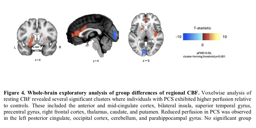
Experts have discovered a mutation in SARS-CoV-2 that plays a key role in its ability to infect brain. This may help in understanding neurological symptoms & mystery of "long COVID," & could one day even lead to specific treatments to protect & clear the virus from brain 1/ Image
Researchers discovered a series of mutations in the SARS-CoV-2 spike protein that enhanced the virus's ability to infect the brains of mice. 2/ Image
They assessed SARS-CoV-2 evolution in the lung versus CNS of infected mice. Higher levels of viral divergence were observed in the CNS than the lung after intranasal challenge with a high frequency of mutations in the spike furin cleavage site (FCS). 3/
Looking at the genomes of viruses found in the brain compared to the lung, the researchers found that viruses with a specific deletion in spike (Furin Cleavage Site) were much better at infecting the brains of these animals. 4/ Image
In this study, researchers infected mice with SARS-CoV-2 and sequenced the genomes of viruses that replicated in the brain versus the lung. In the lung, the spike protein looked very similar to the virus used to infect the mice. 5/
In the brain, however, most viruses had a deletion or mutation in a critical region of spike, i.e. FCS that dictates how it enters a cell. When viruses with this deletion were used to directly infect the brains of mice, it was largely repaired when it traveled to the lungs 6/ Image
In order for the virus to traffic from the lung to the brain, it required changes in the spike protein that are already known to dictate how the virus gets into different types of cells. 7/
Researchers think this region of spike (FCS) is a critical regulator of whether or not the virus gets into the brain, and it could have large implications for the treatment and management of neurological symptoms reported by COVID-19 patients. 8/ Image
It's still not known if #longCOVID is caused by direct infection of cells in the brain or due to some adverse immune response that persists beyond the infection. 9/ Image
If it is caused by infection of cells in the central nervous system, this study suggests there may be specific treatments that could work better than others in clearing the virus from this compartment. 10/10

nature.com/articles/s4156…

• • •

Missing some Tweet in this thread? You can try to force a refresh
 

Keep Current with Vipin M. Vashishtha

Vipin M. Vashishtha Profile picture

Stay in touch and get notified when new unrolls are available from this author!

Read all threads

This Thread may be Removed Anytime!

PDF

Twitter may remove this content at anytime! Save it as PDF for later use!

Try unrolling a thread yourself!

how to unroll video
  1. Follow @ThreadReaderApp to mention us!

  2. From a Twitter thread mention us with a keyword "unroll"
@threadreaderapp unroll

Practice here first or read more on our help page!

More from @vipintukur

Feb 6
New study suggests #LongCOVID may involve disrupted cortisol rhythms, not just inflammation.

Patients showed:
• Reduced morning cortisol
• Elevated evening levels
• Flattened daily cortisol cycle

➡️ Indicating hypothalamic–pituitary–adrenal (HPA) axis dysfunction. 1/ Image
Prospective study of post-COVID patients:

➡️ Compared with healthy controls,
✔ Long COVID patients had blunted morning cortisol peaks
✔ Higher evening cortisol
✔ Loss of normal circadian pattern

Blood cortisol alone failed to detect these changes. 2/ Image
Key insight:

➡️ Salivary cortisol profiling may be a more sensitive marker of stress-system dysfunction in LongCOVID than standard blood tests.

➡️ HPA axis disruption could underlie:
• Fatigue
• Brain fog
• Sleep disturbance
• Dysautonomia. 3/ Image
Read 5 tweets
Feb 5
Brain Fog after COVID-19: What’s driving it?

➡️ New review highlights that persistent cognitive symptoms in COVID survivors are strongly linked to pro-inflammatory cytokines and blood–brain barrier (BBB) dysfunction.

➡️ Key culprits include IL-6, TNF-α, IL-1β, IL-8, IL-13 and MCP-1 — many remain elevated months after infection.

🔥 COVID-19 is not just a respiratory disease.

➡️ Evidence suggests cognitive impairment can occur due to:

• Systemic inflammation
• Neuroinflammation
• Microvascular injury
• Persistent immune activation
• BBB disruption

➡️ These mechanisms may explain prolonged attention, memory & executive dysfunction. 1/Image
Cytokine signature of cognitive impairment in #LongCOVID:

🔹 Acute phase → IL-6, IL-1β, CXCL10 rise
🔹 Post-acute → Persistent IL-6, TNF-α, MCP-1
🔹 Long phase (>6 months) → IL-6, IL-13, IL-8 linked with “brain fog”

Inflammation clearly outlives the infection.

➡️ Blood–brain barrier disruption appears central in post-COVID cognitive decline.

Markers suggesting BBB injury:
• GFAP
• Neurofilament light chain
• MMP-9
• S100β

➡️ BBB leakage may persist in patients with cognitive symptoms even >1 year. 2/Image
Blood–brain barrier disruption appears central in post-COVID cognitive decline.

➡️ Markers suggesting BBB injury:
• GFAP
• Neurofilament light chain
• MMP-9
• S100β

➡️ BBB leakage may persist in patients with cognitive symptoms even >1 year.

Post-COVID cognitive deficits often affect:

✔ Attention
✔ Working memory
✔ Executive function
✔ Processing speed

➡️ Deficits may start as inflammatory-driven dysfunction but can evolve into persistent neuronal/glial injury.

Early cognitive rehabilitation may be crucial. 3/Image
Read 4 tweets
Jan 24
Post-COVID fatigue isn’t just subjective.
Using advanced MRI, researchers found real changes in brain blood flow and oxygen metabolism in people with Post-COVID-19 Syndrome (PCS) after mild infection.

➡️ Key finding:

PCS patients showed increased oxygen metabolism in the hippocampus (memory hub) but reduced metabolism in the anterior cingulate cortex (ACC) — despite no visible brain atrophy. 1/Image
Why this matters:

➡️ Higher hippocampal metabolism was linked to better cognitive performance, suggesting a compensatory response to maintain thinking and memory in PCS. 2/ Image
In contrast, lower anterior cingulate cortex (ACC) metabolism correlated with:

• depressive symptoms
• reduced motivation
• higher inflammatory & glial markers (TNF-α, GFAP)
➡️ pointing to immune-driven neurovascular uncoupling. 3/ Image
Image
Read 4 tweets
Jan 22
Why do some people feel exhausted long after COVID-19?

➡️ New brain-imaging research shows that even after mild COVID, people with persistent fatigue can have subtle but real changes in brain structure.

➡️ These changes are not large or widespread, but tend to appear in connected brain networks, especially areas involved in attention, decision-making, and sensory processing. 1/Image
Image
Importantly, the brain regions affected overlap with areas that naturally express TMPRSS2, a protein that helps SARS-CoV-2 enter cells — suggesting certain brain circuits may be more vulnerable to the virus. 2/ Image
The study also links these changes to brain chemical systems involved in mood, energy, and cognition (serotonin, acetylcholine, glutamate, and cannabinoids). 3/ Image
Read 4 tweets
Jan 19
COVID-19 doesn’t just affect the lungs — it can disrupt how cells produce energy. New research shows that COVID-19 alters the genetic “switches” that control mitochondria, the structures that power our cells. 1/ Image
By comparing people who died from severe COVID-19, those who recovered, and healthy individuals, researchers found lasting changes in how mitochondrial genes are regulated. These changes were most prominent in genes involved in energy production and metabolism. 2/ Image
Importantly, people with COVID-19 showed abnormally high levels of proteins that control mitochondrial structure and stress responses, suggesting long-term damage to the cell’s energy system. 3/ Image
Read 5 tweets
Dec 26, 2025
#LongCOVID (LC) shares striking symptom overlap with hypermobility spectrum disorders (HSD/hEDS): fatigue, brain fog, dysautonomia, pain—especially in women.

➡️ A new case series explores whether some “intractable” LC may reflect undiagnosed hypermobility disorders.

➡️ Five women with persistent LC symptoms were evaluated at an hEDS/HSD clinic.
All met Beighton score criteria for hypermobility.

➡️ 4 diagnosed with hEDS, 1 with HSD
➡️ 3 had dysautonomia

None had prior hypermobility diagnoses. 1/Image
All patients carried MTHFR polymorphisms (C677T or A1298C)—recently linked to hEDS/HSD.

➡️ Several also showed features of mast cell activation, suggesting immune dysregulation may unmask latent connective tissue disorders after SARS-CoV-2 infection.

➡️ Targeted management (physical therapy, methylfolate/B12, mast cell stabilization, pain interventions) led to clinical improvement in all cases.

🔑 Takeaway: Consider hEDS/HSD in women with refractory Long COVID, especially with multisystem pain and dysautonomia. 2/Image
This case series suggests that some patients with severe, persistent #LongCOVID—especially women—may have previously undiagnosed hypermobility disorders (hEDS/HSD).

➡️ Five women with refractory LongCOVID symptoms were found to meet criteria for hypermobility, often with dysautonomia, mast cell–related features, and MTHFR polymorphisms.

➡️ Targeted management led to clinical improvement, highlighting the need to consider hEDS/HSD in patients with intractable Long COVID symptoms. 3/
Read 4 tweets

Did Thread Reader help you today?

Support us! We are indie developers!


This site is made by just two indie developers on a laptop doing marketing, support and development! Read more about the story.

Become a Premium Member ($3/month or $30/year) and get exclusive features!

Become Premium

Don't want to be a Premium member but still want to support us?

Make a small donation by buying us coffee ($5) or help with server cost ($10)

Donate via Paypal

Or Donate anonymously using crypto!

Ethereum

0xfe58350B80634f60Fa6Dc149a72b4DFbc17D341E copy

Bitcoin

3ATGMxNzCUFzxpMCHL5sWSt4DVtS8UqXpi copy

Thank you for your support!

Follow Us!

:(