Vipin M. Vashishtha Profile picture
Aug 28, 2024 7 tweets 2 min read Read on X
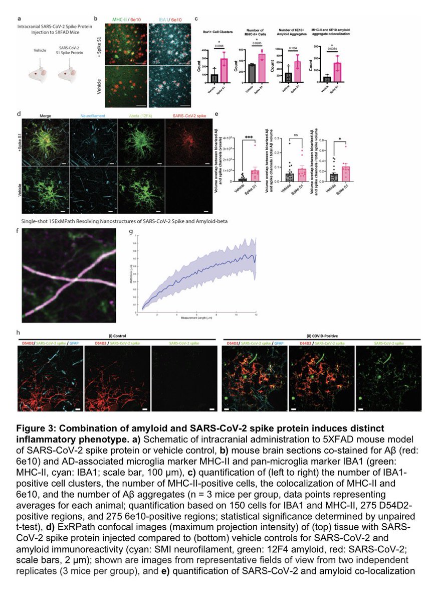
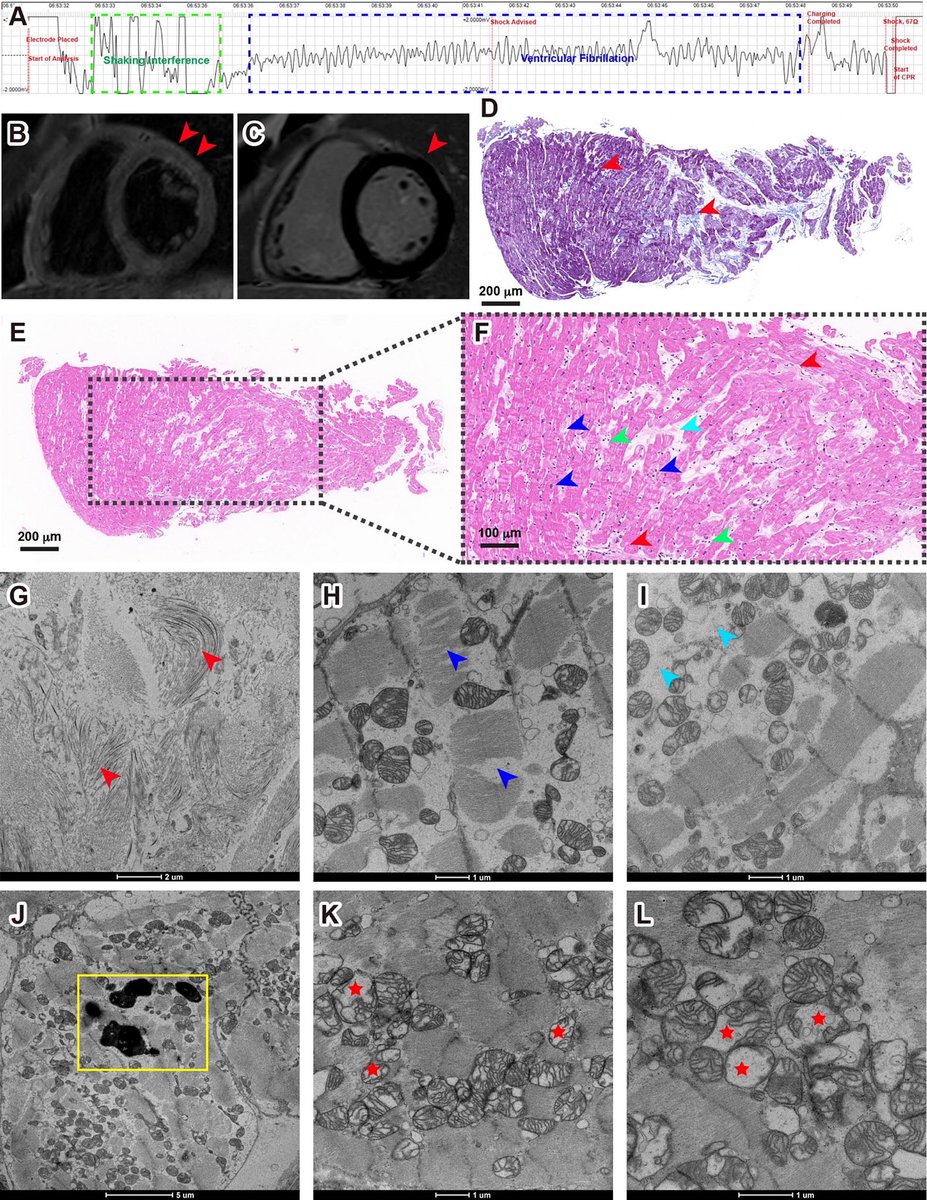
In a study that reshapes what we know about COVID19, scientists have discovered that coagulation protein fibrin causes unusual clotting & inflammation that have become hallmarks of the disease, while also suppressing the body's ability to clear virus.

nature.com/articles/s4158…
Image
Importantly, the team also identified a new antibody therapy to combat all of these deleterious effects. The study by overturns the prevailing theory that blood clotting is merely a consequence of inflammation in COVID-19. Image
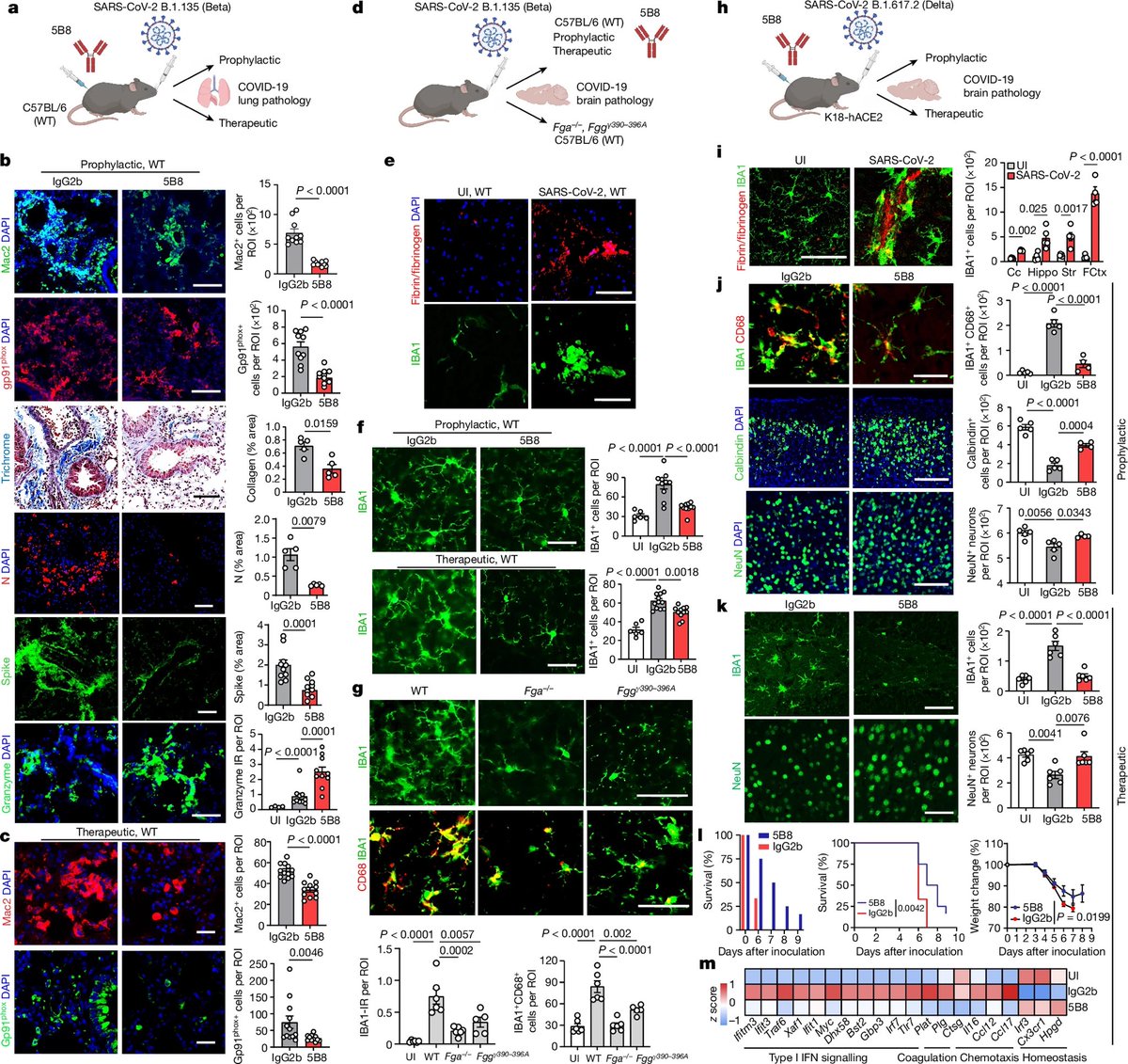
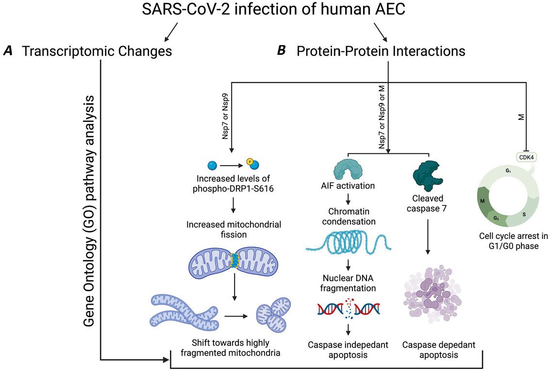
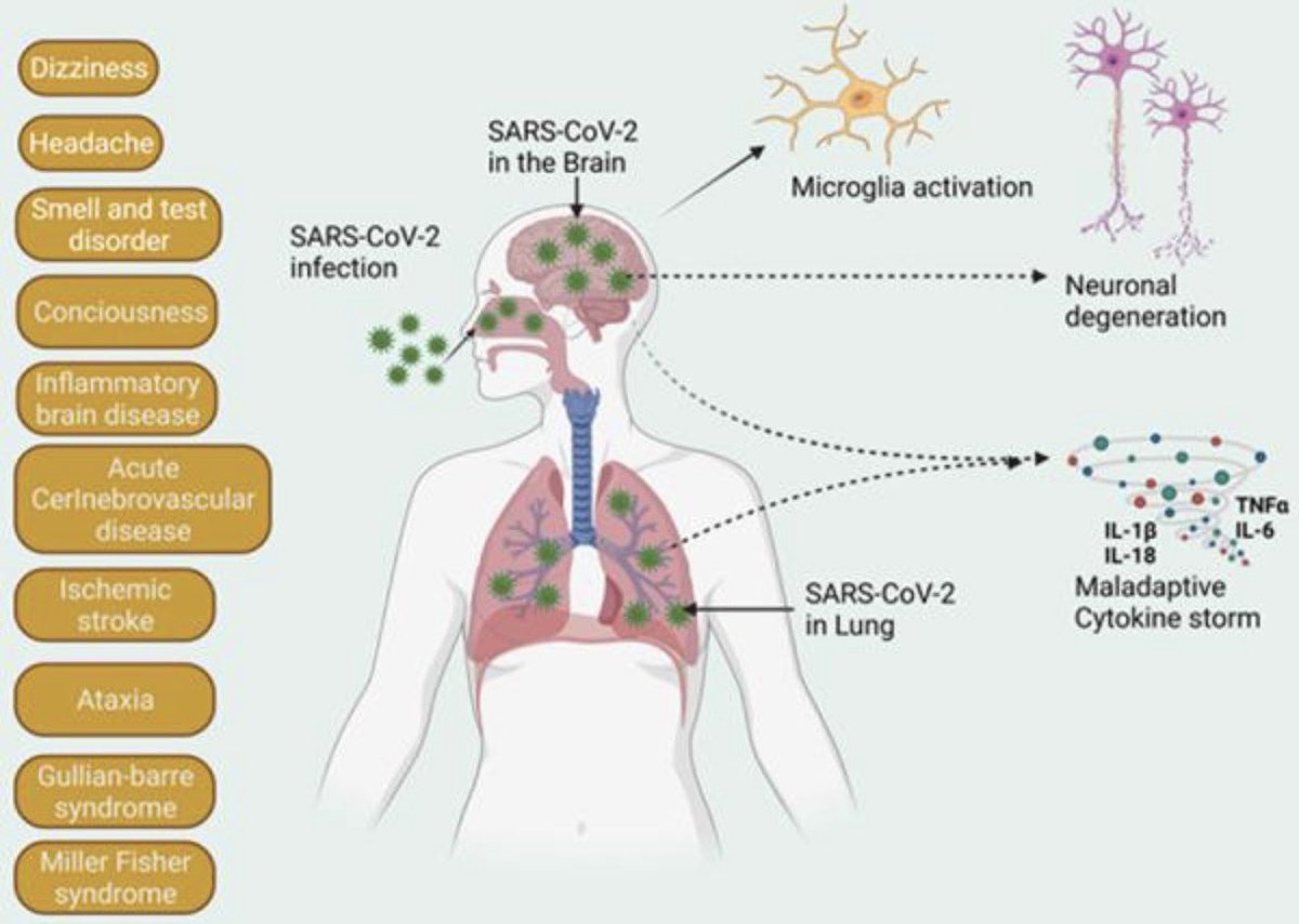
Through experiments in the lab and with mice, the researchers show that blood clotting is instead a primary effect, driving other problems—including toxic inflammation, impaired viral clearance, and neurological symptoms prevalent in those with COVID-19 and long COVID.
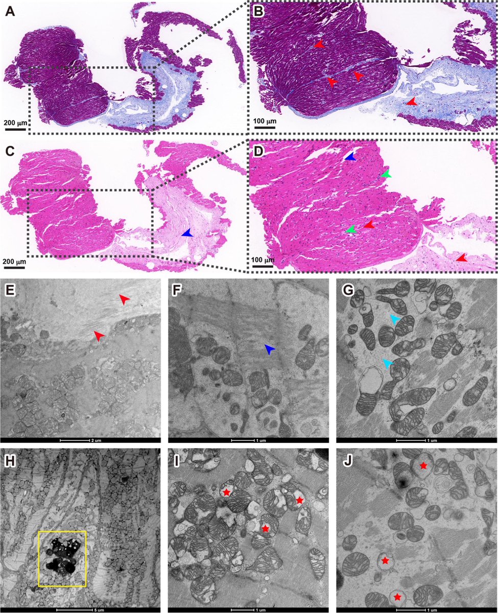
In this study, scientists found that fibrin becomes even more toxic in COVID-19 as it binds to both the virus and immune cells, creating unusual clots that lead to inflammation, fibrosis, and loss of neurons.
Knowing that fibrin is instigator of inflammation & neurological symptoms, we can build a new path forward for treating the disease at the root. In their experiments, neutralizing blood toxicity with fibrin antibody therapy can protect the brain and body after COVID infection.
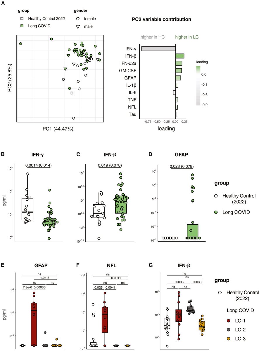
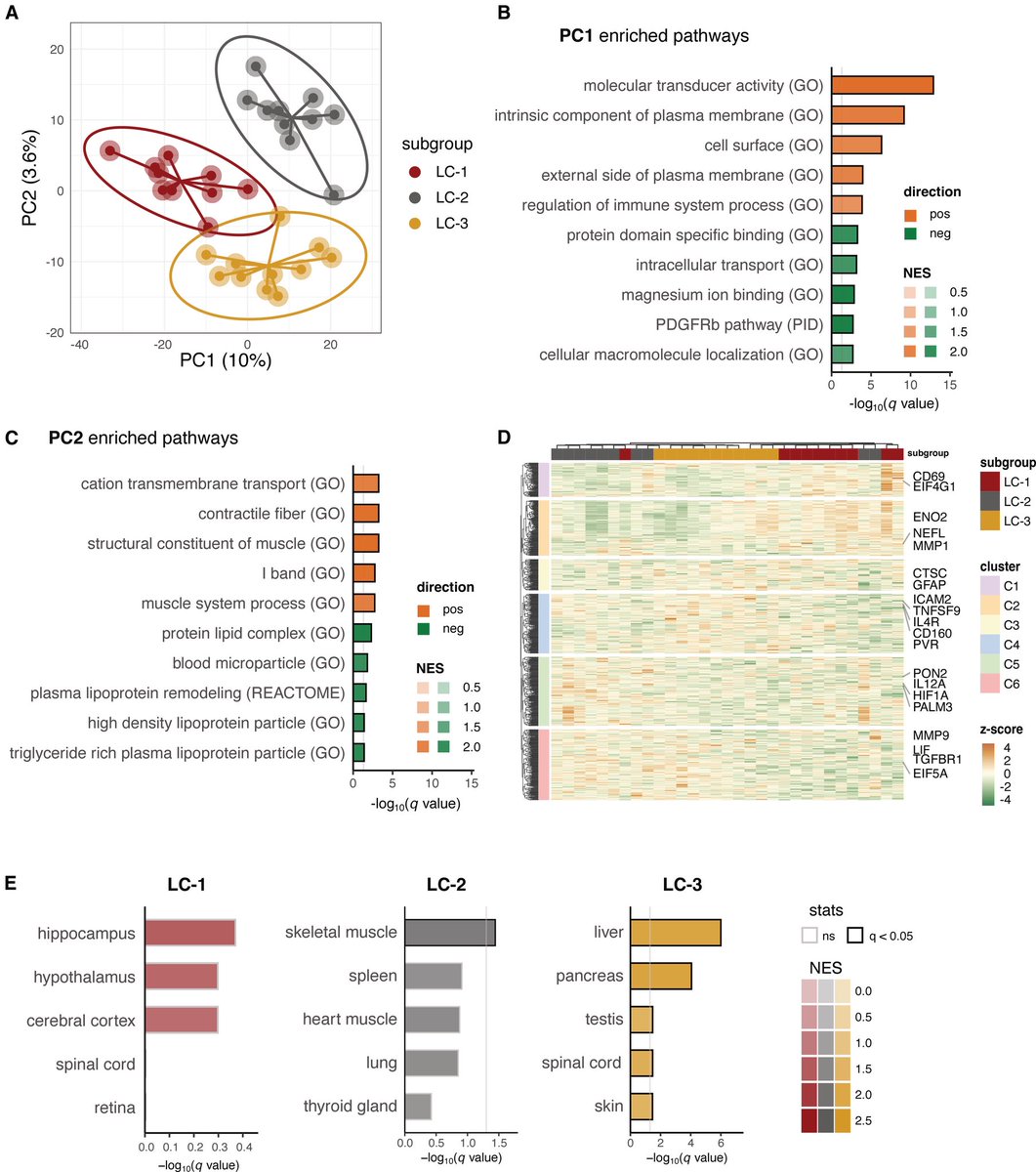
As fibrinogen plasma levels in acute COVID-19 are a predictive biomarker for cognitive impairment in longCOVID, it could be used to stratify patients as candidates for entry into phase 2 trials. Image
Fibrin immunotherapy can be tested for its potential to reduce adverse health outcomes due to long COVID as part of a multipronged approach with prevention and vaccination measures.

nature.com/articles/s4158…

• • •

Missing some Tweet in this thread? You can try to force a refresh
 

Keep Current with Vipin M. Vashishtha

Vipin M. Vashishtha Profile picture

Stay in touch and get notified when new unrolls are available from this author!

Read all threads

This Thread may be Removed Anytime!

PDF

Twitter may remove this content at anytime! Save it as PDF for later use!

Try unrolling a thread yourself!

how to unroll video
  1. Follow @ThreadReaderApp to mention us!

  2. From a Twitter thread mention us with a keyword "unroll"
@threadreaderapp unroll

Practice here first or read more on our help page!

More from @vipintukur

Feb 6
New study suggests #LongCOVID may involve disrupted cortisol rhythms, not just inflammation.

Patients showed:
• Reduced morning cortisol
• Elevated evening levels
• Flattened daily cortisol cycle

➡️ Indicating hypothalamic–pituitary–adrenal (HPA) axis dysfunction. 1/ Image
Prospective study of post-COVID patients:

➡️ Compared with healthy controls,
✔ Long COVID patients had blunted morning cortisol peaks
✔ Higher evening cortisol
✔ Loss of normal circadian pattern

Blood cortisol alone failed to detect these changes. 2/ Image
Key insight:

➡️ Salivary cortisol profiling may be a more sensitive marker of stress-system dysfunction in LongCOVID than standard blood tests.

➡️ HPA axis disruption could underlie:
• Fatigue
• Brain fog
• Sleep disturbance
• Dysautonomia. 3/ Image
Read 5 tweets
Feb 5
Brain Fog after COVID-19: What’s driving it?

➡️ New review highlights that persistent cognitive symptoms in COVID survivors are strongly linked to pro-inflammatory cytokines and blood–brain barrier (BBB) dysfunction.

➡️ Key culprits include IL-6, TNF-α, IL-1β, IL-8, IL-13 and MCP-1 — many remain elevated months after infection.

🔥 COVID-19 is not just a respiratory disease.

➡️ Evidence suggests cognitive impairment can occur due to:

• Systemic inflammation
• Neuroinflammation
• Microvascular injury
• Persistent immune activation
• BBB disruption

➡️ These mechanisms may explain prolonged attention, memory & executive dysfunction. 1/Image
Cytokine signature of cognitive impairment in #LongCOVID:

🔹 Acute phase → IL-6, IL-1β, CXCL10 rise
🔹 Post-acute → Persistent IL-6, TNF-α, MCP-1
🔹 Long phase (>6 months) → IL-6, IL-13, IL-8 linked with “brain fog”

Inflammation clearly outlives the infection.

➡️ Blood–brain barrier disruption appears central in post-COVID cognitive decline.

Markers suggesting BBB injury:
• GFAP
• Neurofilament light chain
• MMP-9
• S100β

➡️ BBB leakage may persist in patients with cognitive symptoms even >1 year. 2/Image
Blood–brain barrier disruption appears central in post-COVID cognitive decline.

➡️ Markers suggesting BBB injury:
• GFAP
• Neurofilament light chain
• MMP-9
• S100β

➡️ BBB leakage may persist in patients with cognitive symptoms even >1 year.

Post-COVID cognitive deficits often affect:

✔ Attention
✔ Working memory
✔ Executive function
✔ Processing speed

➡️ Deficits may start as inflammatory-driven dysfunction but can evolve into persistent neuronal/glial injury.

Early cognitive rehabilitation may be crucial. 3/Image
Read 4 tweets
Jan 24
Post-COVID fatigue isn’t just subjective.
Using advanced MRI, researchers found real changes in brain blood flow and oxygen metabolism in people with Post-COVID-19 Syndrome (PCS) after mild infection.

➡️ Key finding:

PCS patients showed increased oxygen metabolism in the hippocampus (memory hub) but reduced metabolism in the anterior cingulate cortex (ACC) — despite no visible brain atrophy. 1/Image
Why this matters:

➡️ Higher hippocampal metabolism was linked to better cognitive performance, suggesting a compensatory response to maintain thinking and memory in PCS. 2/ Image
In contrast, lower anterior cingulate cortex (ACC) metabolism correlated with:

• depressive symptoms
• reduced motivation
• higher inflammatory & glial markers (TNF-α, GFAP)
➡️ pointing to immune-driven neurovascular uncoupling. 3/ Image
Image
Read 4 tweets
Jan 22
Why do some people feel exhausted long after COVID-19?

➡️ New brain-imaging research shows that even after mild COVID, people with persistent fatigue can have subtle but real changes in brain structure.

➡️ These changes are not large or widespread, but tend to appear in connected brain networks, especially areas involved in attention, decision-making, and sensory processing. 1/Image
Image
Importantly, the brain regions affected overlap with areas that naturally express TMPRSS2, a protein that helps SARS-CoV-2 enter cells — suggesting certain brain circuits may be more vulnerable to the virus. 2/ Image
The study also links these changes to brain chemical systems involved in mood, energy, and cognition (serotonin, acetylcholine, glutamate, and cannabinoids). 3/ Image
Read 4 tweets
Jan 19
COVID-19 doesn’t just affect the lungs — it can disrupt how cells produce energy. New research shows that COVID-19 alters the genetic “switches” that control mitochondria, the structures that power our cells. 1/ Image
By comparing people who died from severe COVID-19, those who recovered, and healthy individuals, researchers found lasting changes in how mitochondrial genes are regulated. These changes were most prominent in genes involved in energy production and metabolism. 2/ Image
Importantly, people with COVID-19 showed abnormally high levels of proteins that control mitochondrial structure and stress responses, suggesting long-term damage to the cell’s energy system. 3/ Image
Read 5 tweets
Dec 26, 2025
#LongCOVID (LC) shares striking symptom overlap with hypermobility spectrum disorders (HSD/hEDS): fatigue, brain fog, dysautonomia, pain—especially in women.

➡️ A new case series explores whether some “intractable” LC may reflect undiagnosed hypermobility disorders.

➡️ Five women with persistent LC symptoms were evaluated at an hEDS/HSD clinic.
All met Beighton score criteria for hypermobility.

➡️ 4 diagnosed with hEDS, 1 with HSD
➡️ 3 had dysautonomia

None had prior hypermobility diagnoses. 1/Image
All patients carried MTHFR polymorphisms (C677T or A1298C)—recently linked to hEDS/HSD.

➡️ Several also showed features of mast cell activation, suggesting immune dysregulation may unmask latent connective tissue disorders after SARS-CoV-2 infection.

➡️ Targeted management (physical therapy, methylfolate/B12, mast cell stabilization, pain interventions) led to clinical improvement in all cases.

🔑 Takeaway: Consider hEDS/HSD in women with refractory Long COVID, especially with multisystem pain and dysautonomia. 2/Image
This case series suggests that some patients with severe, persistent #LongCOVID—especially women—may have previously undiagnosed hypermobility disorders (hEDS/HSD).

➡️ Five women with refractory LongCOVID symptoms were found to meet criteria for hypermobility, often with dysautonomia, mast cell–related features, and MTHFR polymorphisms.

➡️ Targeted management led to clinical improvement, highlighting the need to consider hEDS/HSD in patients with intractable Long COVID symptoms. 3/
Read 4 tweets

Did Thread Reader help you today?

Support us! We are indie developers!


This site is made by just two indie developers on a laptop doing marketing, support and development! Read more about the story.

Become a Premium Member ($3/month or $30/year) and get exclusive features!

Become Premium

Don't want to be a Premium member but still want to support us?

Make a small donation by buying us coffee ($5) or help with server cost ($10)

Donate via Paypal

Or Donate anonymously using crypto!

Ethereum

0xfe58350B80634f60Fa6Dc149a72b4DFbc17D341E copy

Bitcoin

3ATGMxNzCUFzxpMCHL5sWSt4DVtS8UqXpi copy

Thank you for your support!

Follow Us!

:(