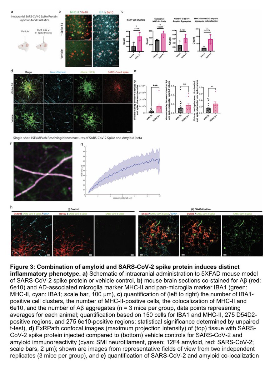
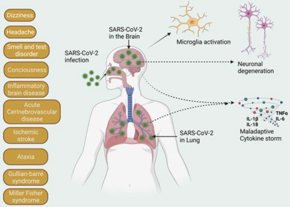
The molecular origins of neuro-COVID, and how it contributes to #LongCovid (PASC) are unknown, however a growing body of research highlights that the self-assembly of protein fragments from SARS-CoV-2 into amyloid nanofibrils may play a causative role. 1/
Earlier, researchers identified two fragments from the SARS-CoV-2 proteins, ORF 6 & ORF10, that self-assemble into neurotoxic amyloid assemblies.
Here they show the self-assembly mechanisms & nano-architectures formed by these fragments and their biological responses. 2/
By solubilising the peptides in a fluorinated solvent, they eliminated insoluble aggregates in the starting materials (seeds) that change the polymorphic landscape of the assemblies. 3/
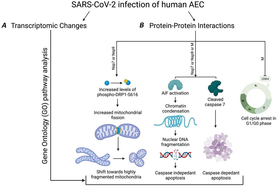
The resultant assemblies are dominated by structures with higher free energies (e.g. ribbons and amorphous aggregates) that are less toxic to cultured neurons but do affect their mitochondrial respiration. 4/
The researchers also show the first direct evidence of cellular uptake of viral amyloids. 5/
This work highlights the importance of understanding the polymorphic behaviour of amyloids and the correlation to neurotoxicity, particularly in the context of neuro-COVID & PASC. 6/6
Post-COVID fatigue isn’t just subjective.
Using advanced MRI, researchers found real changes in brain blood flow and oxygen metabolism in people with Post-COVID-19 Syndrome (PCS) after mild infection.
➡️ Key finding:
PCS patients showed increased oxygen metabolism in the hippocampus (memory hub) but reduced metabolism in the anterior cingulate cortex (ACC) — despite no visible brain atrophy. 1/
Why this matters:
➡️ Higher hippocampal metabolism was linked to better cognitive performance, suggesting a compensatory response to maintain thinking and memory in PCS. 2/
In contrast, lower anterior cingulate cortex (ACC) metabolism correlated with:
Why do some people feel exhausted long after COVID-19?
➡️ New brain-imaging research shows that even after mild COVID, people with persistent fatigue can have subtle but real changes in brain structure.
➡️ These changes are not large or widespread, but tend to appear in connected brain networks, especially areas involved in attention, decision-making, and sensory processing. 1/
Importantly, the brain regions affected overlap with areas that naturally express TMPRSS2, a protein that helps SARS-CoV-2 enter cells — suggesting certain brain circuits may be more vulnerable to the virus. 2/
The study also links these changes to brain chemical systems involved in mood, energy, and cognition (serotonin, acetylcholine, glutamate, and cannabinoids). 3/
COVID-19 doesn’t just affect the lungs — it can disrupt how cells produce energy. New research shows that COVID-19 alters the genetic “switches” that control mitochondria, the structures that power our cells. 1/
By comparing people who died from severe COVID-19, those who recovered, and healthy individuals, researchers found lasting changes in how mitochondrial genes are regulated. These changes were most prominent in genes involved in energy production and metabolism. 2/
Importantly, people with COVID-19 showed abnormally high levels of proteins that control mitochondrial structure and stress responses, suggesting long-term damage to the cell’s energy system. 3/
#LongCOVID (LC) shares striking symptom overlap with hypermobility spectrum disorders (HSD/hEDS): fatigue, brain fog, dysautonomia, pain—especially in women.
➡️ A new case series explores whether some “intractable” LC may reflect undiagnosed hypermobility disorders.
➡️ Five women with persistent LC symptoms were evaluated at an hEDS/HSD clinic.
All met Beighton score criteria for hypermobility.
➡️ 4 diagnosed with hEDS, 1 with HSD
➡️ 3 had dysautonomia
None had prior hypermobility diagnoses. 1/
All patients carried MTHFR polymorphisms (C677T or A1298C)—recently linked to hEDS/HSD.
➡️ Several also showed features of mast cell activation, suggesting immune dysregulation may unmask latent connective tissue disorders after SARS-CoV-2 infection.
➡️ Targeted management (physical therapy, methylfolate/B12, mast cell stabilization, pain interventions) led to clinical improvement in all cases.
🔑 Takeaway: Consider hEDS/HSD in women with refractory Long COVID, especially with multisystem pain and dysautonomia. 2/
This case series suggests that some patients with severe, persistent #LongCOVID—especially women—may have previously undiagnosed hypermobility disorders (hEDS/HSD).
➡️ Five women with refractory LongCOVID symptoms were found to meet criteria for hypermobility, often with dysautonomia, mast cell–related features, and MTHFR polymorphisms.
➡️ Targeted management led to clinical improvement, highlighting the need to consider hEDS/HSD in patients with intractable Long COVID symptoms. 3/
🔥 A landmark study challenges the long-held belief that Alzheimer’s disease (AD) is irreversible.
➡️ Using advanced mouse models that mimic human AD pathology, researchers found that restoring and maintaining healthy levels of NAD⁺, a key cellular energy molecule, can not only prevent but also reverse advanced Alzheimer’s pathology and fully restore cognitive function in mice. 1/
The team showed that NAD⁺ deficiency is a central driver of AD pathology—leading to blood-brain barrier breakdown, neuroinflammation, oxidative damage, and impaired neurogenesis. 2/
➡️ By administering a compound that rebalances NAD⁺ (P7C3-A20), all these pathological features were reversed, and memory and cognitive function were recovered.
➡️ These effects were seen in both amyloid-driven and tau-driven models, with supporting evidence from human AD brain samples suggesting disrupted NAD⁺ homeostasis in patients. 3/
As we age, our immune system becomes less effective, partly because key cells called CD8⁺ T-cells have trouble forming long-lasting memory.
A new study shows that a process called autophagy — the cell’s way of cleaning out old or damaged components — plays a central role in this problem. 1/
When a T-cell divides, it can make two daughter cells with different future roles: one becomes a long-lived ‘memory T cell’ that helps protect against future infections, and the other becomes a short-lived ‘effector T cell’ that fights the immediate infection.
For this to happen, the cell must sort its internal parts unevenly during division. 2/
The researchers found that #autophagy helps clear out old mitochondria before division, allowing daughter cells to inherit different mitochondrial content.
➡️ This asymmetric inheritance is crucial for creating a mix of T-cells with distinct fates — including memory cells.
➡️ Without autophagy, old mitochondria aren’t cleared, the inheritance becomes symmetric, and the diversity in T-cell fates is lost.
➡️ This has major implications for understanding why immune memory weakens with age and may inform new strategies to boost T-cell immunity. 3/