1/Raise your hand if you’re confused by the BRACHIAL PLEXUS!
I could never seem to remember or understand it—but now I do & I’ll show you how!
A thread so you will never fear brachial plexus anatomy again!
I could never seem to remember or understand it—but now I do & I’ll show you how!
A thread so you will never fear brachial plexus anatomy again!

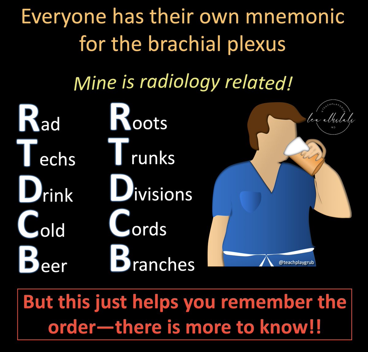
2/Everyone has a mnemonic to remember brachial plexus anatomy.
I’m a radiologist, so I remember one about Rad Techs.
But just remembering the names & their order isn’t enough.
That is just the starting point--let’s really understand it
I’m a radiologist, so I remember one about Rad Techs.
But just remembering the names & their order isn’t enough.
That is just the starting point--let’s really understand it
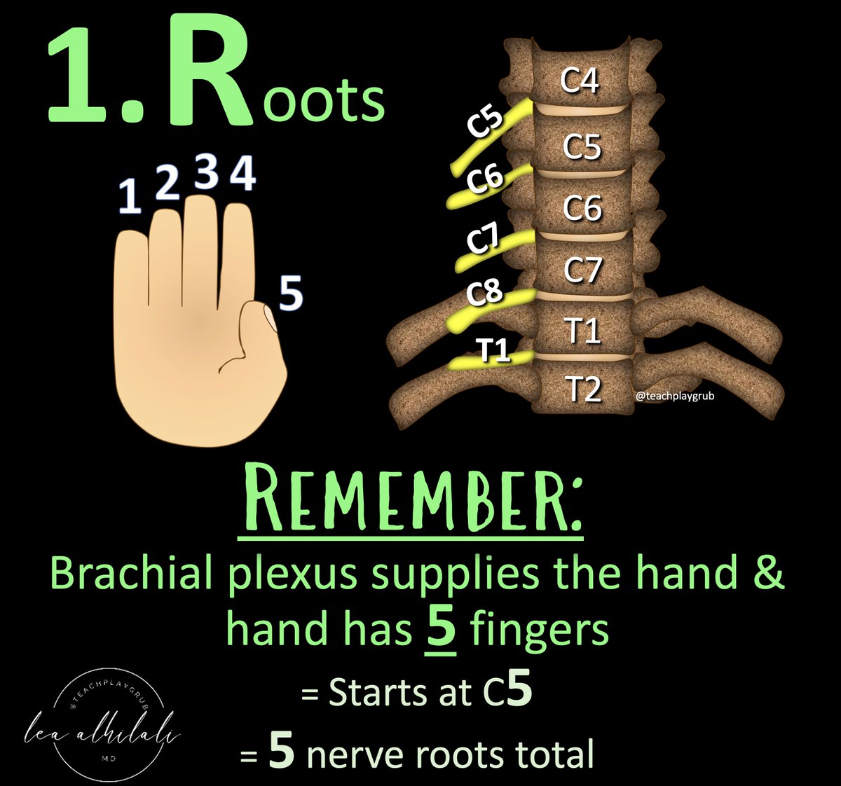
3/From the mnemonic, we start with the roots—the cervical nerve roots.
I remember which roots make up the brachial plexus by remembering that it supplies the hand.
You have 5 fingers on your hand so we start with C5 & we take 5 nerve roots (C5-T1).
I remember which roots make up the brachial plexus by remembering that it supplies the hand.
You have 5 fingers on your hand so we start with C5 & we take 5 nerve roots (C5-T1).
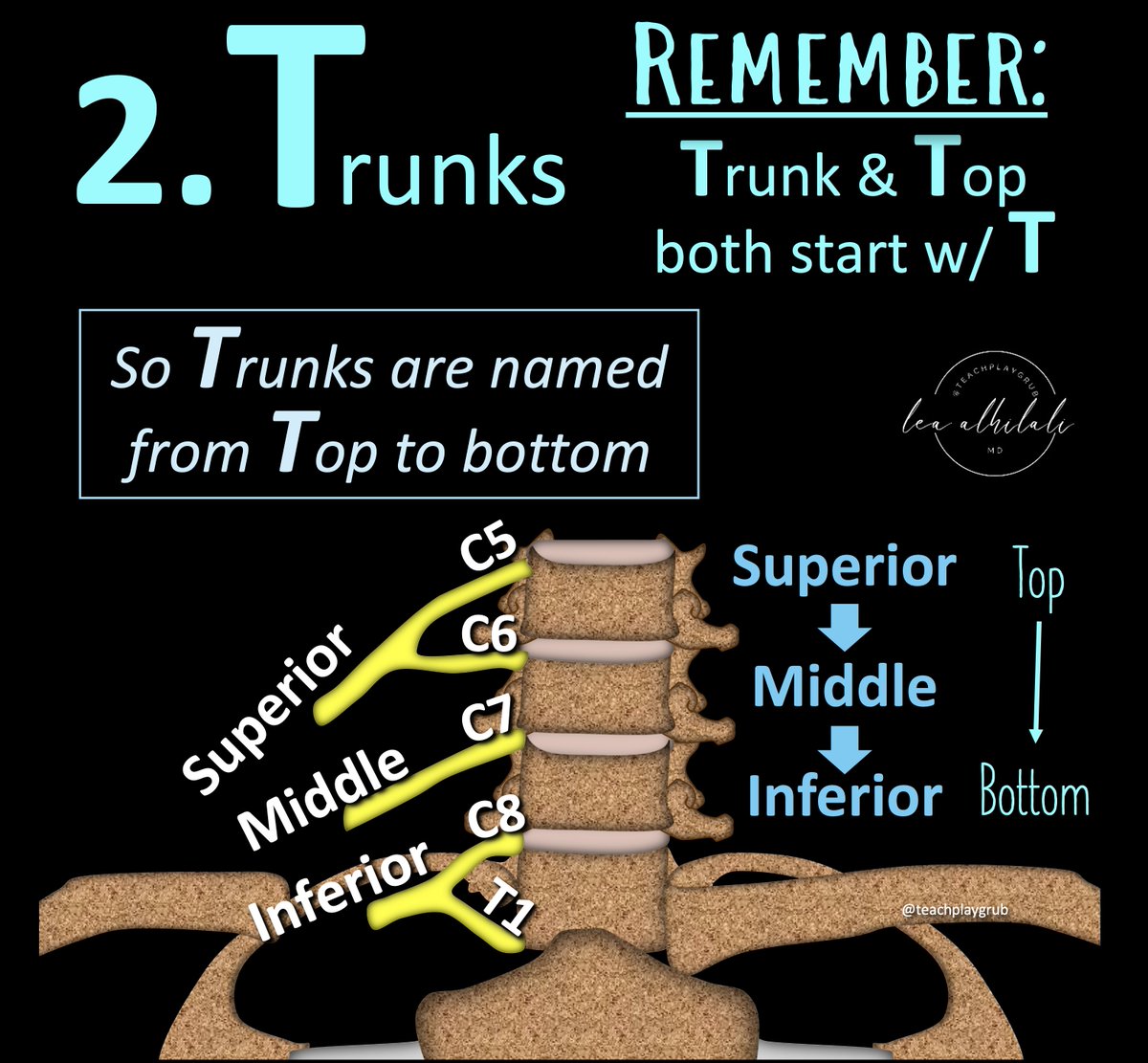
4/Next in the mnemonic are the Trunks.
Bc Trunks starts w/T, I can remember how they are named.
T is Top to bottom. Trunks are named top to bottom: Superior, Middle, and Inferior.
But how to remember which nerve roots combine to give you which trunks?
Bc Trunks starts w/T, I can remember how they are named.
T is Top to bottom. Trunks are named top to bottom: Superior, Middle, and Inferior.
But how to remember which nerve roots combine to give you which trunks?
5/Pairing of the nerve roots into the trunks is like pairing off at a dance when there is an odd number
Everyone immediately turns to the person next to them & the person in the middle is left out.
For the roots, C7 is in the middle & has to go it alone as the middle trunk
Everyone immediately turns to the person next to them & the person in the middle is left out.
For the roots, C7 is in the middle & has to go it alone as the middle trunk

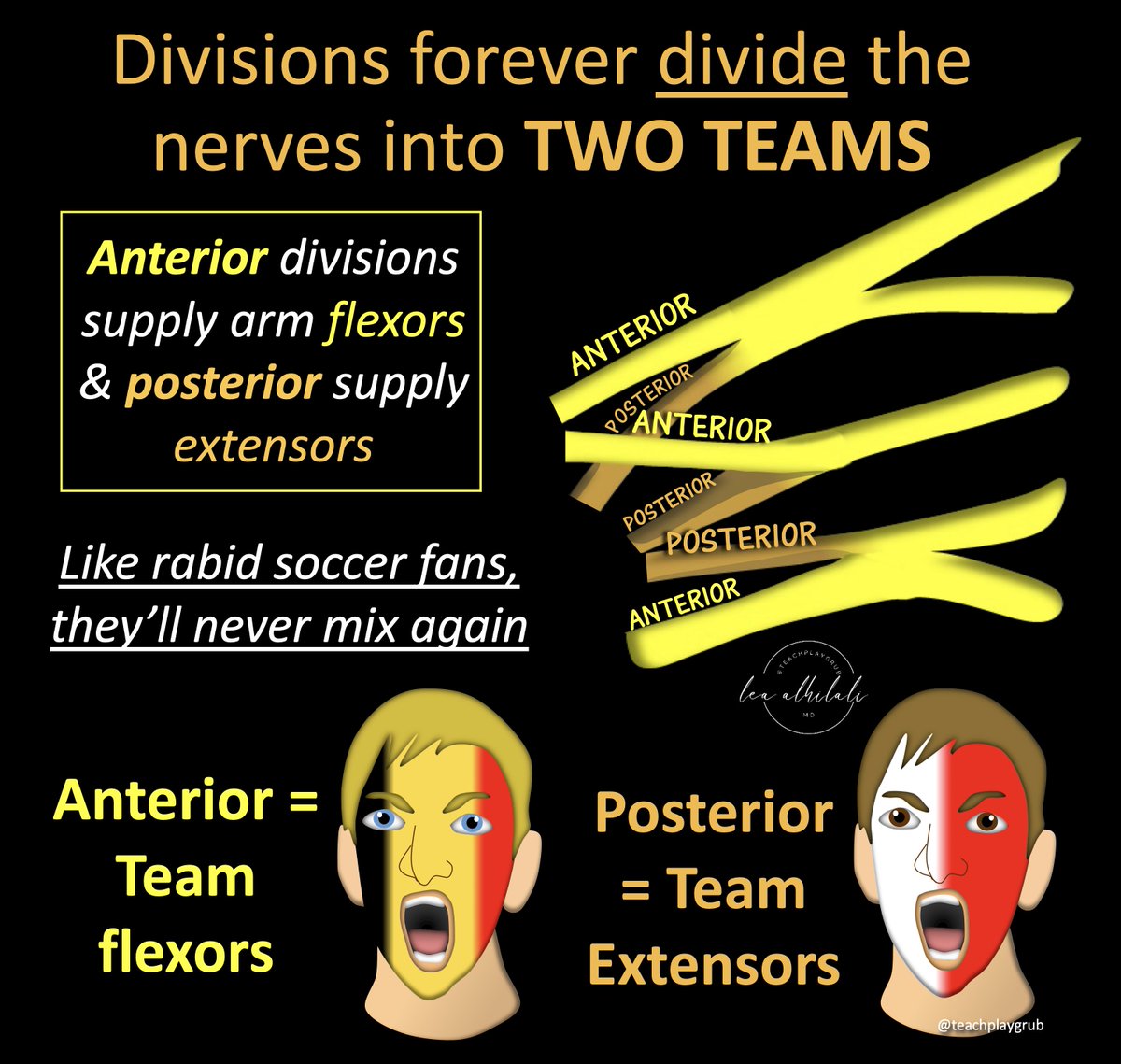
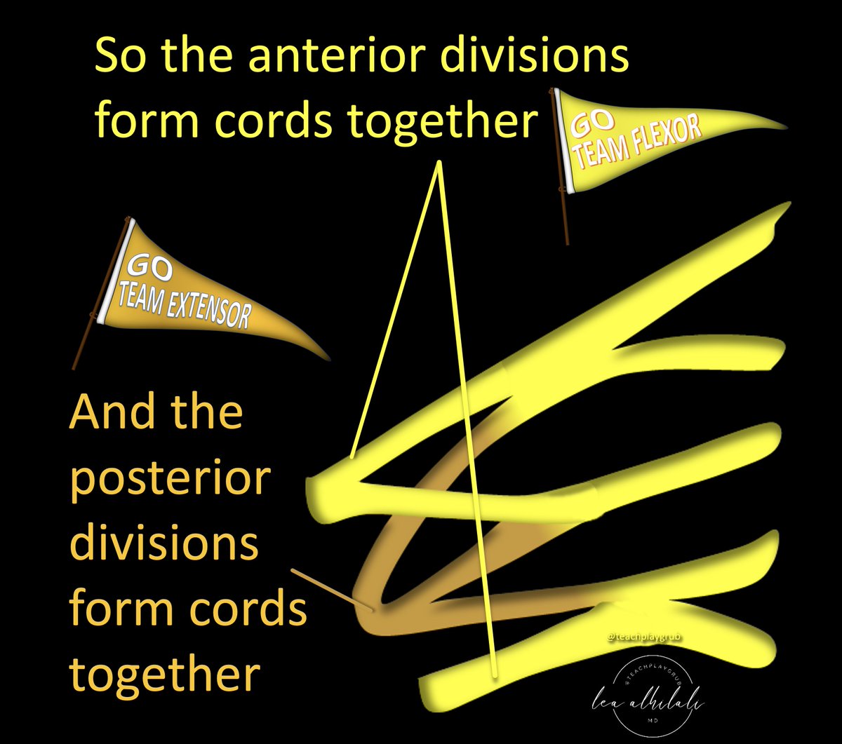
6/Next in the mnemonic are the Divisions.
Divisions do what their name implies—they divide the trunks.
Each trunk is split or DIVIDED into an anterior & posterior division.
Divisions will look like scissors coming off the trunks, helping you to remember they are splitting.
Divisions do what their name implies—they divide the trunks.
Each trunk is split or DIVIDED into an anterior & posterior division.
Divisions will look like scissors coming off the trunks, helping you to remember they are splitting.

7/This division results in a fundamental change in the nerves—anterior divisions will supply flexors & posterior divisions will supply the extensors.
This is an important dividing line.
Like rabid soccer fans, once they have chosen a team, they will never mix w/fans of the other team
This is an important dividing line.
Like rabid soccer fans, once they have chosen a team, they will never mix w/fans of the other team
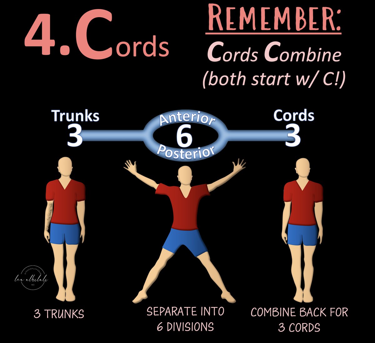
8/After the split of the divisions, the nerves come back together as the cords. It is kind of like doing jumping jacks—they open up and then close back up again
I remember that they come back together as the Cords bc Cords and Combine both start with C
I remember that they come back together as the Cords bc Cords and Combine both start with C
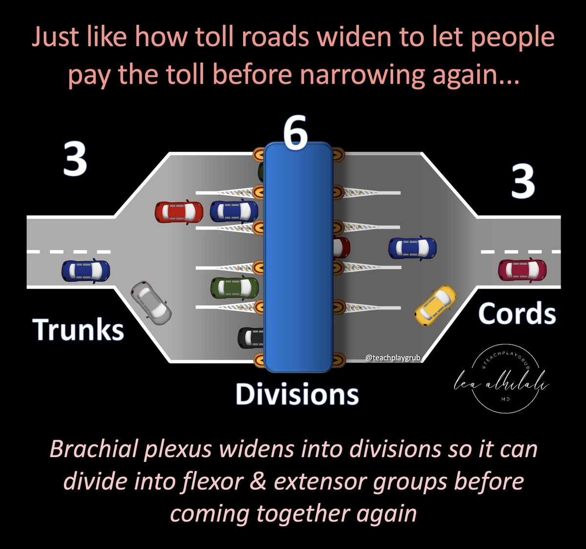
9/It’s like a toll road.
The road widens to let more cars get to the toll booths. Once they have paid the toll, road narrows again
This is what happens w/the divisions—but instead of paying a toll, they are organizing into flexor & extensor groups & coming back together again.
The road widens to let more cars get to the toll booths. Once they have paid the toll, road narrows again
This is what happens w/the divisions—but instead of paying a toll, they are organizing into flexor & extensor groups & coming back together again.
10/But it’s more like going through a worm hole than toll booth.
When you go through a wormhole, you are fundamentally changed when you come out the other side (or so I read on the internet).
Once cords emerge from divisions, they’re either team flexor or extensor & can’t go back
When you go through a wormhole, you are fundamentally changed when you come out the other side (or so I read on the internet).
Once cords emerge from divisions, they’re either team flexor or extensor & can’t go back
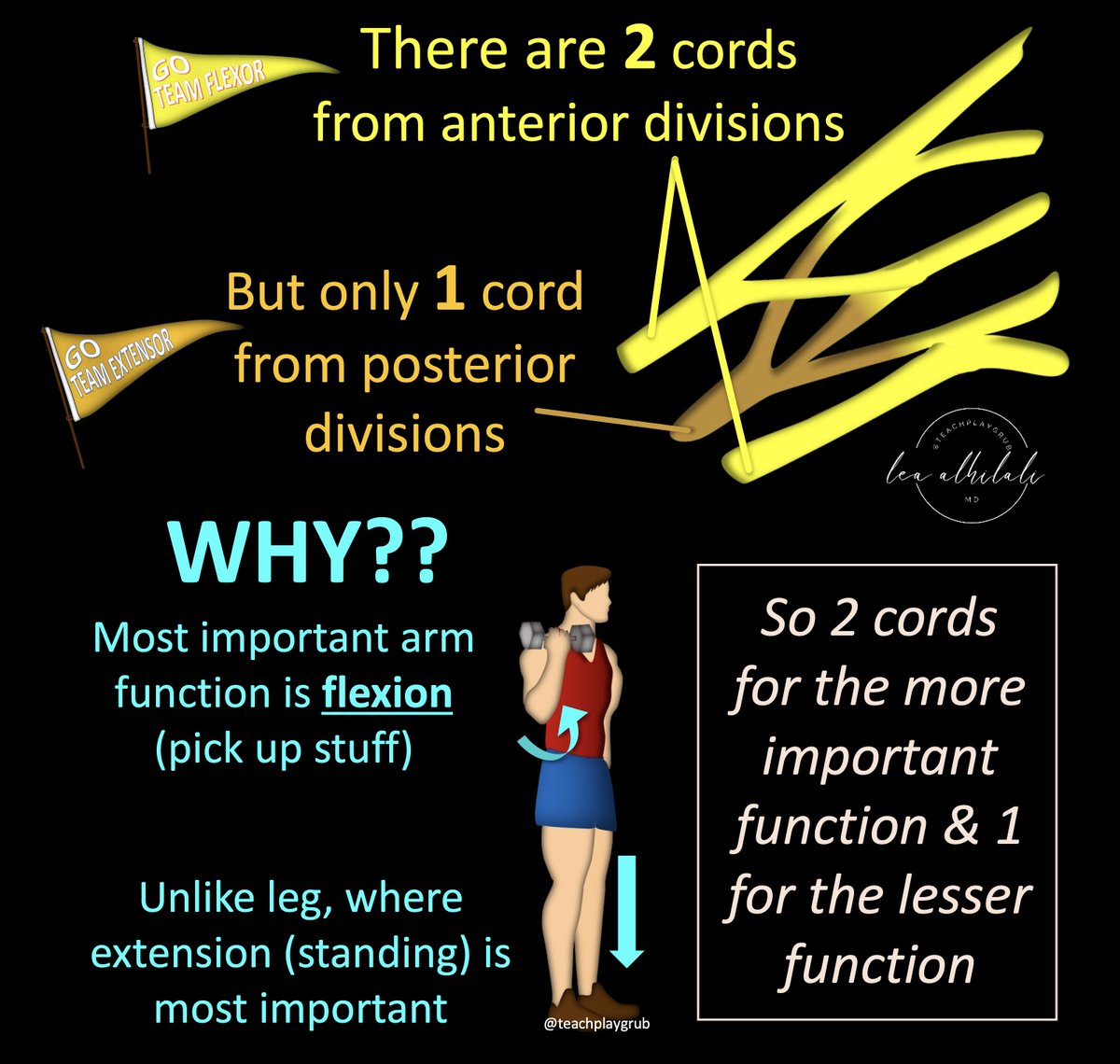
11/Divisions combine to form 2 ant. cords & 1 post. cord. Why the inequality?
Well, the fundamental purpose of the arm is to flex (pick up things), unlike the leg (which is to extend/stand up).
So bc it’s more important to flex, remember 2 cords to flexors & 1 to extensors
Well, the fundamental purpose of the arm is to flex (pick up things), unlike the leg (which is to extend/stand up).
So bc it’s more important to flex, remember 2 cords to flexors & 1 to extensors
12/All post divisions go to the 1 post cord.
How do you remember which ant divisions go into which cord?
How do you remember which ant divisions go into which cord?
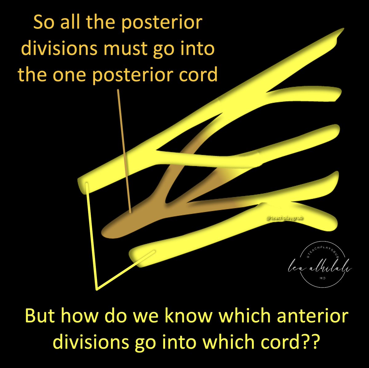
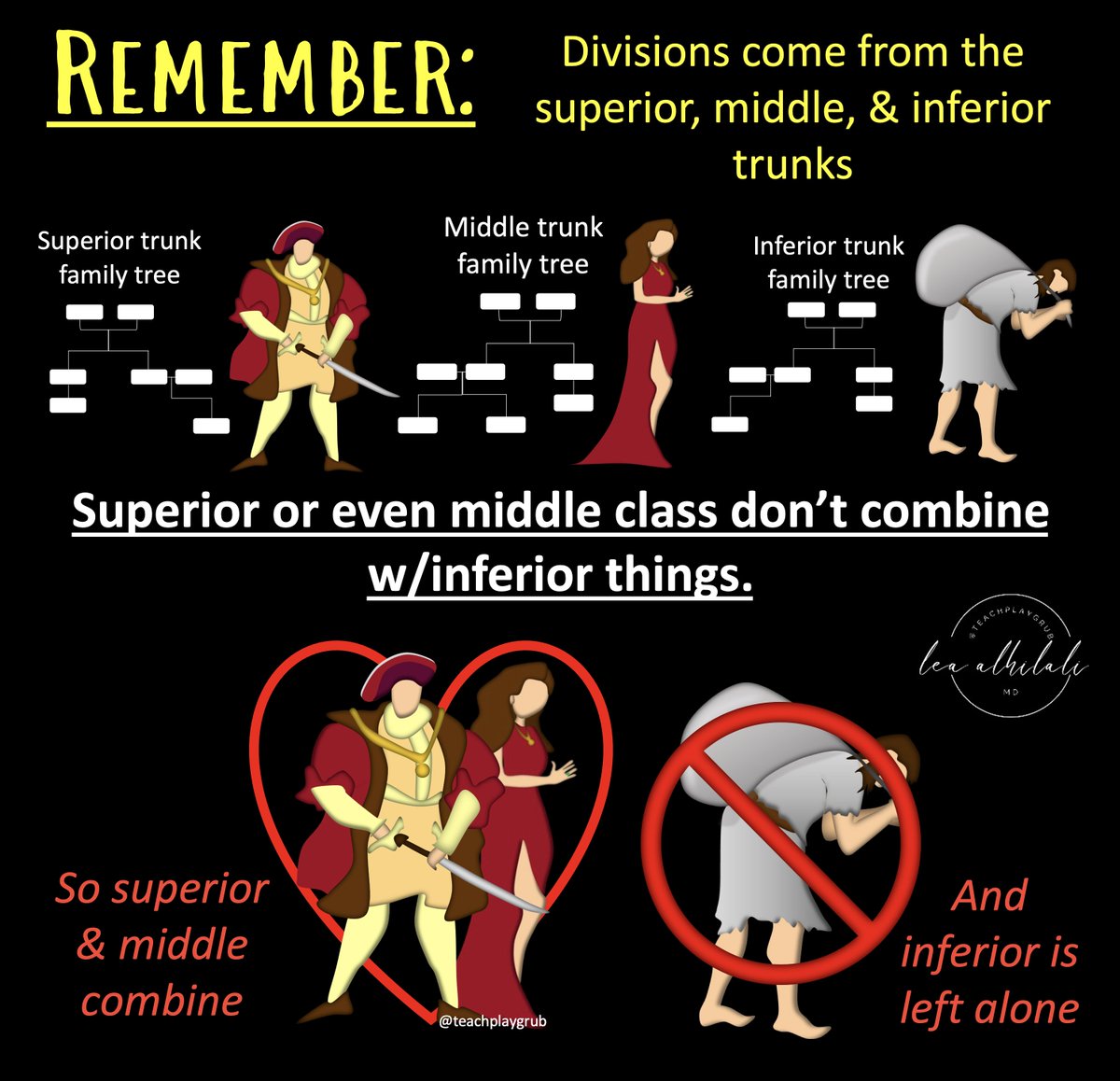
13/Remember, divisions come from the superior, middle, & inferior trunks.
Superior or even middle class don’t combine w/inferior things.
So superior & middle combine. Poor inferior is left alone
Superior or even middle class don’t combine w/inferior things.
So superior & middle combine. Poor inferior is left alone
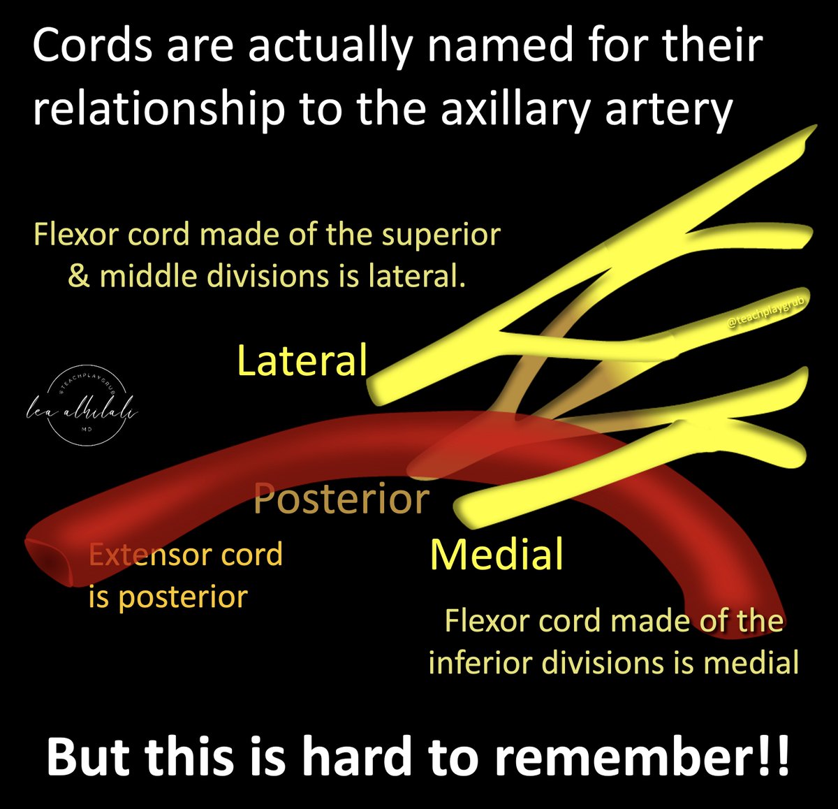
14/Names of the cords are based on their relationship to the axillary artery.
Posterior cord (extensors) is posterior to it.
The flexor cord made of the superior & middle divisions is lateral.
Flexor cord made from the lonely inferior division is medial
But this is hard to remember!
Posterior cord (extensors) is posterior to it.
The flexor cord made of the superior & middle divisions is lateral.
Flexor cord made from the lonely inferior division is medial
But this is hard to remember!
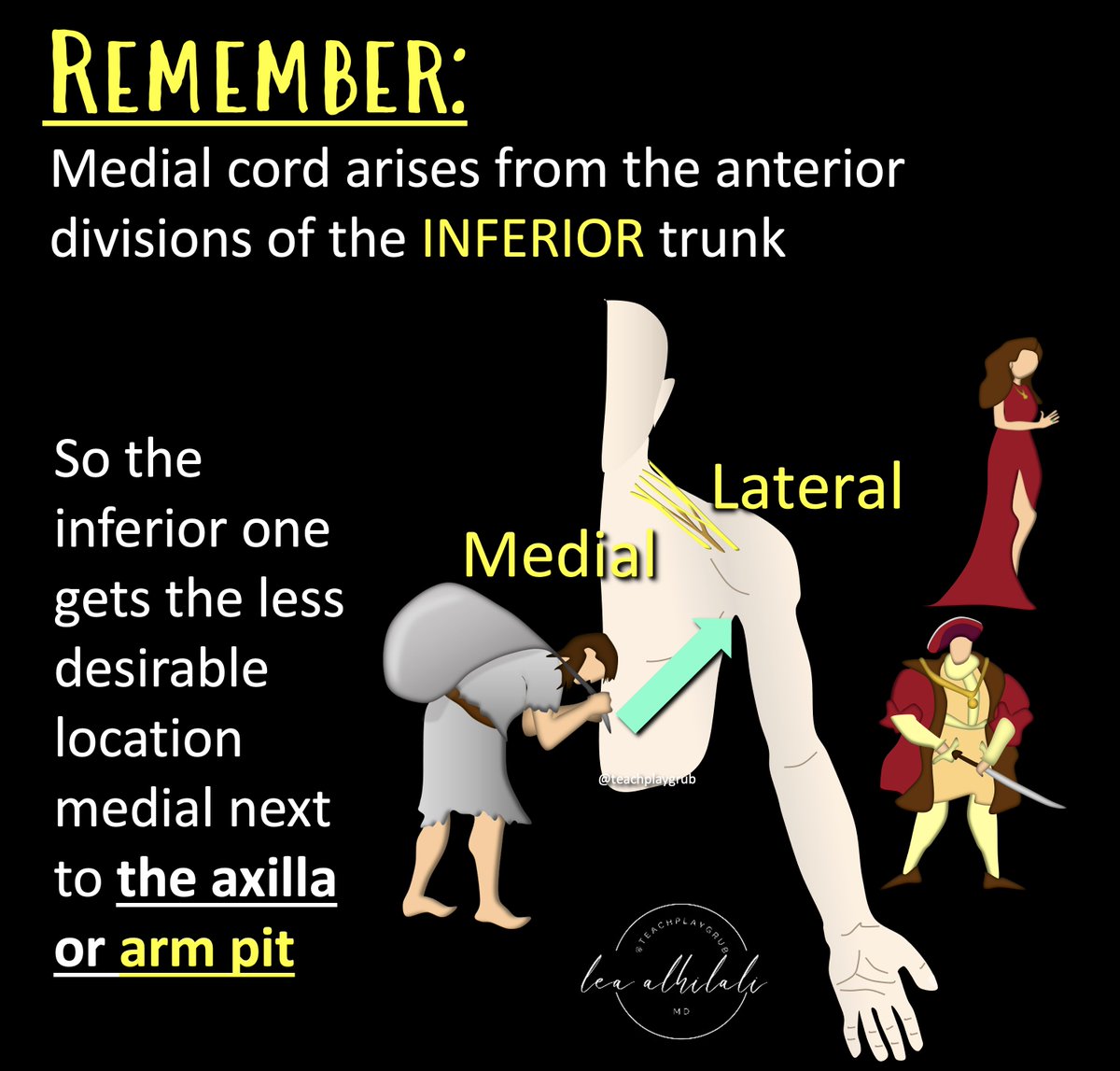
15/But unless you have an axillary artery in front of you, this is hard to remember.
So I remember that the flexor cord made from the poor inferior division is looked down upon, so it is given the worst seat—at the arm pit.
In anatomic positioning, closest to the arm pit is medial, so it is the medial cord.
So I remember that the flexor cord made from the poor inferior division is looked down upon, so it is given the worst seat—at the arm pit.
In anatomic positioning, closest to the arm pit is medial, so it is the medial cord.
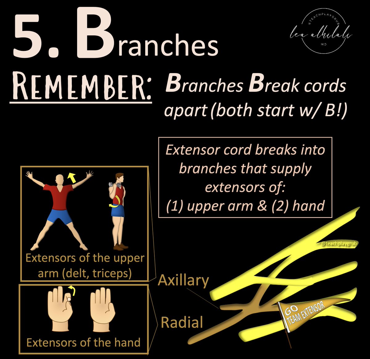
16/Now the final division into branches.
Remember posterior cord only supplies extensors & is the only extensor cord.
So when it branches, it needs to innervate extensors all along the arm (elbow, forearm, hand).
So it gives off axillary to the upper arm & radial to the lower arm
Remember posterior cord only supplies extensors & is the only extensor cord.
So when it branches, it needs to innervate extensors all along the arm (elbow, forearm, hand).
So it gives off axillary to the upper arm & radial to the lower arm
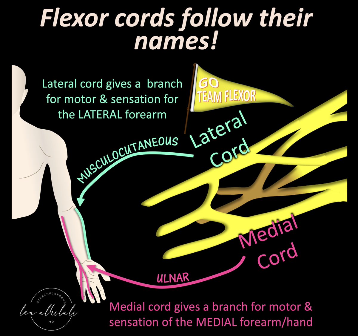
17/Now the branches of the flexor cords
As expected from the names, MEDIAL cord gives a branch for flexors/sensation to MEDIAL forearm/hand (in anatomic position = PINKY side, so ulnar nerve)
LATERAL cord gives a branch for motor/sensation to LATERAL forearm (musculocutaneous)
As expected from the names, MEDIAL cord gives a branch for flexors/sensation to MEDIAL forearm/hand (in anatomic position = PINKY side, so ulnar nerve)
LATERAL cord gives a branch for motor/sensation to LATERAL forearm (musculocutaneous)
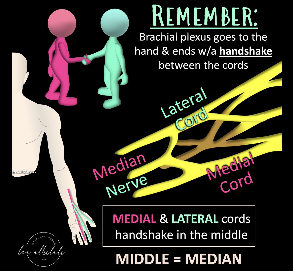
18/Now the brachial plexus goes to the hand & ends w/a handshake!
Medial & lateral cords meet at the end in a handshake in the middle.
And what do you call something in middle: MEDIAN
So medial & lateral cords handshake in the middle to make the median nerve
Medial & lateral cords meet at the end in a handshake in the middle.
And what do you call something in middle: MEDIAN
So medial & lateral cords handshake in the middle to make the median nerve
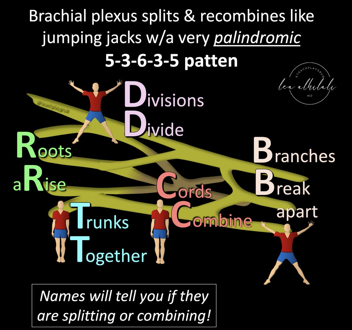
19/Now move beyond mneumonics!
Remember, brachial plexus splits & recombines like jumping jacks w/a very palindromic 5-3-6-3-5 pattern
The names tell you if they are splitting or combining (Trunk=Together, Division=Divide, Cord=Combine, Branch=break)
Remember, brachial plexus splits & recombines like jumping jacks w/a very palindromic 5-3-6-3-5 pattern
The names tell you if they are splitting or combining (Trunk=Together, Division=Divide, Cord=Combine, Branch=break)
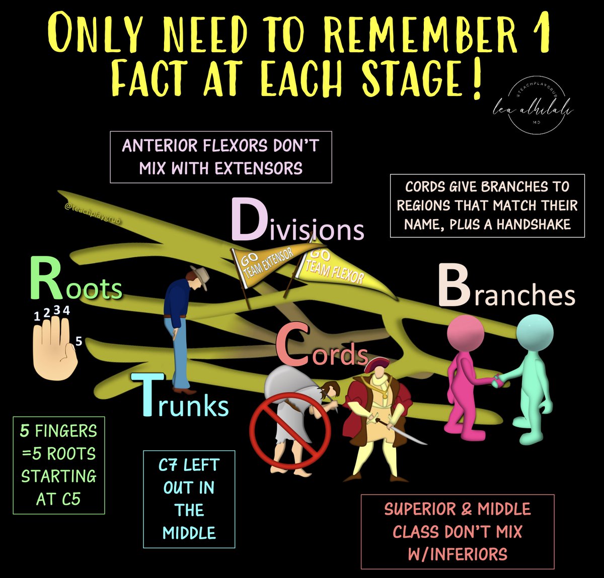
20/Now all you need is to recall 1 fact at each stage!
Trunk: C7 is left out
Div: Ant flexors don’t mix w/post extensors
Cord: Sup/mid class don’t mix w/inferiors
Br: Each cord gives a branch to region its name describes (post, med, lat) & a handshake in the middle!
Trunk: C7 is left out
Div: Ant flexors don’t mix w/post extensors
Cord: Sup/mid class don’t mix w/inferiors
Br: Each cord gives a branch to region its name describes (post, med, lat) & a handshake in the middle!
• • •
Missing some Tweet in this thread? You can try to
force a refresh