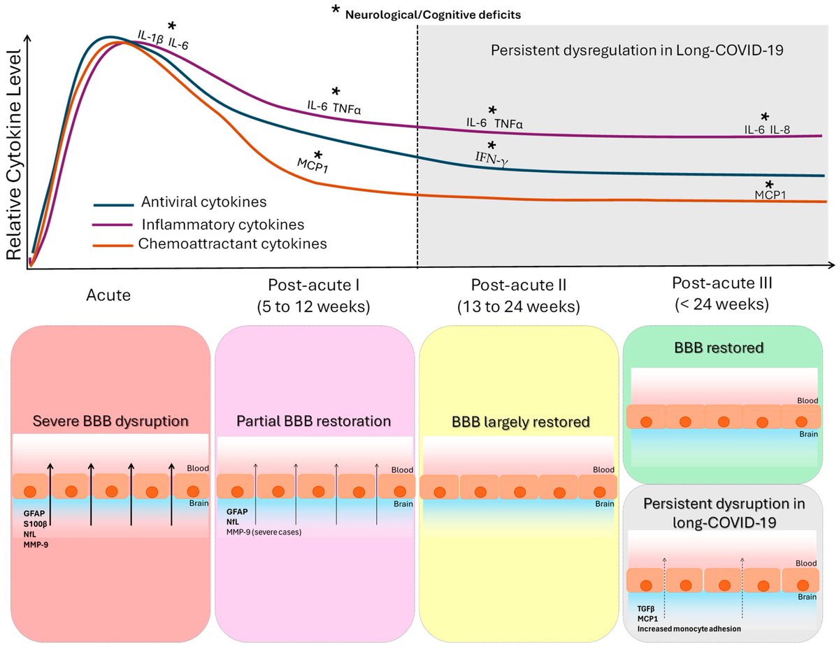
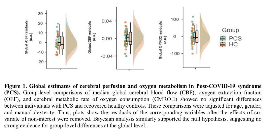
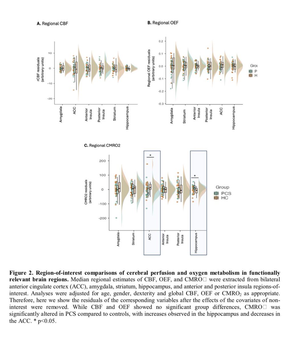
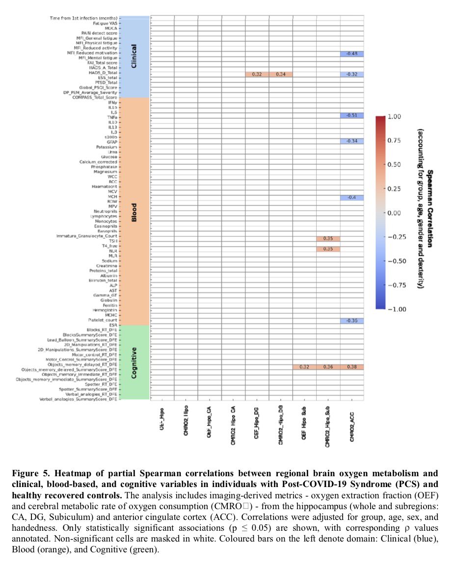
Clinical evidence suggests that SARS-CoV-2 directly disrupts vascular homeostasis, with perfusion abnormalities observed in various tissues. The pancreatic islet, a key endocrine mini-organ reliant on its microvasculature for optimal function, may be particularly vulnerable. 1/
Studies have proposed a link between SARS-CoV-2 infection and islet dysfunction, but the mechanisms remain unclear.
Here, researchers investigated how SARS-CoV-2 spike S1 protein affects human islet microvascular function. 2/
Using confocal microscopy and living pancreas slices from non-diabetic organ donors, they show that a SARS-CoV-2 spike S1 recombinant protein activates pericytes — key regulators of islet capillary diameter and beta cell function—and induces capillary constriction. 3/
These effects are driven by a loss of ACE2 from pericytes’ plasma membrane, impairing ACE2 activity and increasing local angiotensin II levels. 4/
These findings highlight islet pericyte dysfunction as a potential contributor to the diabetogenic effects of SARS-CoV-2 and offer new insights into the mechanisms linking COVID-19, vascular dysfunction and diabetes. 5/5
➡️ Compared with healthy controls,
✔ Long COVID patients had blunted morning cortisol peaks
✔ Higher evening cortisol
✔ Loss of normal circadian pattern
Blood cortisol alone failed to detect these changes. 2/
Key insight:
➡️ Salivary cortisol profiling may be a more sensitive marker of stress-system dysfunction in LongCOVID than standard blood tests.
➡️ HPA axis disruption could underlie:
• Fatigue
• Brain fog
• Sleep disturbance
• Dysautonomia. 3/
➡️ New review highlights that persistent cognitive symptoms in COVID survivors are strongly linked to pro-inflammatory cytokines and blood–brain barrier (BBB) dysfunction.
➡️ Key culprits include IL-6, TNF-α, IL-1β, IL-8, IL-13 and MCP-1 — many remain elevated months after infection.
🔥 COVID-19 is not just a respiratory disease.
➡️ Evidence suggests cognitive impairment can occur due to:
Post-COVID fatigue isn’t just subjective.
Using advanced MRI, researchers found real changes in brain blood flow and oxygen metabolism in people with Post-COVID-19 Syndrome (PCS) after mild infection.
➡️ Key finding:
PCS patients showed increased oxygen metabolism in the hippocampus (memory hub) but reduced metabolism in the anterior cingulate cortex (ACC) — despite no visible brain atrophy. 1/
Why this matters:
➡️ Higher hippocampal metabolism was linked to better cognitive performance, suggesting a compensatory response to maintain thinking and memory in PCS. 2/
In contrast, lower anterior cingulate cortex (ACC) metabolism correlated with:
Why do some people feel exhausted long after COVID-19?
➡️ New brain-imaging research shows that even after mild COVID, people with persistent fatigue can have subtle but real changes in brain structure.
➡️ These changes are not large or widespread, but tend to appear in connected brain networks, especially areas involved in attention, decision-making, and sensory processing. 1/
Importantly, the brain regions affected overlap with areas that naturally express TMPRSS2, a protein that helps SARS-CoV-2 enter cells — suggesting certain brain circuits may be more vulnerable to the virus. 2/
The study also links these changes to brain chemical systems involved in mood, energy, and cognition (serotonin, acetylcholine, glutamate, and cannabinoids). 3/