Vipin M. Vashishtha Profile picture
Jan 31, 2025 12 tweets 4 min read Read on X
Researchers found a link between COVID-19 & blood markers linked to faulty proteins in the brain. They found people who had previously had COVID-19 were more likely to have increased levels of biomarkers linked to faulty amyloid proteins—a hallmark for Alzheimer's disease. 1/ Image
On average, the effects were comparable to 4 years of aging with the greatest effects seen in those hospitalized with severe COVID-19 or with underlying risk factors for dementia such as smoking or high blood pressure. 2/ Image
The findings suggest that mild or moderate COVID may accelerate biological processes that contribute to buildup of disease-promoting amyloid in brain. This raises possibility that COVID-19 might contribute to an increase in later risks of developing Alzheimer's disease. 3/ Image
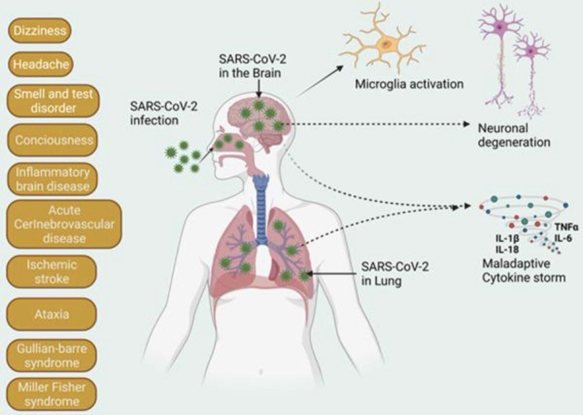
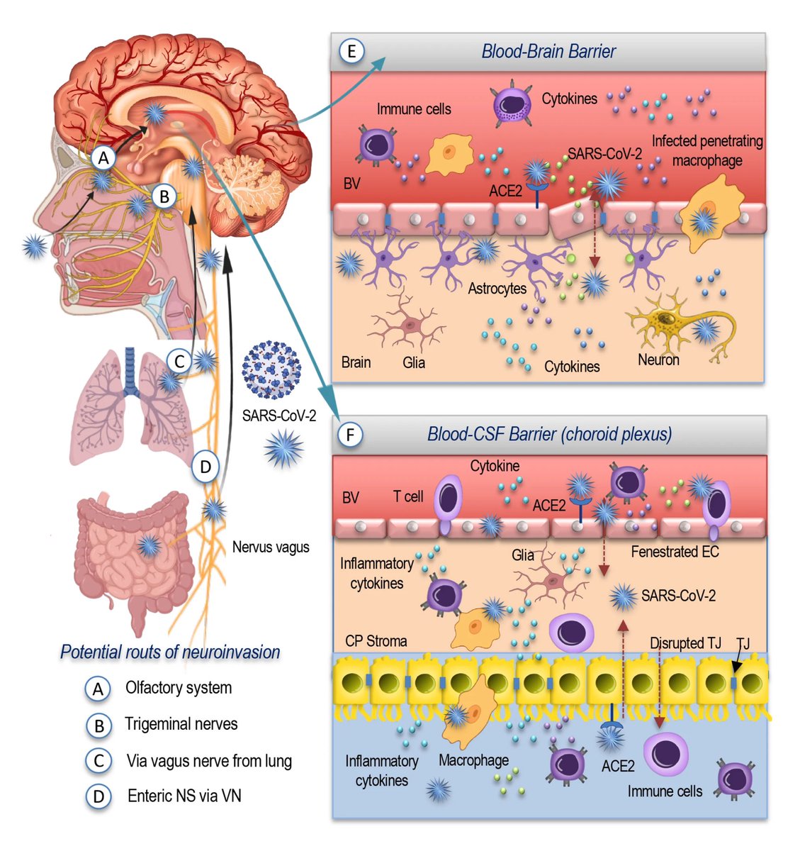
These findings suggest COVID-19 may drive changes which contribute to neurodegenerative disease. This may be due to the inflammation triggered by the disease, although how this inflammation might impact the brain and changes to amyloid is not yet fully clear. 4/ Image
However, the researchers can’t say that catching the SARS-CoV-2 virus directly causes these changes, or if it does, by how much a single episode of infection increases someone's risk. 5/
But these findings do suggest that COVID-19 may increase the risk of Alzheimer's in the future—as has been suggested in the past for other kinds of infections—especially among people with pre-existing risk factors. 6/ Image
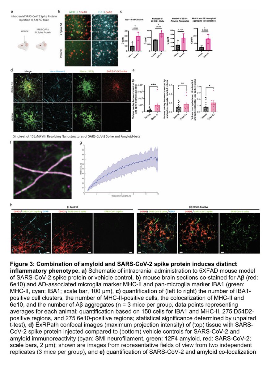
Amyloid is a common protein with a range of functions in the body. But the buildup of an abnormal form of the protein, called beta amyloid (Aβ), is a key component of many diseases. 7/ Image
Aβ forms the characteristic clumps seen in the brains of patients with Alzheimer's disease, which are thought to cause damage to the neurons in the brain, leading to changes in cognition and behavior. 8/ Image
In this study, the researchers found SARS-CoV-2 infection was associated w/ changes in several blood proteins previously linked to brain Aβ pathology. The magnitude of changes was similar to that associated w/ a well-known genetic risk factor for AD, a genetic variant: APOE4 9/ Image
Greater changes found in older participants & those hospitalized with COVID-19-19 or had a history of hypertension. These correlated with poorer cognitive test scores & measures of overall health as well as changes in brain imaging patterns associated w/neurodegeneration 10/ Image
More studies now are needed to prove any causal links. Ultimately, the more we know about factors that contribute to dementia risk—whether they are directly under our control, like lifestyle or diet, or modifiable by vaccines or early treatment for infectious diseases. 11/11 Image
Here is the link to the study👇

nature.com/articles/s4159…

• • •

Missing some Tweet in this thread? You can try to force a refresh
 

Keep Current with Vipin M. Vashishtha

Vipin M. Vashishtha Profile picture

Stay in touch and get notified when new unrolls are available from this author!

Read all threads

This Thread may be Removed Anytime!

PDF

Twitter may remove this content at anytime! Save it as PDF for later use!

Try unrolling a thread yourself!

how to unroll video
  1. Follow @ThreadReaderApp to mention us!

  2. From a Twitter thread mention us with a keyword "unroll"
@threadreaderapp unroll

Practice here first or read more on our help page!

More from @vipintukur

Apr 22
COVID-19 may be, in part, a mitochondrial disease.

➡️ A Cambridge review shows SARS-CoV-2 disrupts mitochondrial function in lung cells—driving inflammation and worsening pneumonia.

➡️ Emerging studies suggest even after the active infection is resolved, residual viral proteins, particularly SARS-CoV-2 spike protein, may linger and continue to cause damage to the mitochondria by increasing oxidative stress and disrupting energy metabolism, offering a plausible mechanism for #LongCOVID. 1/

H/T: @CatchTheBabyImage
COVID-19 is not just viral—it’s metabolic.

SARS-CoV-2 hijacks mitochondria →
↓ Energy production
↑ Inflammatory signaling

A key pathway worsening lung injury. 2/ Image
Mitochondria may link acute COVID → #LongCOVID.

Viral disruption of mitochondrial function can persist, sustaining oxidative stress and immune dysregulation even after infection. 3/ Image
Read 5 tweets
Apr 16
How does COVID affect the brain?

➡️ New research highlights a key player: astrocytes—the brain’s support cells.

👉 SARS-CoV-2 can disrupt their function, with downstream effects on neurons. 1/ Image
Key mechanism:

➡️ The virus can infect or impair astrocytes, which normally:

• Support neurons
• Regulate metabolism
• Maintain brain homeostasis

➡️ Disruption → neuronal dysfunction 2/ Image
What happens next?

➡️ Altered astrocytes can:

• Trigger inflammation
• Impair energy supply to neurons
• Contribute to neuronal injury or death 3/ Image
Read 6 tweets
Apr 10
New study shows SARS-CoV-2 directly damages heart cell mitochondria—key energy engines—offering a mechanistic link to #LongCOVID cardiovascular symptoms. 1/ Image
#LongCOVID may be a mitochondrial disease: electron microscopy reveals structural damage & myofilament breakdown in cardiomyocytes. 2/ Image
Biopsies from LongCOVID patients confirm myocarditis with mitochondrial disruption—mirrored in infected animal models. Strong biological plausibility for persistent cardiac symptoms. 3/ Image
Read 5 tweets
Mar 24
Autoantibodies as drivers of #LongCOVID

➡️ Compelling new evidence strengthens the autoimmune hypothesis of long COVID.

Transfer of patient-derived IgG induces pain-associated behaviours in mice—suggesting a causal, not associative, role.

Key experiment:

➡️ Total IgG from long COVID patients → injected into mice

➡️ Result: mechanical hypersensitivity (allodynia)

This recapitulates a core clinical feature—chronic pain.

➡️ Strikingly, pathogenicity is durable:
IgG collected 2 years later from persistently symptomatic patients
→ still induces pain in vivo

Implies long-term stability of autoreactive clones. 1/Image
Not all LongCOVID is the same.

➡️ Patients stratified using:
• GFAP
• Neurofilament light chain (NFL)
• IFN-β

➡️ Distinct biomarker-defined subgroups with different pathogenic pathways.

Proteome-wide profiling reveals:

➡️ Subgroup-specific autoantibody signatures
➡️ Persistent over time
➡️ Independently validated

Supports biological heterogeneity rather than a single syndrome. 2/Image
Conceptually aligns with conditions like fibromyalgia:

👉 Chronic symptoms driven by functional autoantibodies
👉 Neuro-immune interface involvement

➡️ Clinical implications:

• Identifying pathogenic IgG could enable risk stratification
• Opens avenues for targeted immunomodulation (e.g., IVIG, plasmapheresis, B-cell therapies?)

➡️ Methodological strength:

-Functional transfer model (human → mouse)
-Longitudinal sampling
-Multi-omics support

➡️ Moves the field from correlation → causation. 3/Image
Image
Read 4 tweets
Mar 13
New research finds that SARS-CoV-2 spike protein can persist in the gut of people with #LongCOVID, even months after infection.

➡️ This persistent viral antigen may drive ongoing immune changes in intestinal tissue.

➡️ Scientists detected viral spike RNA and protein in colon and ileum biopsies from Long COVID patients.

➡️ In these regions, genes linked to inflammation, immune dysfunction, and tissue stress were altered. 1/Image
Persistent spike-positive areas in the colon showed increased immune cell activity, including:

• Macrophages
• Plasma cells
• Regulatory T cells

Suggesting an active local immune response in the gut.

➡️ Researchers also found disrupted expression of key immune-signaling genes, indicating impaired immune coordination and chronic inflammation in gut tissues. 2/Image
SARS-CoV-2 persistence is a proposed driver of Long COVID (LC), but the in-situ relationship between residual viral antigen and immune dysregulation remains poorly defined.

➡️ This NEW study provides robust evidence that persistent SARS-CoV-2 Spike protein detection in the gut is not immunologically inert.

➡️ Instead, it is actively associated with distinct, immune cell composition shifts and a dysfunctional pro-inflammatory transcriptional profile, supporting the hypothesis that retained viral antigen drives chronic immune dysregulation in tissue of LongCOVID subjects. 3/Image
Read 4 tweets
Mar 10
New research suggests that gut bacteria may contribute to neurological symptoms in #LongCOVID.

➡️ Small particles released by gut microbes—called extracellular vesicles—may trigger inflammation affecting both the body and the brain.

➡️ Scientists found that people with Long COVID and neurological symptoms show a persistent imbalance in gut microbiota.

➡️ This altered microbiome may disrupt the intestinal barrier and promote systemic inflammation. 1/Image
In experiments, transferring gut microbes from LongCOVID patients into mice caused intestinal barrier damage and signs of brain inflammation.

➡️ This suggests a biological link between the gut and neurological symptoms. 2/ Image
Gut microbe–derived vesicles were shown to activate inflammatory pathways and immune cells, including microglia in the brain.

➡️ These processes may contribute to symptoms such as brain fog. 3/ Image
Read 4 tweets

Did Thread Reader help you today?

Support us! We are indie developers!


This site is made by just two indie developers on a laptop doing marketing, support and development! Read more about the story.

Become a Premium Member ($3/month or $30/year) and get exclusive features!

Become Premium

Don't want to be a Premium member but still want to support us?

Make a small donation by buying us coffee ($5) or help with server cost ($10)

Donate via Paypal

Or Donate anonymously using crypto!

Ethereum

0xfe58350B80634f60Fa6Dc149a72b4DFbc17D341E copy

Bitcoin

3ATGMxNzCUFzxpMCHL5sWSt4DVtS8UqXpi copy

Thank you for your support!

Follow Us!

:(