Vipin M. Vashishtha Profile picture
May 28, 2025 13 tweets 4 min read Read on X
A significant discovery in the fight against #LongCovid!

➡️ Researchers have identified the epipharynx, a part of the pharynx, as a key site for chronic inflammation driven by residual SARS-CoV-2 RNA. 1/ Image
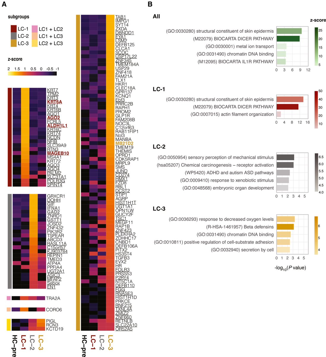
Using a next-generation molecular mapping technology called Visium HD spatial transcriptomics, researchers from Japan provided the world's first high-resolution spatial gene expression analysis of the epipharynx in patients with longCOVID. 2/ Image
According to the study, the viral RNA from SARS-CoV-2 can persist in the epipharynx for more than six months post-infection, and here they activate local immune signals in specialized cells like B cells, plasmacytoid dendritic cells, and ciliated epithelial cells. 3/ Image
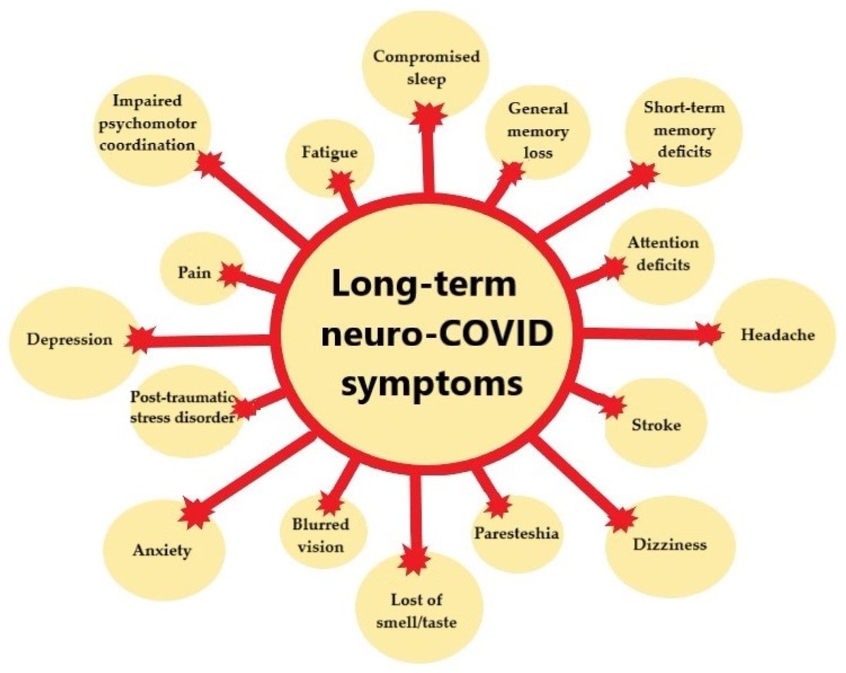
This signaling potentially contributes to the chronic symptoms experienced by patients with long COVID, which include fatigue, persistent cough, dizziness, and cognitive issues continuing for months after the acute phase of infection. 4/ Image
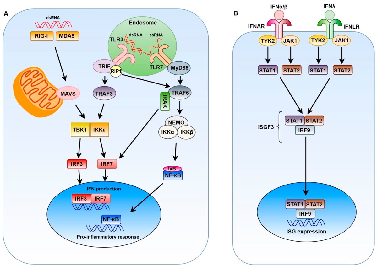
Using advanced technique, the researchers found that the residual viral RNA isn't merely leftover debris; instead, it actively triggers immune responses and inflammation. 5/ Image
The epipharynx is one of the most important sites for SARS-CoV-2 infection, yet its anatomical location makes it difficult to observe w/out endoscopic evaluation. This region will gain more research attention as a potential target in understanding and treating longCOVID 6/ Image
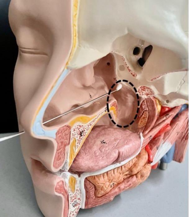
To address the issue, the team explored epipharyngeal abrasive therapy (EAT) as a treatment. EAT is a treatment for chronic epipharyngitis that has been practiced in since the 1960s, involving the swabbing of the epipharynx with a 1% zinc chloride solution. 7/ Image
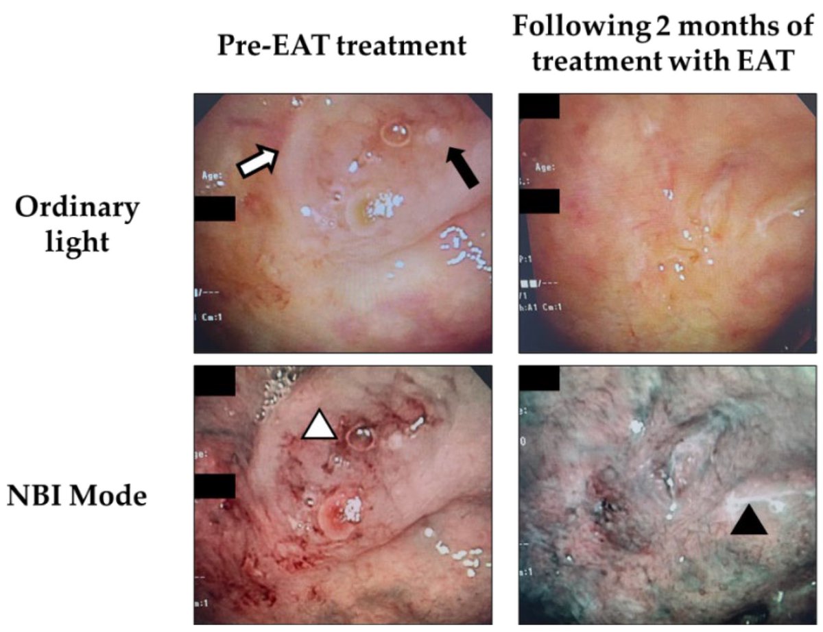
After three months of weekly EAT treatment, the patients showed a remarkable improvement in symptoms. 8/ Image
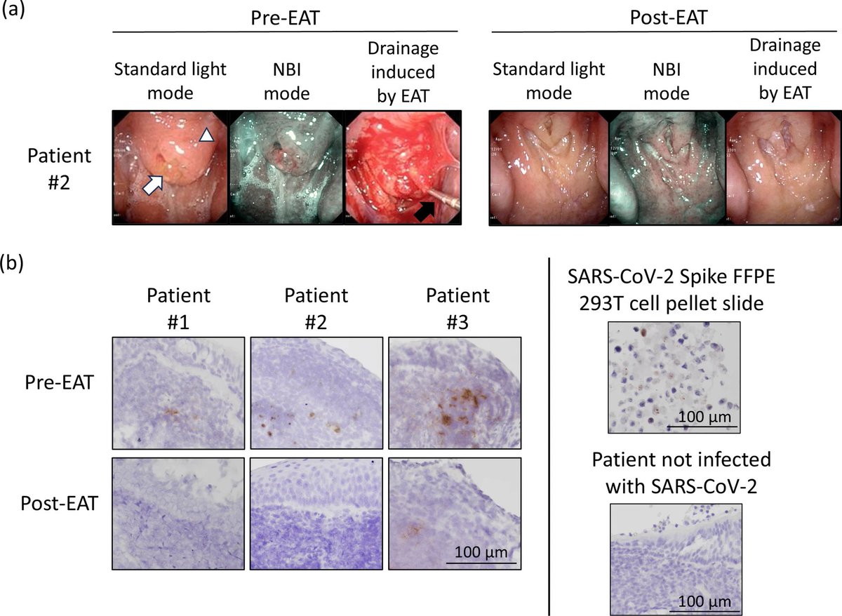
On a closer analysis, the researchers observed a significant reduction in the viral RNA and a suppression of inflammatory responses marked by a decrease in expression of signaling molecules like pro-inflammatory cytokines and antibody-related genes. 9/
The spatial gene analysis post-treatment revealed that EAT promotes the removal of damaged ciliated epithelium. Additionally, it also downregulates the overactive immune pathways, underscoring its promising role in immune modulation and tissue repair. 10/ Image
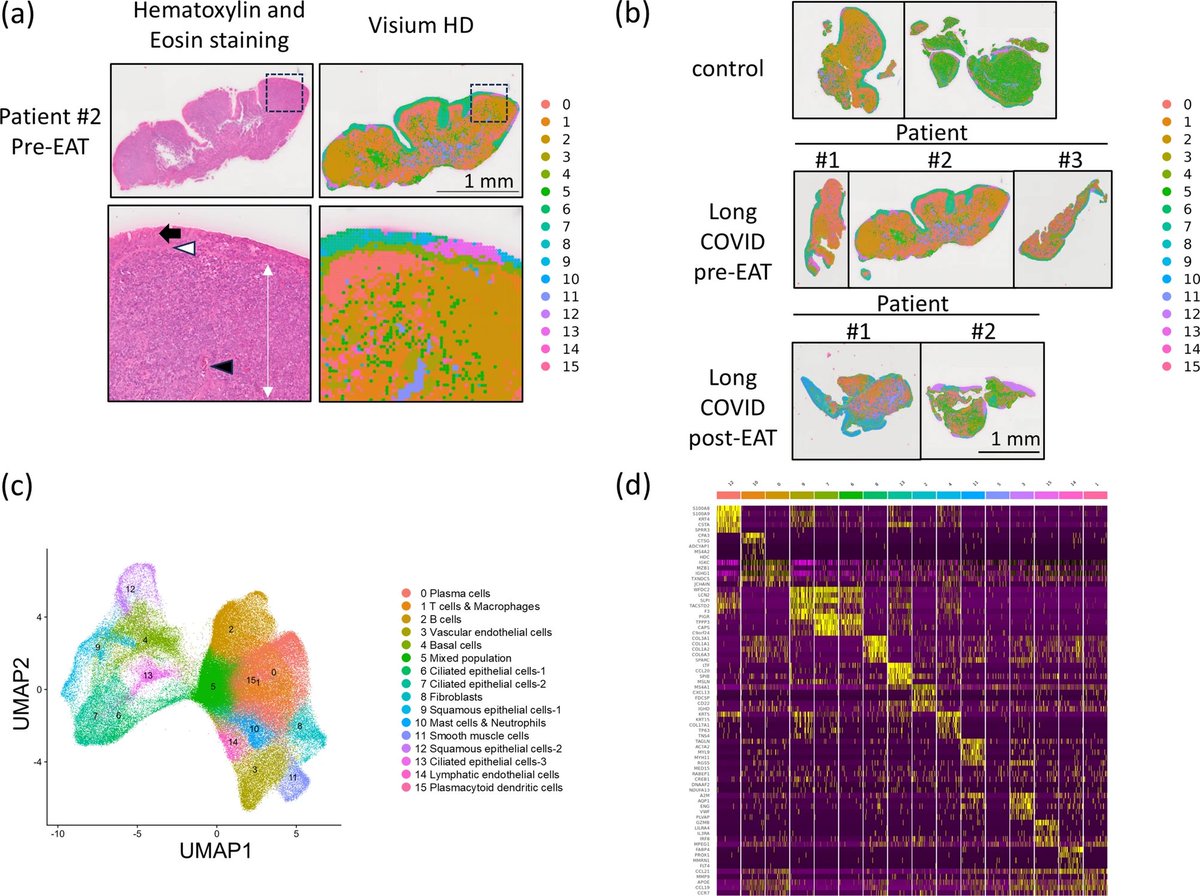
Spatial transcriptomic analysis has enabled a deeper understanding of the biology of longCOVID from a completely different perspective than conventional approaches, including the potential application of a treatment already in clinical use in Japan. 11/
While most existing treatments focus on managing these symptoms, this study goes deeper, identifying the epipharynx as a hidden reservoir of viral RNA and immune disruption, thereby opening new avenues for more targeted therapies. 12/
The study marks a significant leap in medical science, especially in light of estimates showing that 31–69% of recovered COVID-19 patients continue to suffer lingering symptoms. 13/13

nature.com/articles/s4159…

• • •

Missing some Tweet in this thread? You can try to force a refresh
 

Keep Current with Vipin M. Vashishtha

Vipin M. Vashishtha Profile picture

Stay in touch and get notified when new unrolls are available from this author!

Read all threads

This Thread may be Removed Anytime!

PDF

Twitter may remove this content at anytime! Save it as PDF for later use!

Try unrolling a thread yourself!

how to unroll video
  1. Follow @ThreadReaderApp to mention us!

  2. From a Twitter thread mention us with a keyword "unroll"
@threadreaderapp unroll

Practice here first or read more on our help page!

More from @vipintukur

May 17
SARS-CoV-2 spike protein may directly amplify brain inflammation.

➡️ Researchers found that spike proteins can colocalize with amyloid-β (Aβ) and trigger distinct inflammatory responses in microglia — the brain’s immune cells.

➡️ This raises important questions about potential long-term neurodegenerative consequences of COVID-19. 1/Image
Researchers developed advanced “expansion microscopy” techniques that physically enlarge human brain tissue, allowing scientists to see disease-related structures at near-nanoscale resolution using ordinary microscopes. 2/ Image
Applying this method to brains from some COVID-19 patients revealed tiny amyloid-like protein clusters closely associated with SARS-CoV-2 particles in a small subset of cases, suggesting a possible link between COVID-19, neuroinflammation, and abnormal protein aggregation in the brain.

The study highlights how ultra-high-resolution imaging could uncover previously hidden mechanisms of neurological disease. 3/Image
Read 4 tweets
May 12
#LongCOVID is increasingly emerging as an immune-mediated disorder driven by:

➡️ Viral persistence
➡️ Chronic inflammation
➡️ Immune dysregulation
➡️ Tissue remodeling

👉 The lungs may remain biologically altered long after acute infection resolves. 1/ Image
A new review highlights how persistent immune activation in LongCOVID may lead to:
• Fibrosis-like lung changes
• Endothelial dysfunction
• Microvascular injury
• Ongoing respiratory symptoms

COVID may end clinically—but not biologically.
#LongCOVID #Pulmonology 2/ Image
LongCOVID respiratory sequelae may result from a “perfect storm” of:

➡️ Aberrant immune signaling
➡️ Residual viral antigens
➡️ Microvascular dysfunction
➡️ Dysregulated tissue repair

👉 A unifying pathophysiology is slowly taking shape. 3/ Image
Read 9 tweets
Apr 22
COVID-19 may be, in part, a mitochondrial disease.

➡️ A Cambridge review shows SARS-CoV-2 disrupts mitochondrial function in lung cells—driving inflammation and worsening pneumonia.

➡️ Emerging studies suggest even after the active infection is resolved, residual viral proteins, particularly SARS-CoV-2 spike protein, may linger and continue to cause damage to the mitochondria by increasing oxidative stress and disrupting energy metabolism, offering a plausible mechanism for #LongCOVID. 1/

H/T: @CatchTheBabyImage
COVID-19 is not just viral—it’s metabolic.

SARS-CoV-2 hijacks mitochondria →
↓ Energy production
↑ Inflammatory signaling

A key pathway worsening lung injury. 2/ Image
Mitochondria may link acute COVID → #LongCOVID.

Viral disruption of mitochondrial function can persist, sustaining oxidative stress and immune dysregulation even after infection. 3/ Image
Read 5 tweets
Apr 16
How does COVID affect the brain?

➡️ New research highlights a key player: astrocytes—the brain’s support cells.

👉 SARS-CoV-2 can disrupt their function, with downstream effects on neurons. 1/ Image
Key mechanism:

➡️ The virus can infect or impair astrocytes, which normally:

• Support neurons
• Regulate metabolism
• Maintain brain homeostasis

➡️ Disruption → neuronal dysfunction 2/ Image
What happens next?

➡️ Altered astrocytes can:

• Trigger inflammation
• Impair energy supply to neurons
• Contribute to neuronal injury or death 3/ Image
Read 6 tweets
Apr 10
New study shows SARS-CoV-2 directly damages heart cell mitochondria—key energy engines—offering a mechanistic link to #LongCOVID cardiovascular symptoms. 1/ Image
#LongCOVID may be a mitochondrial disease: electron microscopy reveals structural damage & myofilament breakdown in cardiomyocytes. 2/ Image
Biopsies from LongCOVID patients confirm myocarditis with mitochondrial disruption—mirrored in infected animal models. Strong biological plausibility for persistent cardiac symptoms. 3/ Image
Read 5 tweets
Mar 24
Autoantibodies as drivers of #LongCOVID

➡️ Compelling new evidence strengthens the autoimmune hypothesis of long COVID.

Transfer of patient-derived IgG induces pain-associated behaviours in mice—suggesting a causal, not associative, role.

Key experiment:

➡️ Total IgG from long COVID patients → injected into mice

➡️ Result: mechanical hypersensitivity (allodynia)

This recapitulates a core clinical feature—chronic pain.

➡️ Strikingly, pathogenicity is durable:
IgG collected 2 years later from persistently symptomatic patients
→ still induces pain in vivo

Implies long-term stability of autoreactive clones. 1/Image
Not all LongCOVID is the same.

➡️ Patients stratified using:
• GFAP
• Neurofilament light chain (NFL)
• IFN-β

➡️ Distinct biomarker-defined subgroups with different pathogenic pathways.

Proteome-wide profiling reveals:

➡️ Subgroup-specific autoantibody signatures
➡️ Persistent over time
➡️ Independently validated

Supports biological heterogeneity rather than a single syndrome. 2/Image
Conceptually aligns with conditions like fibromyalgia:

👉 Chronic symptoms driven by functional autoantibodies
👉 Neuro-immune interface involvement

➡️ Clinical implications:

• Identifying pathogenic IgG could enable risk stratification
• Opens avenues for targeted immunomodulation (e.g., IVIG, plasmapheresis, B-cell therapies?)

➡️ Methodological strength:

-Functional transfer model (human → mouse)
-Longitudinal sampling
-Multi-omics support

➡️ Moves the field from correlation → causation. 3/Image
Image
Read 4 tweets

Did Thread Reader help you today?

Support us! We are indie developers!


This site is made by just two indie developers on a laptop doing marketing, support and development! Read more about the story.

Become a Premium Member ($3/month or $30/year) and get exclusive features!

Become Premium

Don't want to be a Premium member but still want to support us?

Make a small donation by buying us coffee ($5) or help with server cost ($10)

Donate via Paypal

Or Donate anonymously using crypto!

Ethereum

0xfe58350B80634f60Fa6Dc149a72b4DFbc17D341E copy

Bitcoin

3ATGMxNzCUFzxpMCHL5sWSt4DVtS8UqXpi copy

Thank you for your support!

Follow Us!

:(