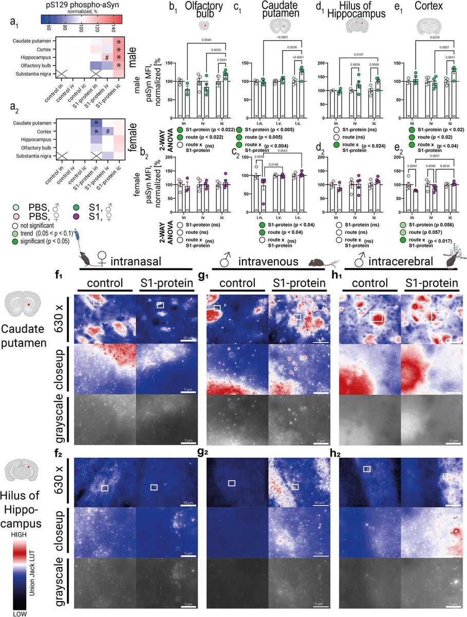
A new study from Germany found that intravenous administration of the SARS-CoV-2 spike protein in mice led to neuroinflammation and accumulation of alpha-synuclein in brain regions associated with Parkinson’s disease. 1/
Authors also discovered “sex-dependent alterations in astrocyte reactivity and parvalbumin-positive interneurons.” 2/
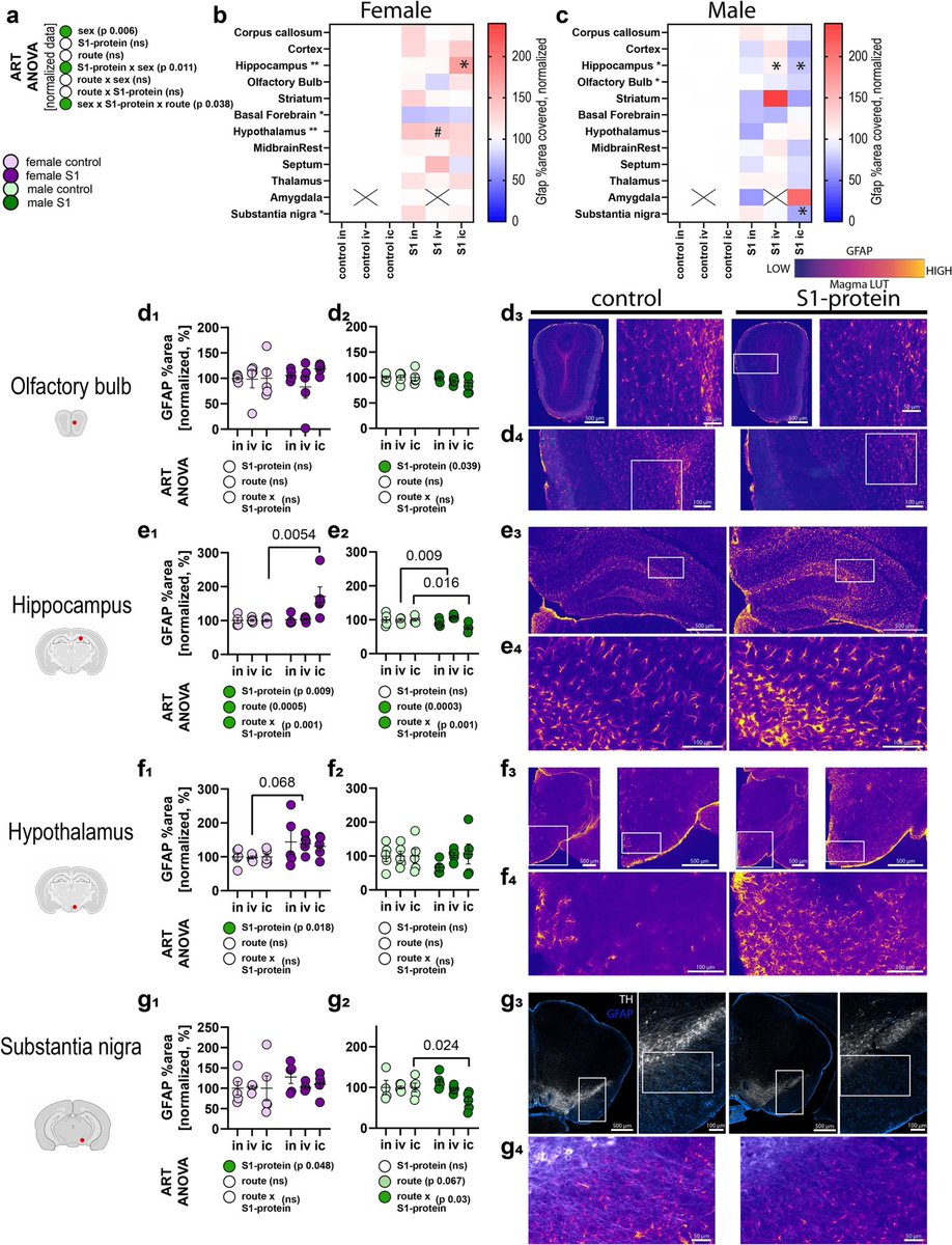
These findings suggest that exposure to the spike protein alone, without full viral infection, may contribute to neurodegenerative processes linked to Parkinson's, thus highlighting potential long-term neurological risks following COVID infection. 3/
ACE2 and Neuropilin-1 receptor expression altered following S1-protein exposure.
Most pronounced effects occur in brain regions associated with Parkinson’s disease pathology. 4/
The above mentioned study builds upon Ali Erturk’s lab report from December 2024 which showed that persistence of spike protein at the skull-meninges-brain axis induced neurological damage in mice. 5/
Vaccination reduced but did not eliminate spike protein accumulation after infection in mice. These findings suggest persistent spike protein at the brain borders may contribute to lasting neurological sequelae of COVID-19. 6/6
• • •
Missing some Tweet in this thread? You can try to
force a refresh
➡️ Compared with healthy controls,
✔ Long COVID patients had blunted morning cortisol peaks
✔ Higher evening cortisol
✔ Loss of normal circadian pattern
Blood cortisol alone failed to detect these changes. 2/
Key insight:
➡️ Salivary cortisol profiling may be a more sensitive marker of stress-system dysfunction in LongCOVID than standard blood tests.
➡️ HPA axis disruption could underlie:
• Fatigue
• Brain fog
• Sleep disturbance
• Dysautonomia. 3/
➡️ New review highlights that persistent cognitive symptoms in COVID survivors are strongly linked to pro-inflammatory cytokines and blood–brain barrier (BBB) dysfunction.
➡️ Key culprits include IL-6, TNF-α, IL-1β, IL-8, IL-13 and MCP-1 — many remain elevated months after infection.
🔥 COVID-19 is not just a respiratory disease.
➡️ Evidence suggests cognitive impairment can occur due to:
Post-COVID fatigue isn’t just subjective.
Using advanced MRI, researchers found real changes in brain blood flow and oxygen metabolism in people with Post-COVID-19 Syndrome (PCS) after mild infection.
➡️ Key finding:
PCS patients showed increased oxygen metabolism in the hippocampus (memory hub) but reduced metabolism in the anterior cingulate cortex (ACC) — despite no visible brain atrophy. 1/
Why this matters:
➡️ Higher hippocampal metabolism was linked to better cognitive performance, suggesting a compensatory response to maintain thinking and memory in PCS. 2/
In contrast, lower anterior cingulate cortex (ACC) metabolism correlated with:
Why do some people feel exhausted long after COVID-19?
➡️ New brain-imaging research shows that even after mild COVID, people with persistent fatigue can have subtle but real changes in brain structure.
➡️ These changes are not large or widespread, but tend to appear in connected brain networks, especially areas involved in attention, decision-making, and sensory processing. 1/
Importantly, the brain regions affected overlap with areas that naturally express TMPRSS2, a protein that helps SARS-CoV-2 enter cells — suggesting certain brain circuits may be more vulnerable to the virus. 2/
The study also links these changes to brain chemical systems involved in mood, energy, and cognition (serotonin, acetylcholine, glutamate, and cannabinoids). 3/
COVID-19 doesn’t just affect the lungs — it can disrupt how cells produce energy. New research shows that COVID-19 alters the genetic “switches” that control mitochondria, the structures that power our cells. 1/
By comparing people who died from severe COVID-19, those who recovered, and healthy individuals, researchers found lasting changes in how mitochondrial genes are regulated. These changes were most prominent in genes involved in energy production and metabolism. 2/
Importantly, people with COVID-19 showed abnormally high levels of proteins that control mitochondrial structure and stress responses, suggesting long-term damage to the cell’s energy system. 3/
#LongCOVID (LC) shares striking symptom overlap with hypermobility spectrum disorders (HSD/hEDS): fatigue, brain fog, dysautonomia, pain—especially in women.
➡️ A new case series explores whether some “intractable” LC may reflect undiagnosed hypermobility disorders.
➡️ Five women with persistent LC symptoms were evaluated at an hEDS/HSD clinic.
All met Beighton score criteria for hypermobility.
➡️ 4 diagnosed with hEDS, 1 with HSD
➡️ 3 had dysautonomia
None had prior hypermobility diagnoses. 1/
All patients carried MTHFR polymorphisms (C677T or A1298C)—recently linked to hEDS/HSD.
➡️ Several also showed features of mast cell activation, suggesting immune dysregulation may unmask latent connective tissue disorders after SARS-CoV-2 infection.
➡️ Targeted management (physical therapy, methylfolate/B12, mast cell stabilization, pain interventions) led to clinical improvement in all cases.
🔑 Takeaway: Consider hEDS/HSD in women with refractory Long COVID, especially with multisystem pain and dysautonomia. 2/
This case series suggests that some patients with severe, persistent #LongCOVID—especially women—may have previously undiagnosed hypermobility disorders (hEDS/HSD).
➡️ Five women with refractory LongCOVID symptoms were found to meet criteria for hypermobility, often with dysautonomia, mast cell–related features, and MTHFR polymorphisms.
➡️ Targeted management led to clinical improvement, highlighting the need to consider hEDS/HSD in patients with intractable Long COVID symptoms. 3/