1~ A THREAD EVERY HUMAN BEING NEEDS TO READ & WILL AFFECT EVERY PERSON ON THIS PLANET REGARDLESS OF VACCINE STATUS ~
~> Amyloidogenic Fibrin Microclotting Following Prenatal mRNA Vaccination Exposure <~
*** HOUSTON, WE HAVE A PROBLEM ***
(@KevinMcCairnPhD)
05.24.25 at 20:59
~> Amyloidogenic Fibrin Microclotting Following Prenatal mRNA Vaccination Exposure <~
*** HOUSTON, WE HAVE A PROBLEM ***
(@KevinMcCairnPhD)
05.24.25 at 20:59

2~ PREAMBLE: Houston, WE HAVE A PROBLEM!
•Scientific investigations involving emerging & potentially paradigm-shifting findings often walk a difficult line between the need for caution & the imperative to inform
• While early publication of case studies carries inherent risks—such as overinterpretation of individual data points or lack of statistical power—it also provides critical, time-sensitive insights that can drive new lines of inquiry & inform ongoing clinical & public health departments
• This report forms part of a robust, real-time investigation into the proteopathic & vascular consequences of prenatal exposure to mRNA-based SARS-COV-2 “vaccines”
• The intention is not to draw definitive epidemiological conclusions at this stage, but to publicly document the emergence of novel findings as they occur
• This transparent approach is particularly important in areas where existing safety literature has not yet integrated proteomic misfolding or amyloidogenic biomarker screening into its framework
• This investigative format mirrors the best practices seen in real-time pathogen tracking & pharmacovigilance
• In such contexts, timelines & transparency are essential for mitigating long-term risk & prompting refinement of public health frameworks
(@KevinMcCairnPhD)
•Scientific investigations involving emerging & potentially paradigm-shifting findings often walk a difficult line between the need for caution & the imperative to inform
• While early publication of case studies carries inherent risks—such as overinterpretation of individual data points or lack of statistical power—it also provides critical, time-sensitive insights that can drive new lines of inquiry & inform ongoing clinical & public health departments
• This report forms part of a robust, real-time investigation into the proteopathic & vascular consequences of prenatal exposure to mRNA-based SARS-COV-2 “vaccines”
• The intention is not to draw definitive epidemiological conclusions at this stage, but to publicly document the emergence of novel findings as they occur
• This transparent approach is particularly important in areas where existing safety literature has not yet integrated proteomic misfolding or amyloidogenic biomarker screening into its framework
• This investigative format mirrors the best practices seen in real-time pathogen tracking & pharmacovigilance
• In such contexts, timelines & transparency are essential for mitigating long-term risk & prompting refinement of public health frameworks
(@KevinMcCairnPhD)


3~ *Clinical Case Summary*
SUBJECT
• Patient B3
MATERNAL VACCINATION
• Pfizer
(BNT162b2)
• mother received 2 doses
• 1 dose
(at 32 weeks gestation)
• 1 dose
(at 34 weeks gestation)
Gestational age at BIRTH
• 35 Weeks
(preterm)
AGE AT SAMPLING
• 3 years old
HISTORY
• Premature delivery
• resuscitated at birth
(reuired CPR)
• dead on arrival
(DOA equals no VS)
• immune dysregulation (tonsillectomy, repeated ear infection/surgery
• congenital heart murmur
SAMPLE INTEGRITY
• Excellent
METHODOLOGY
• Thioflavin T staining
• autofluorescence imaging • UV Light Microscopy
(4X, scale bar 50 um scale)
• samples preserved
(for SEM/EDX)
(@KevinMcCairnPhD)
SUBJECT
• Patient B3
MATERNAL VACCINATION
• Pfizer
(BNT162b2)
• mother received 2 doses
• 1 dose
(at 32 weeks gestation)
• 1 dose
(at 34 weeks gestation)
Gestational age at BIRTH
• 35 Weeks
(preterm)
AGE AT SAMPLING
• 3 years old
HISTORY
• Premature delivery
• resuscitated at birth
(reuired CPR)
• dead on arrival
(DOA equals no VS)
• immune dysregulation (tonsillectomy, repeated ear infection/surgery
• congenital heart murmur
SAMPLE INTEGRITY
• Excellent
METHODOLOGY
• Thioflavin T staining
• autofluorescence imaging • UV Light Microscopy
(4X, scale bar 50 um scale)
• samples preserved
(for SEM/EDX)
(@KevinMcCairnPhD)


3~ Microscopic Findings ~
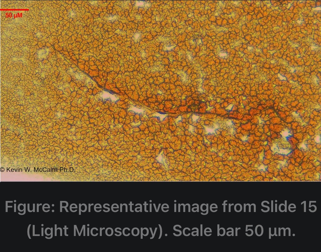
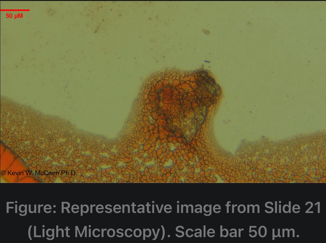
• A total of 31 micrographs were generated from one glass slide sample
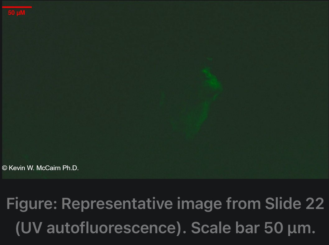
• The majority were imaged using autofluorescence under UV excitation only to avoid chemical interference & preserve structural integrity for scanning electron Microcopy (SEM), energy-dispersive X-ray spectroscopy (EDX) analysis, & Raman Spectroscopy
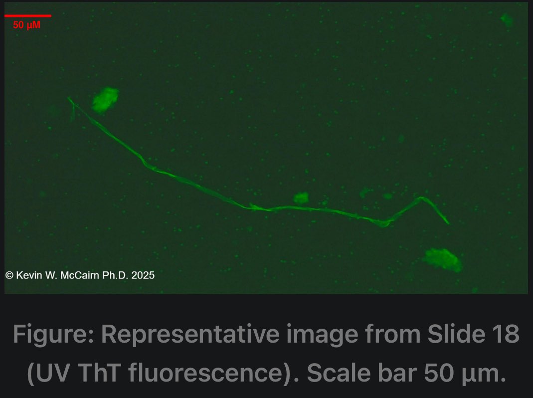
• Selected slides were stained with Thioflavin T (ThT) for amyloid-specific visualization, by microinjecting 5-10 ul of 10 um if ThT onto identified inclusions from light microscopy visualization
Observed Pathological Features Include
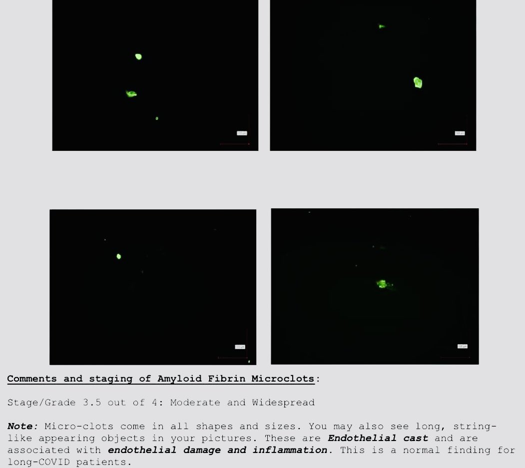
1• autofluorescent fibrillar & spheroid structures consistent with beta-sheet morphology
2• amyloid-positive fibrillar, spheroid, & microclot domains in ThT-stained slides
3• Persistent dense clotting patterns, observable without staining, suggestive of intrinsic fibrin fluorescence & misfolded conformational architecture
(@KevinMcCairnPhD)
• A total of 31 micrographs were generated from one glass slide sample
• The majority were imaged using autofluorescence under UV excitation only to avoid chemical interference & preserve structural integrity for scanning electron Microcopy (SEM), energy-dispersive X-ray spectroscopy (EDX) analysis, & Raman Spectroscopy
• Selected slides were stained with Thioflavin T (ThT) for amyloid-specific visualization, by microinjecting 5-10 ul of 10 um if ThT onto identified inclusions from light microscopy visualization
Observed Pathological Features Include
1• autofluorescent fibrillar & spheroid structures consistent with beta-sheet morphology
2• amyloid-positive fibrillar, spheroid, & microclot domains in ThT-stained slides
3• Persistent dense clotting patterns, observable without staining, suggestive of intrinsic fibrin fluorescence & misfolded conformational architecture
(@KevinMcCairnPhD)


4~ Representative Microscopic Images ~
(@KevinMcCairnPhD)
(@KevinMcCairnPhD)


5~ Representative Microscopic Findings ~
(@KevinMcCairnPhD)
(@KevinMcCairnPhD)
6~ Interpretation & Broader Implications ~
• The presence of intrinsic UV-reactive fibrillar microclots, in both stained & unstained slides, suggests a high degree of structural beta-sheet order—indicative of amyloidogenesis
• This finding, in the context of prenatal mRNA vaccine exposure, hints at a potential, novel & understudied vascular proteopathy in pediatric postnatal health
(@KevinMcCairnPhD)
• The presence of intrinsic UV-reactive fibrillar microclots, in both stained & unstained slides, suggests a high degree of structural beta-sheet order—indicative of amyloidogenesis
• This finding, in the context of prenatal mRNA vaccine exposure, hints at a potential, novel & understudied vascular proteopathy in pediatric postnatal health
(@KevinMcCairnPhD)

7~ Discussion: Analytical Gaps In Neonatal Vaccine Safety ~
• Despite broad claims of maternal mRNA vaccine safety, no studies to date have incorporated amyloid-detection methodologies (ThT, Congo Red, autofluorescent fibrin assessment, or proteinic cross-beta sheet confirmation) into perinatal or neonatal evaluations
• Large cohort studies track only macroscopic outcomes (NICU admission, APGAR scores, malformation rates)
• No proteopathic markers are assessed
• No microclot or amyloid analytics are used to evaluate sub clinical vascular dysfunction
• This leaves a substantial analytical blind spot for prenatal vaccine safety, especially when postnatal pathology—like that seen in Patient B3–could arise from proteomic misfolding initiated in-utero
• This case study documents autofluorescent & Thioflavin T-positive microclotting in a pediatric subject exposed prenatally to mRNA vaccination
• The selective use of autofluorescence microscopy ensured preservation for SEM/EDX, allowing further structural interrogation
• This layered methodology should become standard in post-vaccine safety workups, particularly where amyloidogenic mechanisms are suspected
• All images & findings are subject to (@KevinMcCairnPhD), 2025
• Despite broad claims of maternal mRNA vaccine safety, no studies to date have incorporated amyloid-detection methodologies (ThT, Congo Red, autofluorescent fibrin assessment, or proteinic cross-beta sheet confirmation) into perinatal or neonatal evaluations
• Large cohort studies track only macroscopic outcomes (NICU admission, APGAR scores, malformation rates)
• No proteopathic markers are assessed
• No microclot or amyloid analytics are used to evaluate sub clinical vascular dysfunction
• This leaves a substantial analytical blind spot for prenatal vaccine safety, especially when postnatal pathology—like that seen in Patient B3–could arise from proteomic misfolding initiated in-utero
• This case study documents autofluorescent & Thioflavin T-positive microclotting in a pediatric subject exposed prenatally to mRNA vaccination
• The selective use of autofluorescence microscopy ensured preservation for SEM/EDX, allowing further structural interrogation
• This layered methodology should become standard in post-vaccine safety workups, particularly where amyloidogenic mechanisms are suspected
• All images & findings are subject to (@KevinMcCairnPhD), 2025
Here are my Amyloid Fibrin microclots diagnosed by: (@jfvaughnmd09)
(@KevinMcCairnPhD)
(@KevinMcCairnPhD)

• • •
Missing some Tweet in this thread? You can try to
force a refresh