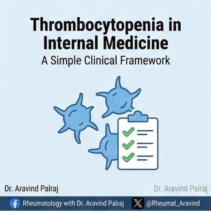
🧵 Clues Your “Arthritis” Patient Doesn’t Actually Have RA
Not all swollen joints are rheumatoid arthritis.
Some look identical—but aren’t.
Here’s how to spot RA mimics before the label sticks forever 👇
@IhabFathiSulima @DrAkhilX @SarahSchaferMD @Janetbirdope #MedTwitter #Rheumatology
1. It’s asymmetric
RA loves symmetry.
If one side is swollen but the other is fine—think again.
2. The wrong joints are involved
RA = MCP, PIP, wrists.
If DIP joints are involved → think OA, psoriatic arthritis.
If only large joints → think reactive, viral, crystal arthritis.
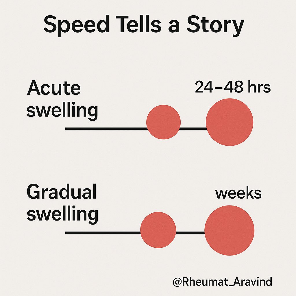
3. Too acute for RA
RA builds over weeks.
If swelling peaks in 24–48 hrs → think gout, pseudogout, septic arthritis, viral arthritis.
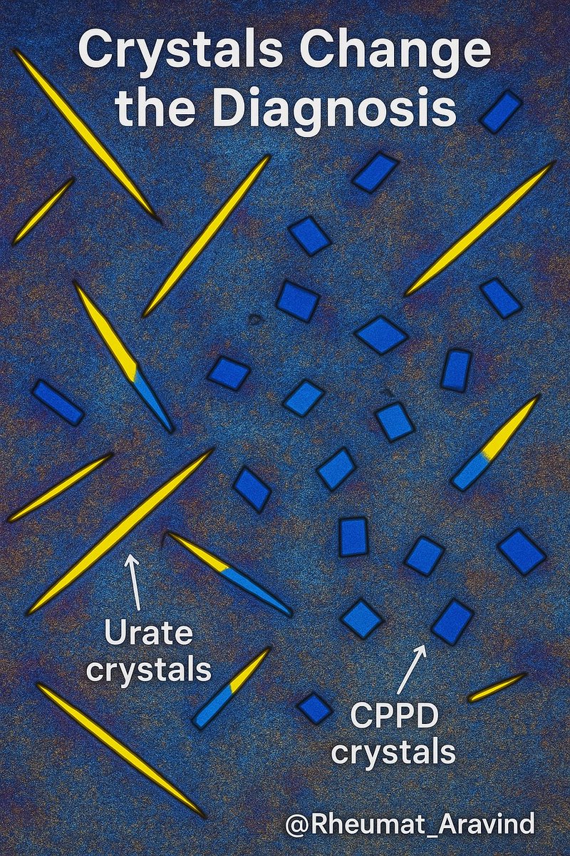
4. Crystal clues
RA won’t give you urate crystals.
If joint aspirate shows crystals → gout or CPPD.
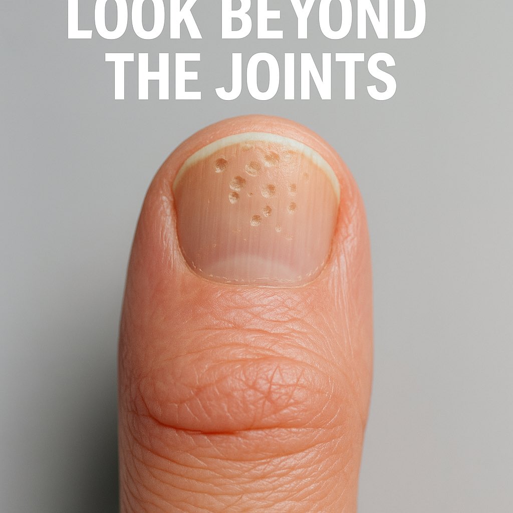
5. Psoriasis changes the game
Skin rash? Nail pitting?
Psoriatic arthritis can look just like RA—until you check the skin.
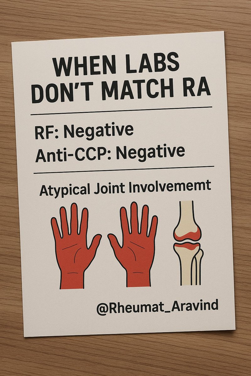
6. Negative RF & anti-CCP + weird features
If both are negative, especially with atypical joint pattern → think seronegative spondyloarthritis, lupus, viral arthritis.
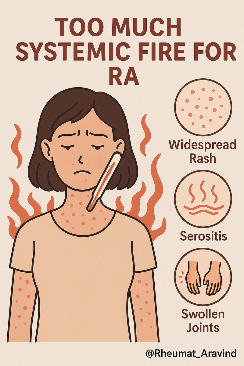
🧵 Giant Cell Arteritis — Save a Sight in 5 Minutes
The vision loss is often permanent—and preventable.
A zero-fluff checklist: who to treat before tests, when ultrasound beats biopsy, steroid start & taper, and the traps (normal ESR/CRP, “PMR only,” jaw pain without headache).
@IhabFathiSulima @DrAkhilX @CelestinoGutirr @Janetbirdope @vascuk #MedTwitter #NEETPG
Why this matters
•GCA is the most common primary vasculitis >50 years
•~15–20% develop vision loss — often before diagnosis
•Half lose the other eye within days if untreated
•Risk drops almost to zero with prompt steroids
🧵 C3 vs C4 — What the Pattern Really Means (in 30 seconds)
We order complements all the time.
But the pattern is the diagnosis.
Here’s the fast way to read C3/C4 without overthinking. 👇
@IhabFathiSulima @DrAkhilX @CelestinoGutirr @DurgaPrasannaM1 @SarahSchaferMD @EMJNephrology #MedTwitter #Rheumatology
1) Quick primer
•C3 = shared hub (alternative + classical).
•C4 = classical pathway marker (C1q → C4).
Pattern > any single value.
2) Both C3 ↓ and C4 ↓ → immune-complex “classical burn”
Think: active SLE, infective endocarditis, serum-sickness/drug IC, mixed cryoglobulinemia.
Next: CH50, anti-dsDNA, C1q binding/anti-C1q, blood cultures if febrile.
🧵 CK Can Lie — Catching Myositis When Creatine Kinase Is Normal
Myalgia + weakness.
CK is normal.
Everyone relaxes.
That’s how dangerous myositis gets missed. Let’s fix it. 👇
@IhabFathiSulima @DrAkhilX @CelestinoGutirr #MedTwitter
1) First principle
Normal CK ≠ no muscle disease. CK reflects muscle necrosis, not strength. Patchy disease, low muscle mass, or perimysial-predominant injury can keep CK normal.
2) When CK is often normal (or only mildly ↑)
•Dermatomyositis (esp. MDA5 phenotype)
•Steroid myopathy (treatment complication, not inflammation)
•Inclusion body myositis (>50 yrs; finger flexors/quads)
•Early/patchy disease, chronic burnt-out myositis
🧵 Drug Combinations That Can Kill — Interactions You Must Never Miss
We prescribe these daily.
Get the combination wrong → bleeding, rhabdomyolysis, bone marrow suppression, cardiac arrest.
Here are the 10 combinations you must always check for 👇
@DrAkhilX @IhabFathiSulima @CelestinoGutirr @Janetbirdope @DurgaPrasannaM1 @SarahSchaferMD @NeuroSjogrens #MedTwitter #RheumTwitter
1) Allopurinol or Febuxostat + Azathioprine or 6-Mercaptopurine
❌ Severe bone marrow suppression (xanthine oxidase inhibition).
✅ Avoid the combination; if unavoidable, drastically reduce azathioprine dose and monitor blood counts closely — but switching is safer.
2) Methotrexate + Trimethoprim–Sulfamethoxazole (Co-trimoxazole)
❌ Pancytopenia, mucositis, acute kidney injury.
✅ Use alternatives such as nitrofurantoin or fosfomycin for urinary tract infections.
🧵 When It’s Not Sepsis – Clues That It’s Actually Autoimmunity
Fever.
Tachycardia.
High CRP.
Looks like sepsis—but cultures stay negative, and antibiotics don’t work.
Let’s break down how to catch autoimmune mimicry of infection—before it’s too late. 👇
@IhabFathiSulima @DrIanWeissman @DrAkhilX @CelestinoGutirr @NeuroSjogrens @SarahSchaferMD @drkeithsiau #MedTwitter #RheumTwitter
1. The classic setup:
Patient has:
✅ Fever
✅ High CRP
✅ High neutrophils
✅ Looks toxic
But…
🧪 Cultures are negative
🧫 Antibiotics fail
🧠 Something’s not adding up
2. When you should pause:
🚩 No response to antibiotics after 48–72 hrs
🚩 Blood cultures negative
🚩 No source on imaging
🚩 Worsening cytopenias
🚩 Rising liver enzymes or ferritin
🚩 Mental status changes