SR in Rheumatology | MMC, Chennai | 🛑 Tweets ≠ Medical Advice | https://t.co/GIsNalCL0w
 Tweet 2 – First Rule
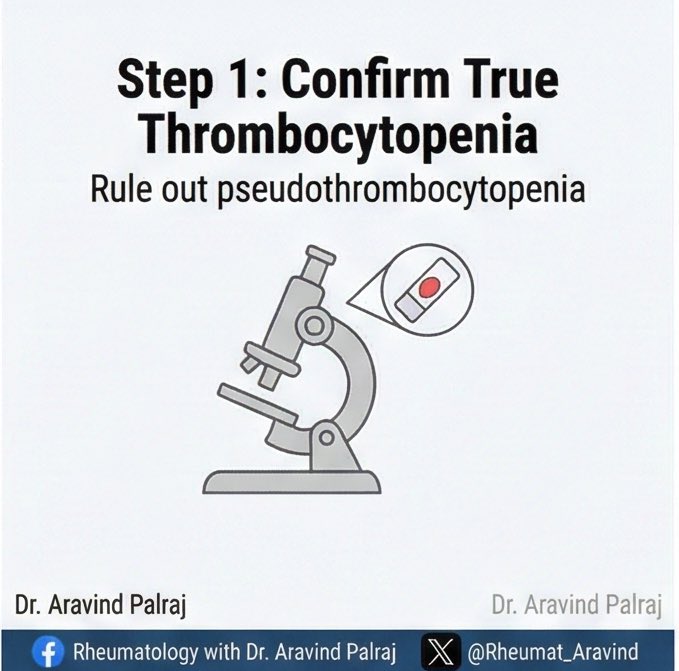
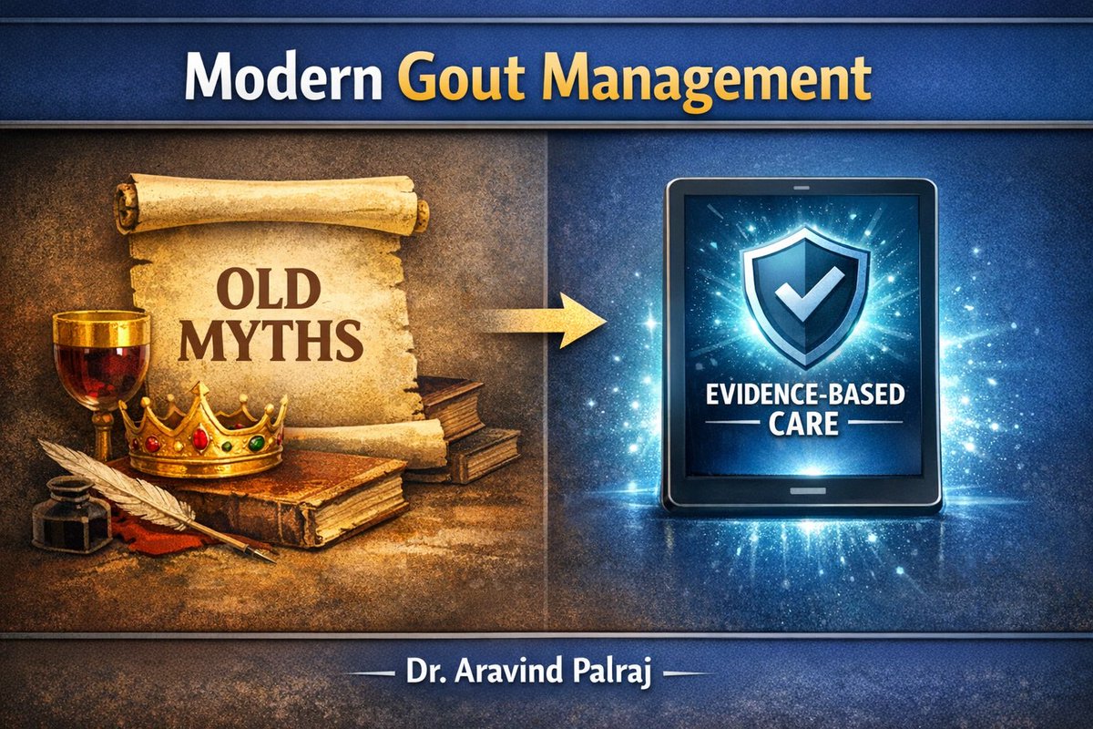 Tweet 2 - The Diet Myth
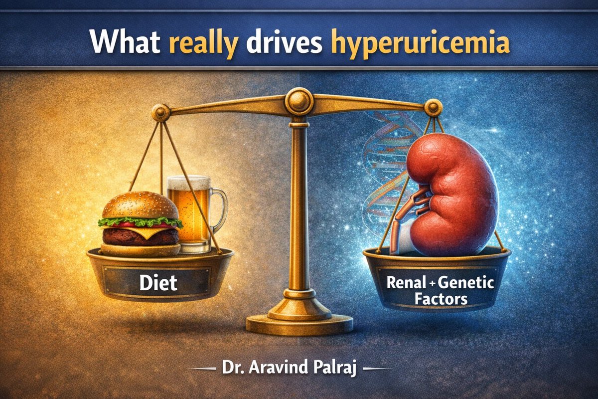
 First, understand the pre-test probability.
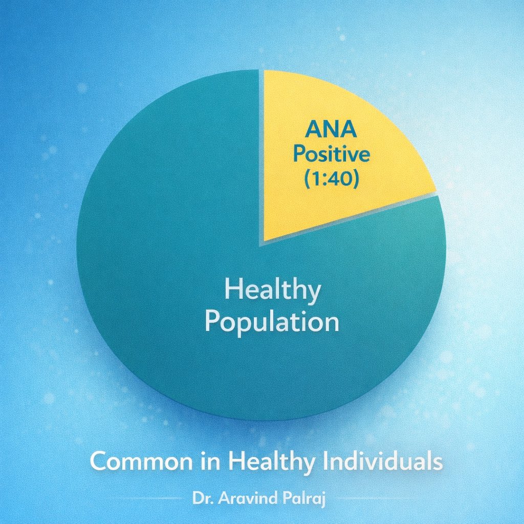
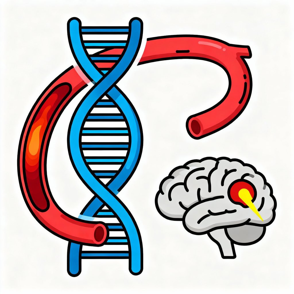
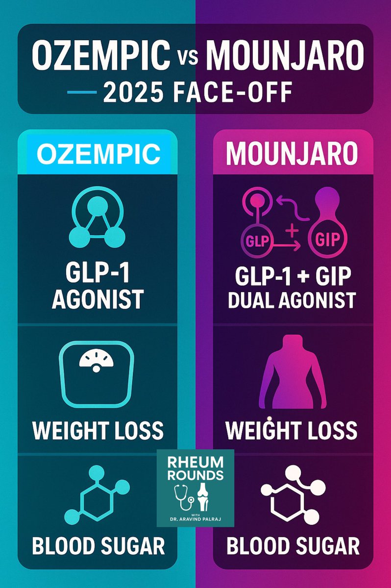 1️⃣ Mechanism
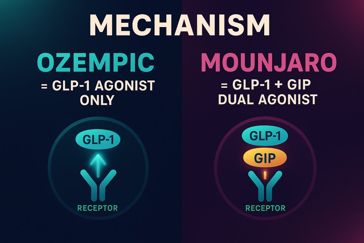
 1️⃣ “ANA is negative, so it’s not lupus.”
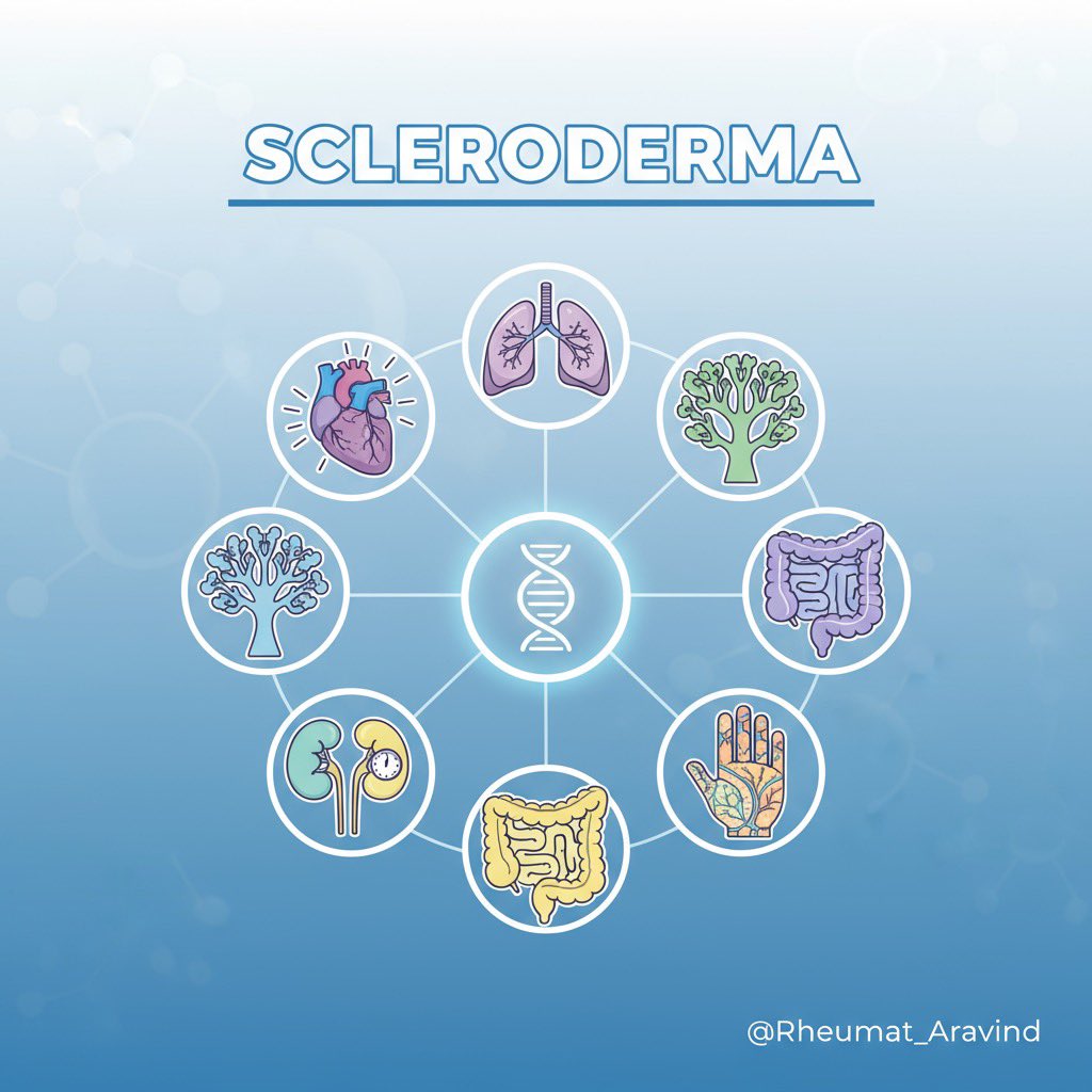
 General and Systemic Signs
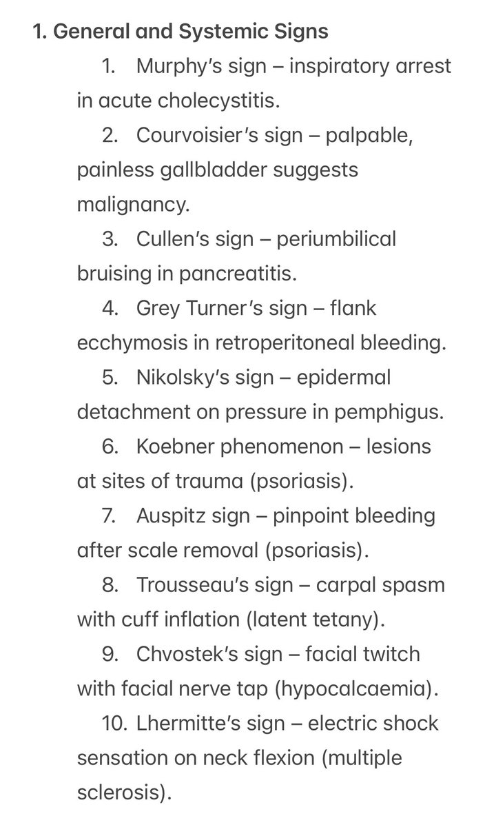
 💬 Tweet 1 – General Principles
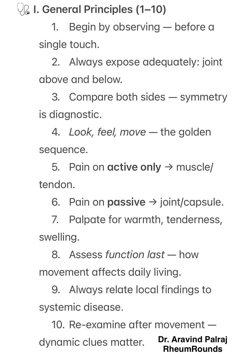
 The Art of Beginning
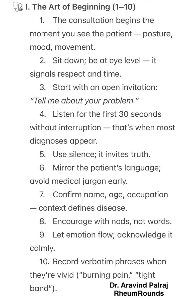
 General Principles
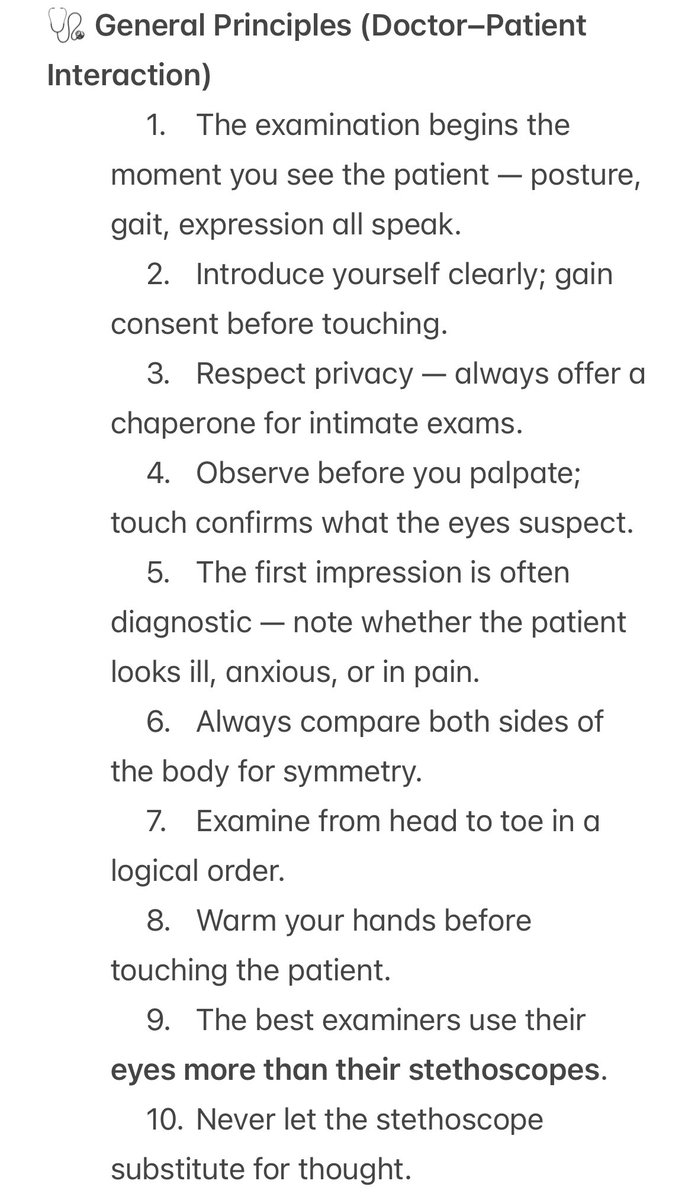
 2/
2/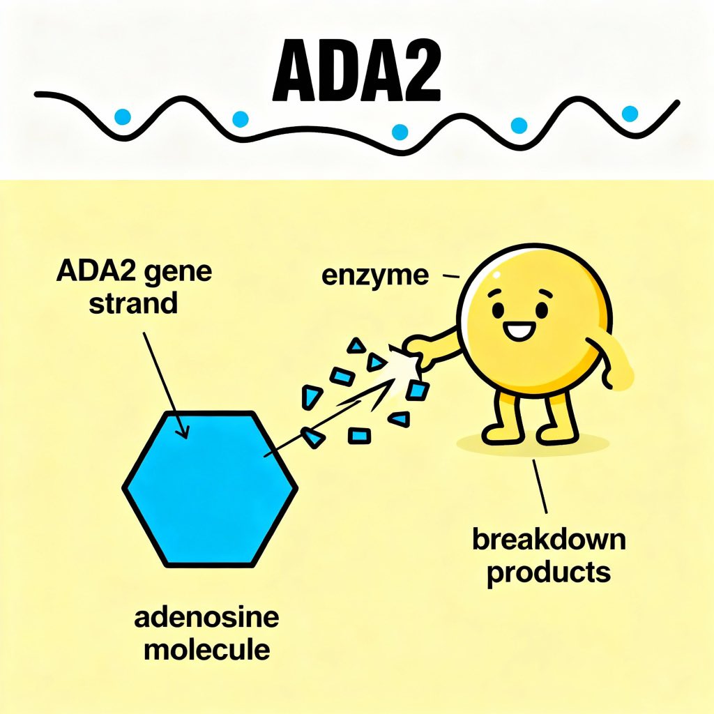
 2/
2/
 Tweet 2:
 Tweet 2:
 Tweet 2:
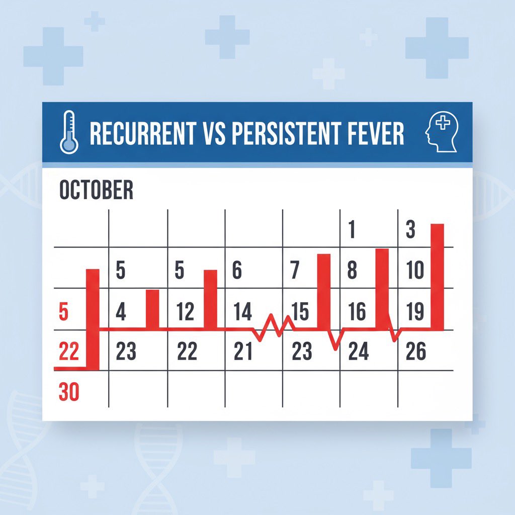
 Tweet 2:
 Tweet 2 (Pearl 1 – MAS in sJIA):
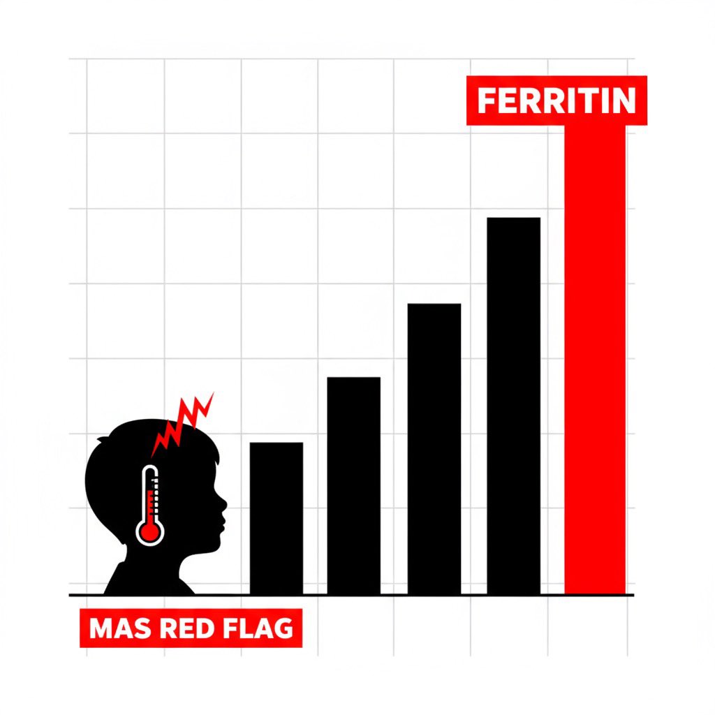
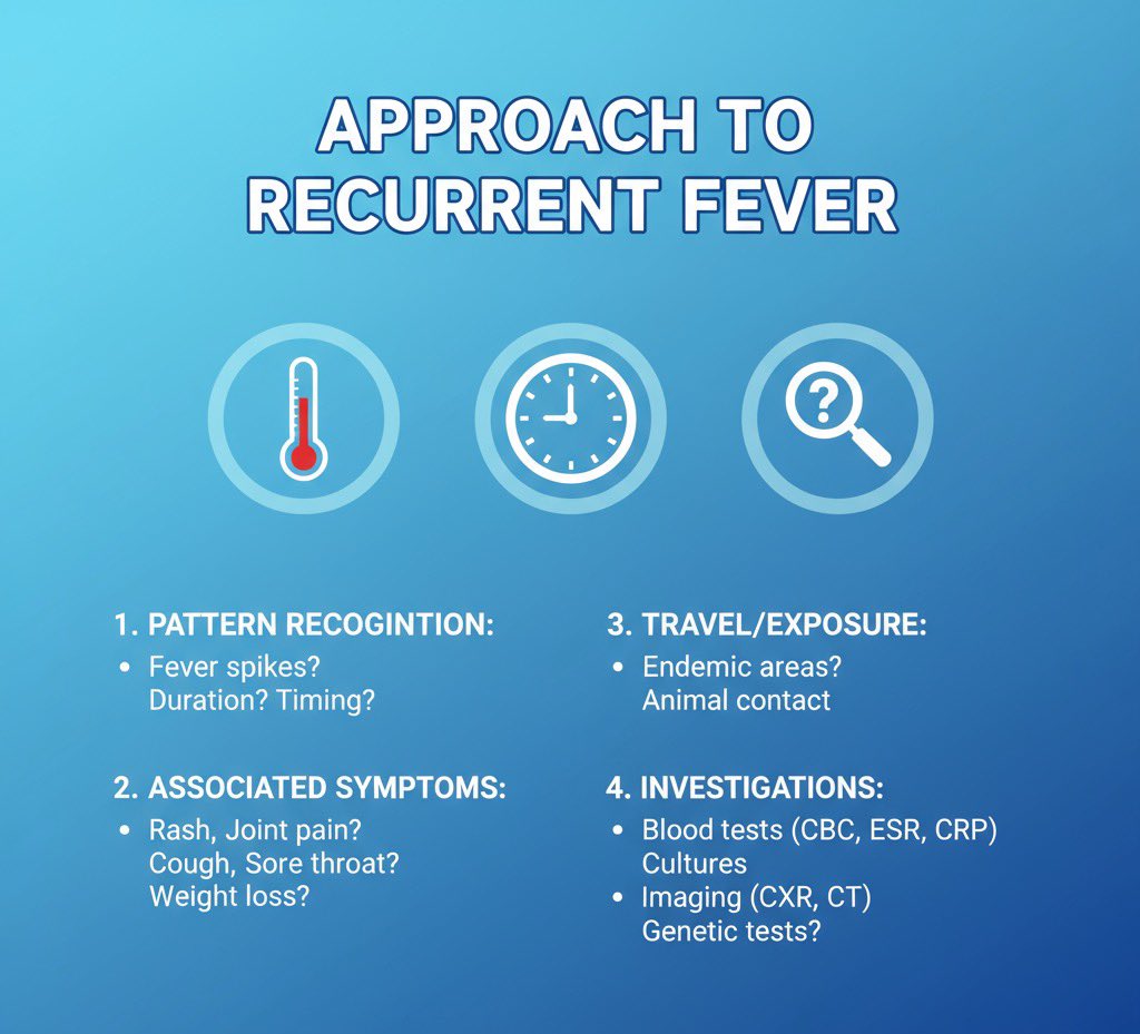
 1. Definition 📌
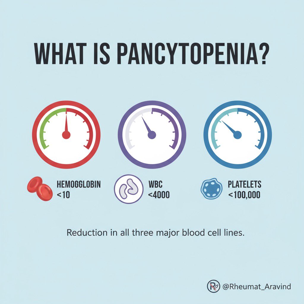
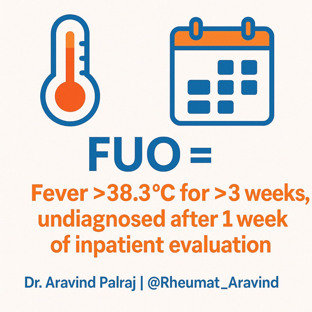 2. Categories of FUO 📂
 2/
2/
 1. Normal Smear
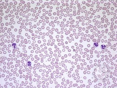
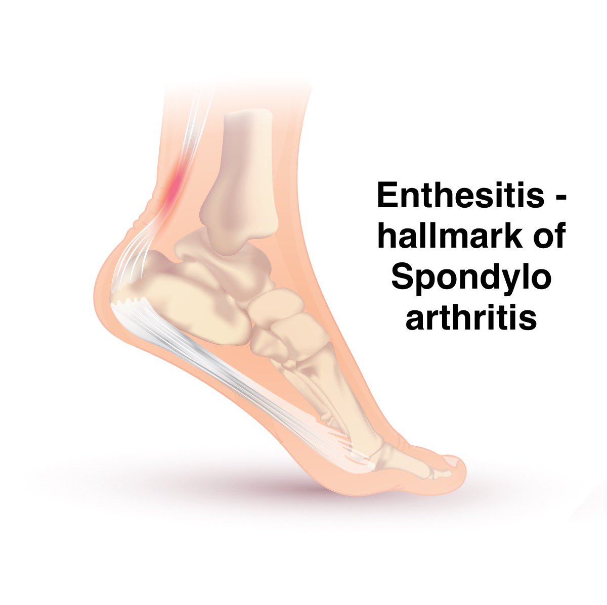
 2️⃣ Enthesitis