Aravind Palraj Profile picture
Aug 10 9 tweets 3 min read Read on X
🧵 C3 vs C4 — What the Pattern Really Means (in 30 seconds)

We order complements all the time.
But the pattern is the diagnosis.
Here’s the fast way to read C3/C4 without overthinking. 👇
@IhabFathiSulima @DrAkhilX @CelestinoGutirr @DurgaPrasannaM1 @SarahSchaferMD @EMJNephrology #MedTwitter #RheumatologyImage
1) Quick primer
•C3 = shared hub (alternative + classical).
•C4 = classical pathway marker (C1q → C4).
Pattern > any single value. Image
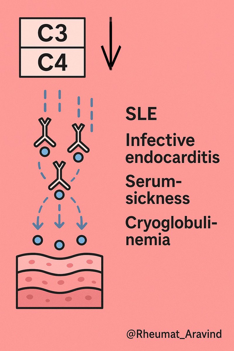
2) Both C3 ↓ and C4 ↓ → immune-complex “classical burn”
Think: active SLE, infective endocarditis, serum-sickness/drug IC, mixed cryoglobulinemia.
Next: CH50, anti-dsDNA, C1q binding/anti-C1q, blood cultures if febrile. Image
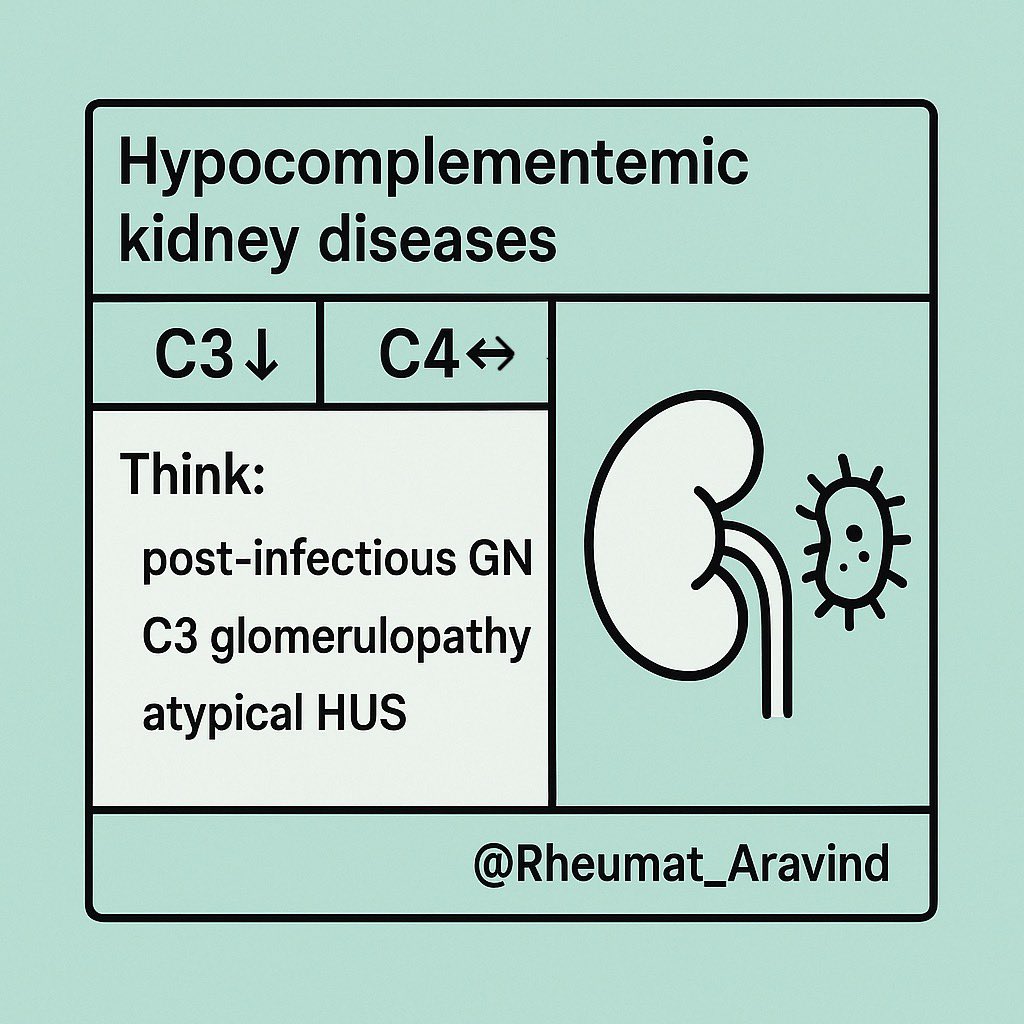
3) C3 ↓ with C4 normal → alternative pathway
Think: post-infectious GN, C3 glomerulopathy, atypical HUS.
Next: AH50, C3 nephritic factor, factor H/I, renal workup. Image
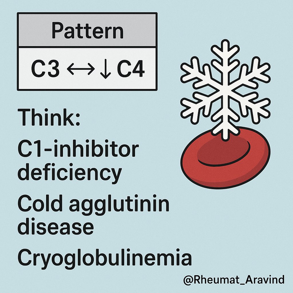
4) C3 normal with C4 ↓ → early classical activation/C1 issues
Think: hereditary or acquired C1-inhibitor deficiency (HAE), cold agglutinin disease, cryoglobulinemia (often C4 ≪ C3).
Next: C1-INH level/function, C1q, hemolysis workup, hepatitis serology. Image
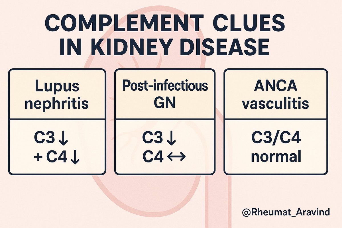
5) Kidneys cheat-sheet
•Lupus nephritis: C3 ↓ + C4 ↓
•Post-infectious GN / C3G: C3 ↓, C4 ↔
•ANCA vasculitis: C3/C4 usually normal
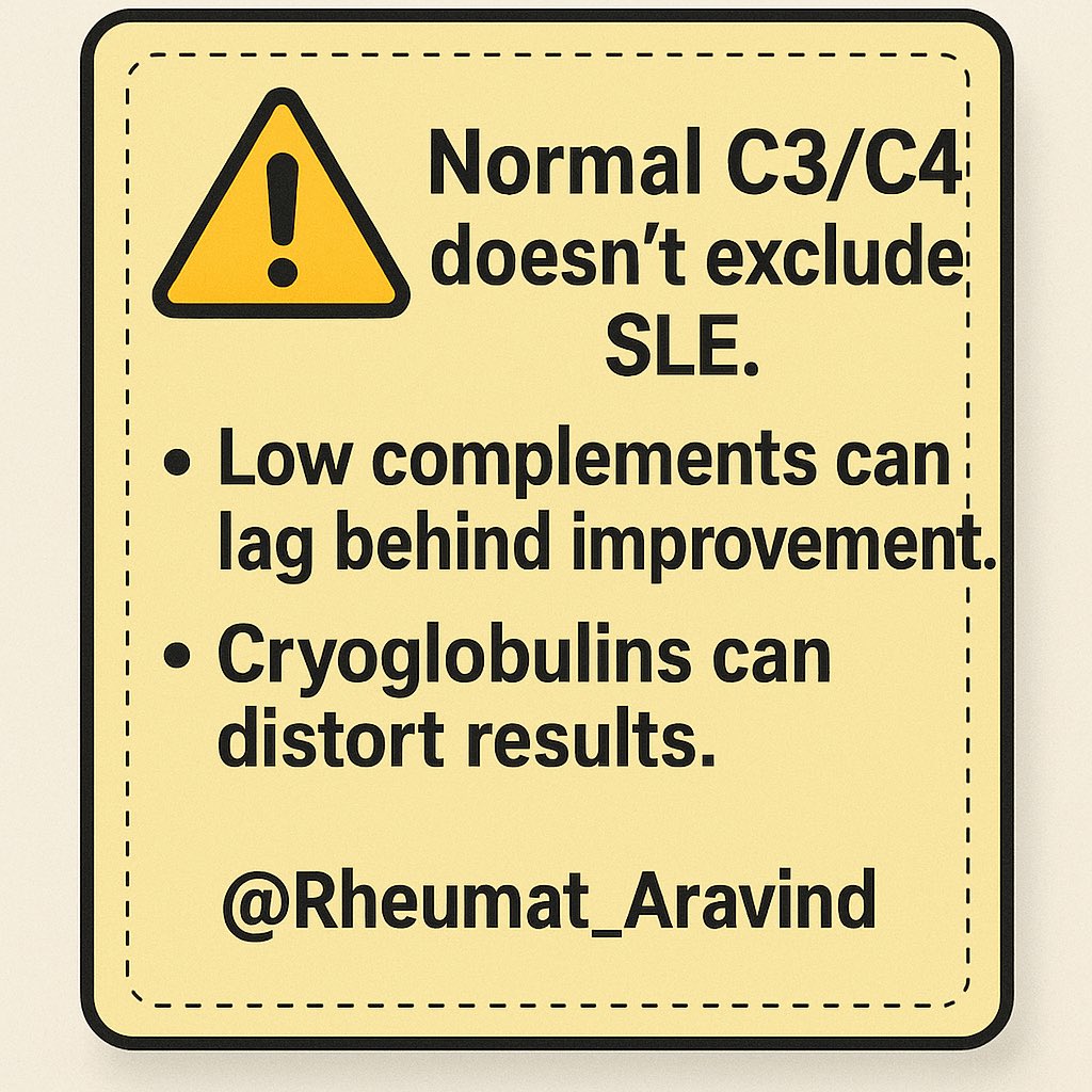
 Image
6) Pitfalls
•Normal complements do not exclude SLE (C3 is an acute-phase reactant).
•Low complements can lag behind clinical improvement.
•Lab interference (cryoglobulins) can distort results—warm the sample. Image
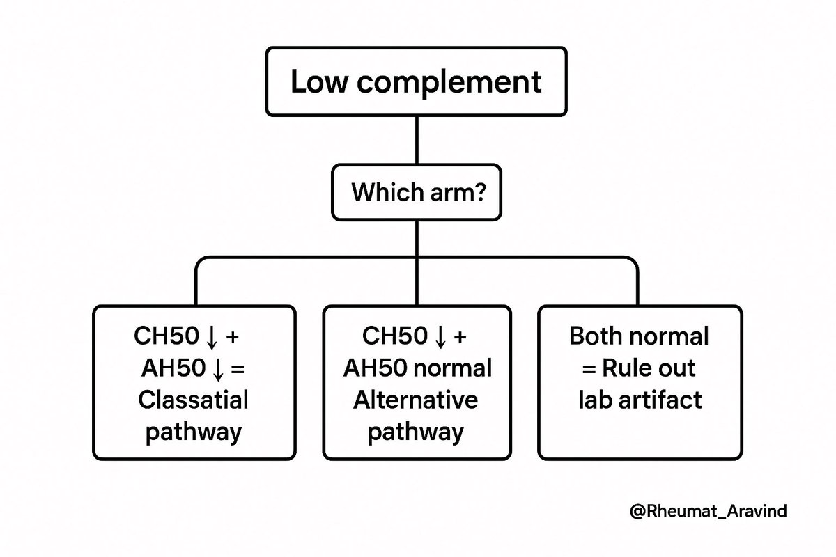
7) What to add when unsure
Order CH50 & AH50 together → tells you which arm is broken.
If angioedema: add C1q + C1-INH level & function.
If GN: add urine protein/Cr, microscopy, serologies, consider biopsy.
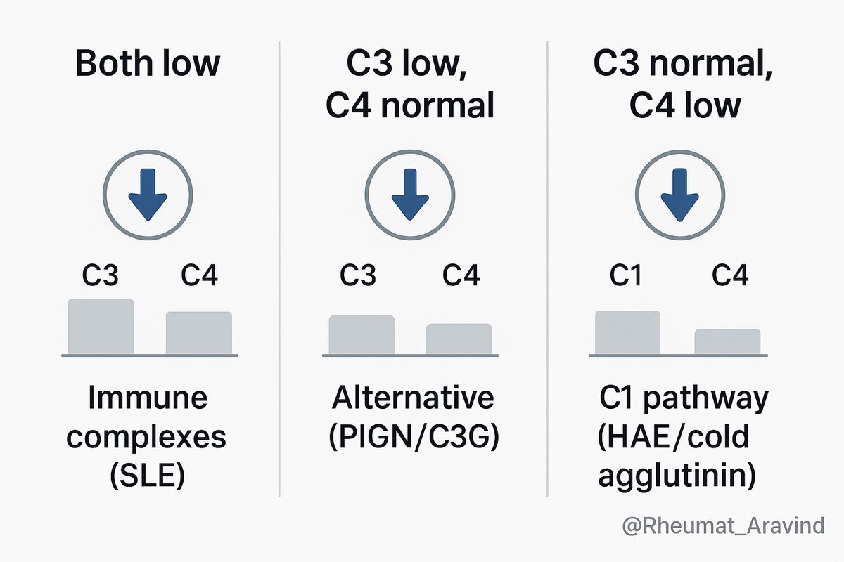
 Image
📌 Takeaway
Read complements like an ECG: pattern first.
•Both low → immune complexes (think SLE).
•C3 low, C4 normal → alternative (think PIGN/C3G).
•C3 normal, C4 low → C1 pathway (think HAE/cold agglutinin).
Context decides the rest.
#neetpg2025 #NEETPG Image

• • •

Missing some Tweet in this thread? You can try to force a refresh
 

Keep Current with Aravind Palraj

Aravind Palraj Profile picture

Stay in touch and get notified when new unrolls are available from this author!

Read all threads

This Thread may be Removed Anytime!

PDF

Twitter may remove this content at anytime! Save it as PDF for later use!

Try unrolling a thread yourself!

how to unroll video
  1. Follow @ThreadReaderApp to mention us!

  2. From a Twitter thread mention us with a keyword "unroll"
@threadreaderapp unroll

Practice here first or read more on our help page!

More from @Rheumat_Aravind

Aug 11
🧵 Steroids Made Safe — 10 Rules in 60 Seconds

We prescribe glucocorticoids often.
Get them right → lifesaving.
Get them wrong → fractures, sepsis, adrenal crisis.

Here’s the no-fluff safety checklist. 👇
@IhabFathiSulima @DrAkhilX @CelestinoGutirr @SarahSchaferMD @NeuroSjogrens #MedTwitter #RheumatologyImage
🧵 Steroids Made Safe — 10 Rules in 60 Seconds

We prescribe glucocorticoids everywhere.
Get them right → lifesaving.
Get them wrong → fractures, sepsis, adrenal crisis.

Here’s the no-fluff safety checklist. 👇 Image
2) Know the equivalence (quick)
Hydrocortisone 20 mg = Prednisolone 5 mg = Dexamethasone 0.75 mg.
Don’t mix potencies when switching. Image
Read 12 tweets
Aug 10
🧵 Giant Cell Arteritis — Save a Sight in 5 Minutes

The vision loss is often permanent—and preventable.
A zero-fluff checklist: who to treat before tests, when ultrasound beats biopsy, steroid start & taper, and the traps (normal ESR/CRP, “PMR only,” jaw pain without headache).
@IhabFathiSulima @DrAkhilX @CelestinoGutirr @Janetbirdope @vascuk #MedTwitter #NEETPGImage
Why this matters
•GCA is the most common primary vasculitis >50 years
•~15–20% develop vision loss — often before diagnosis
•Half lose the other eye within days if untreated
•Risk drops almost to zero with prompt steroids Image
Classic presentation
•Age ≥50
•New headache (often temporal)
•Jaw claudication (highly specific)
•Visual blurring / loss
•Scalp tenderness (pain on combing hair)
•± Polymyalgia rheumatica symptoms Image
Read 10 tweets
Aug 10
🧵 CK Can Lie — Catching Myositis When Creatine Kinase Is Normal

Myalgia + weakness.
CK is normal.
Everyone relaxes.
That’s how dangerous myositis gets missed. Let’s fix it. 👇
@IhabFathiSulima @DrAkhilX @CelestinoGutirr #MedTwitter Image
1) First principle
Normal CK ≠ no muscle disease. CK reflects muscle necrosis, not strength. Patchy disease, low muscle mass, or perimysial-predominant injury can keep CK normal. Image
2) When CK is often normal (or only mildly ↑)
•Dermatomyositis (esp. MDA5 phenotype)
•Steroid myopathy (treatment complication, not inflammation)
•Inclusion body myositis (>50 yrs; finger flexors/quads)
•Early/patchy disease, chronic burnt-out myositis Image
Read 12 tweets
Aug 9
🧵 Drug Combinations That Can Kill — Interactions You Must Never Miss

We prescribe these daily.
Get the combination wrong → bleeding, rhabdomyolysis, bone marrow suppression, cardiac arrest.

Here are the 10 combinations you must always check for 👇
@DrAkhilX @IhabFathiSulima @CelestinoGutirr @Janetbirdope @DurgaPrasannaM1 @SarahSchaferMD @NeuroSjogrens #MedTwitter #RheumTwitterImage
1) Allopurinol or Febuxostat + Azathioprine or 6-Mercaptopurine
❌ Severe bone marrow suppression (xanthine oxidase inhibition).
✅ Avoid the combination; if unavoidable, drastically reduce azathioprine dose and monitor blood counts closely — but switching is safer. Image
2) Methotrexate + Trimethoprim–Sulfamethoxazole (Co-trimoxazole)
❌ Pancytopenia, mucositis, acute kidney injury.
✅ Use alternatives such as nitrofurantoin or fosfomycin for urinary tract infections. Image
Read 12 tweets
Aug 9
🧵 Clues Your “Arthritis” Patient Doesn’t Actually Have RA

Not all swollen joints are rheumatoid arthritis.
Some look identical—but aren’t.
Here’s how to spot RA mimics before the label sticks forever 👇
@IhabFathiSulima @DrAkhilX @SarahSchaferMD @Janetbirdope #MedTwitter #RheumatologyImage
1. It’s asymmetric

RA loves symmetry.
If one side is swollen but the other is fine—think again. Image
2. The wrong joints are involved

RA = MCP, PIP, wrists.
If DIP joints are involved → think OA, psoriatic arthritis.
If only large joints → think reactive, viral, crystal arthritis. Image
Read 8 tweets
Aug 9
🧵 When It’s Not Sepsis – Clues That It’s Actually Autoimmunity

Fever.
Tachycardia.
High CRP.
Looks like sepsis—but cultures stay negative, and antibiotics don’t work.

Let’s break down how to catch autoimmune mimicry of infection—before it’s too late. 👇
@IhabFathiSulima @DrIanWeissman @DrAkhilX @CelestinoGutirr @NeuroSjogrens @SarahSchaferMD @drkeithsiau #MedTwitter #RheumTwitterImage
1. The classic setup:

Patient has:
✅ Fever
✅ High CRP
✅ High neutrophils
✅ Looks toxic

But…
🧪 Cultures are negative
🧫 Antibiotics fail
🧠 Something’s not adding up Image
2. When you should pause:

🚩 No response to antibiotics after 48–72 hrs
🚩 Blood cultures negative
🚩 No source on imaging
🚩 Worsening cytopenias
🚩 Rising liver enzymes or ferritin
🚩 Mental status changes Image
Read 10 tweets

Did Thread Reader help you today?

Support us! We are indie developers!


This site is made by just two indie developers on a laptop doing marketing, support and development! Read more about the story.

Become a Premium Member ($3/month or $30/year) and get exclusive features!

Become Premium

Don't want to be a Premium member but still want to support us?

Make a small donation by buying us coffee ($5) or help with server cost ($10)

Donate via Paypal

Or Donate anonymously using crypto!

Ethereum

0xfe58350B80634f60Fa6Dc149a72b4DFbc17D341E copy

Bitcoin

3ATGMxNzCUFzxpMCHL5sWSt4DVtS8UqXpi copy

Thank you for your support!

Follow Us!

:(