It's often been stated that a particular white matter tract in the human brain, the inferior fronto-occipital fasciculus (IFOF; see below), is altered in gender dysphoria (GD) and may even be the neurological location/substrate for "gender identity".
But what does the data say?
But what does the data say?

The first report of an altered IFOF in GD comes from this 2017 paper (), in which they found reduced white matter structural integrity (FA values) in transgender women (not trans men), and appeared independent of sexual orientation. pmc.ncbi.nlm.nih.gov/articles/PMC57…

Later in 2022, this was "replicated" () in adolescent trans girls (mean age = 15.4). However, no sex-reversal was seen in dysphoric pre-pubescent children (mean age = 10.4).
Importantly, this sample overlapped with the study cited above. pmc.ncbi.nlm.nih.gov/articles/PMC10…
Importantly, this sample overlapped with the study cited above. pmc.ncbi.nlm.nih.gov/articles/PMC10…

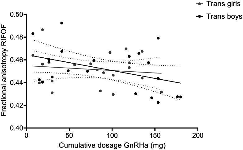
In the previous study, the adolescent trans girls had been exposed to puberty blockers (GnRHa).
The authors noted a negative correlation (pre-multiple comparison correction) between the dosage of GnRHa and the microstructural integrity of the IFOF (i.e., reduced FA value).
The authors noted a negative correlation (pre-multiple comparison correction) between the dosage of GnRHa and the microstructural integrity of the IFOF (i.e., reduced FA value).

The confounded data above make causal conclusions difficult. Further, we don't fully understand regular sex differences in the IFOF to be confident enough that the IFOF in transgender individuals is sex-atypical.
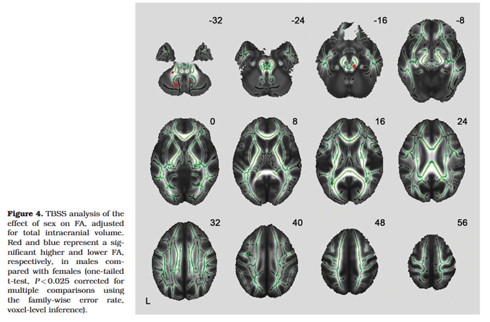
While it's typically assumed that males have greater white matter integrity (increased FA values) compared to females in the majority of white matter tracts, there still lie many inconsistencies.
pubmed.ncbi.nlm.nih.gov/27226438/
pmc.ncbi.nlm.nih.gov/articles/PMC33…
annas-archive.org/scidb/10.1016/…
pubmed.ncbi.nlm.nih.gov/27226438/
pmc.ncbi.nlm.nih.gov/articles/PMC33…
annas-archive.org/scidb/10.1016/…
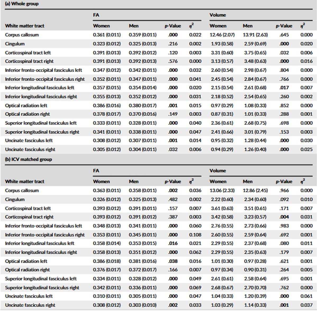
In this paper (), the authors statistically accounted for the sex differences in intracranial volume (ICV) via adding it as a covariate in their statistical model. In doing so, sex differences in all major tracts disappeared. onlinelibrary.wiley.com/doi/epdf/10.10…

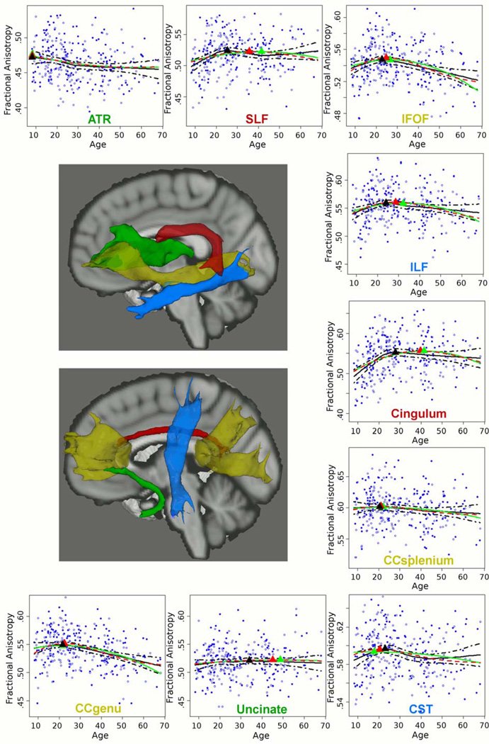
Interestingly, the integrity of the IFOF tends to decrease around the age of 30, and higher FA values during development are associated with greater global cognitive functioning. The impact of age is thus another important consideration.
pmc.ncbi.nlm.nih.gov/articles/PMC44…
pmc.ncbi.nlm.nih.gov/articles/PMC44…

In a larger cohort, and while accounting for both age and brain size via both statistical modelling and/or by using brain-size matched men & women, these authors found that it's actually females who show increased FA in the IFOF:
onlinelibrary.wiley.com/doi/epdf/10.10…
onlinelibrary.wiley.com/doi/epdf/10.10…

It's important to note that in the transgender studies, the authors did not account for brain size, had a small and confounded sample, didn't "replicate" previous data, and the data on regular sex differences in the IFOF are inconsistent (perhaps due to methodological reasons).
Now even consider the fact that other transgender studies investigating white matter integrity (using FA metrics) did not report IFOF sex-reversal.
pmc.ncbi.nlm.nih.gov/articles/PMC46…
pmc.ncbi.nlm.nih.gov/articles/PMC46…
The function of IFOF is much debated (academic.oup.com/brain/article/…), but in transgender individuals some authors have claimed that it's involved in body image distortions (similar reductions in IFOF integrity have also been found in anorexia): pmc.ncbi.nlm.nih.gov/articles/PMC67…
Based on all the data above, I don't find it convincing enough to claim that "gender identity" is located within the IFOF, nor that it being related to sexual dimorphism implicates it in GD aetiology.
Perhaps an overall reduction in IFOF integrity may at the very least 'predispose' one to some form of body distortion (but even then, causal conclusions are a long way away).
• • •
Missing some Tweet in this thread? You can try to
force a refresh


