Sammy Profile picture
PhD student in Neuroscience. Research interests include sex differences in the brain, sexual orientation, and gender dysphoria.
2 subscribers
Mar 21 8 tweets 2 min read
A thread of mis-information here… You make the claim that the hypothalamic nuclei (the BSTc in the cited Zhou et al., 1995 study), in transwomen is similar to that of natal women. However, you fail to mention that the BSTc doesn’t become dimorphic until 30-35 years of age and instead assume causality.
Aug 23, 2024 8 tweets 2 min read
This paper suggests that untreated male-to-female transsexuals (MtF; n = 18) and FtMs (n = 24) show signs of feminisation and masculinisation, respectively, in relation to cortical thickness (CTh).

Let’s take a look: Image 
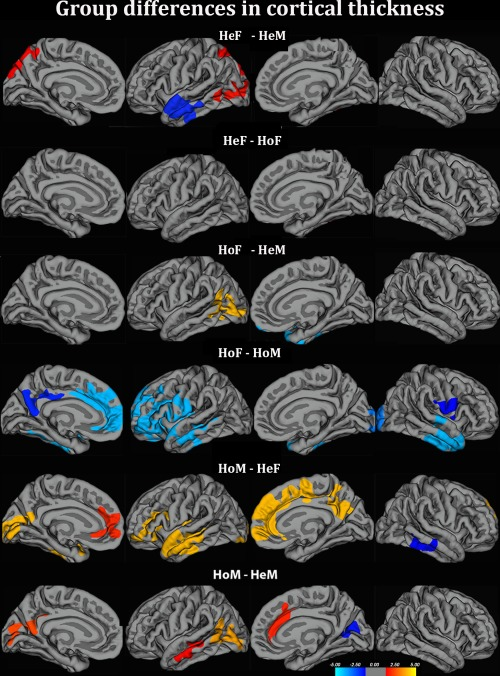
In their whole-brain comparison on CTh in relation to sexual dimorphism, the authors found that FtMs did not differ from female controls (thus having a CTh pattern congruent with their biological sex). Similarly, MtFs also did not differ to female controls (thus having a CTh pattern reflective of the opposite sex).
Dec 7, 2023 9 tweets 3 min read
Brain activation patterns (those involved in self-referential/own body-perception processing) in trans men (n=14) & women (n=16) are similar to those of male (n=15) & female (n=15) controls, except when viewing their bodies morphed to the opposite sex:
pubmed.ncbi.nlm.nih.gov/31813993/
Image Participants wore a skin-tight suit which was virtually morphed to appear more masculine or feminine, representing images that are associated with either their natal sex or the opposite sex.

They then view these images in different increments in an fMRI scanner: Image
Dec 5, 2023 12 tweets 3 min read
NEW STUDY: This is the first attempt to assess the translational potential for utilizing mice as a model for investigating human sex differences in the brain, focusing on regional grey matter volume (GMV).

Let's take a look 👀 Using a large cohort from the Human Connectome Project (597 females & 496 males) and mice (213 females & 216 males), total tissue volume (TTV; i.e., brain size) was assessed.

Human males showed a 13.5% larger TTV, whereas mice showed no such sex difference: Image
Dec 1, 2023 19 tweets 5 min read
@hanksfolly @SimpForSowell @HermanSonofHerm @Anna11Ebiza @RoisinMichaux There is 100% research that demonstrates the impact of homosexuality on the cross-sex shift.

Let’s see some examples: @hanksfolly @SimpForSowell @HermanSonofHerm @Anna11Ebiza @RoisinMichaux In this paper, transgender participants (pre-pubescent and adolescent) smelled a pheromone (androstenedione; AND) which is known to cause a sex difference in hypothalamic activation via fMRI.

ncbi.nlm.nih.gov/pubmed/24904525
Aug 1, 2023 16 tweets 4 min read
This is a thread reviewing two papers that are cited in support of the ‘brain-sex’ theory of gender dysphoria regarding the central subdivision of the bed nucleus of the stria terminalis (BSTc). The two studies in question are Zhou et al., 1995 and Kruijver et al., 2000: The first paper (Zhou et al) investigated the volume of the BSTc via the staining of vasoactive intestinal polypeptide (VIP) fibre innovation arising from the amygdala in 6 male-to-female transsexuals (MtFs):
pubmed.ncbi.nlm.nih.gov/7477289/
Jul 2, 2023 7 tweets 2 min read
Using MRI to assess gray matter volume in trans women (TW; n=33), trans men (TM; n=33), males (n=24), and females (n=25), these authors identified the putamen as a marker associated with “gender identity”:
https://t.co/61TZdmQpuMpubmed.ncbi.nlm.nih.gov/33916288/
Comparing trans participants vs controls, it was observed that the volume of the putamen was greater in the transgender cohort in both the left and right hemispheres:
Jun 11, 2023 6 tweets 2 min read
Incorrect. Specifically, homosexual men & women show less sexually differentiated brains (on average), and this is what's reflected in studies identifying a cross-sex shift in transgender participants. Citations:

pubmed.ncbi.nlm.nih.gov/21467211/
May 27, 2023 6 tweets 2 min read
Large study investigating sex differences in gray matter volume (GMV) in a large sample (n=2,838) of men and women between the ages of 21-90 years old: Image Women, on average, showed larger GMV in various frontal regions, such as the ventrolateral and dorsolateral prefrontal cortex, medial and lateral orbitofrontal cortex, and anterior cingulate cortex.
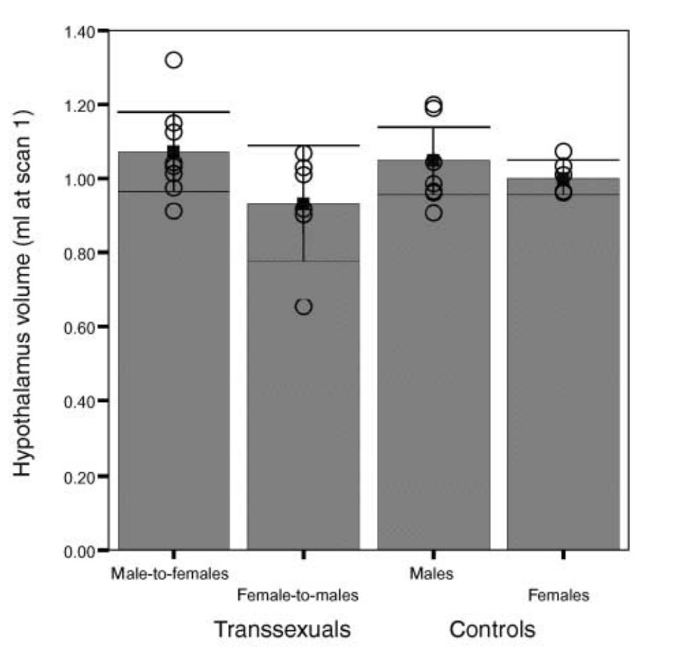
 Image
May 4, 2023 5 tweets 1 min read
Important data to consider when investigating the sociological and biological elements of female-typical gendered behaviour:
researchgate.net/publication/24… Notice how females exposed to the lowest levels of testosterone in utero (top line) show more and more “female-typical” behaviour in adulthood with more and more encouraged femininity from their mothers (sociological effects of increasing certain behaviours) Image
Apr 14, 2023 7 tweets 3 min read
This article is often cited as evidence that the brains of transgender women (TW) resemble the brains of natal women. However, once looking at the data, we see that this may not be the case.
ncbi.nlm.nih.gov/pmc/articles/P… The study included 48 controls (24=male; 24=female) and 24 TW (mean age = 45 for all participants).

All subjects underwent MRI and then the authors implemented a multivariate classifier to these MR images, in which a brain sex index (BSI) was derived. The BSI ranges between “0”… twitter.com/i/web/status/1…
Dec 3, 2022 24 tweets 7 min read
So, here is my response to @ForrestValkai and his video "Sex and Sensibility", breaking down his segment on transgender neurology.
He mentions 3 main sub-cortical structures:

1) The central division of the bed nucleus of the stria terminalis (BSTc)

2) The sexually dimorphic nucleus of the preoptic area (SDN-POA)

3) The vasoactive intestinal polypeptide subdivision of the suprachiasmatic nucleus (VIP-SCN).
Nov 27, 2022 13 tweets 4 min read
Are there differences in certain white matter fiber tracts in the brains of female-to-male transsexuals (FtMs) either pre- or post-androgen treatment?

Let's take a look 👀 The authors of this study implemented diffusion tensor imaging (DTI) to measure fractional anisotropy (FA) in FtMs and male and female controls.

A high FA = high white matter integrity
A low FA = low white matter integrity

pubmed.ncbi.nlm.nih.gov/20562024/
Nov 25, 2022 19 tweets 5 min read
Here is a study investigating sex-atypical hypothalamic responses/activation to androstadienone (AND) in pre-pubertal children and adolescents with gender dysphoria (GD) compared with neurotypical male and female controls.

Let's take a look 👀
pubmed.ncbi.nlm.nih.gov/24904525/ Quick background:

AND is a precursor in the biosynthesis of both testosterone and estrogen (see below), in which its production is increased throughout male puberty and acts as a pheromone in human sweat. Image
Nov 9, 2022 19 tweets 5 min read
Here is a famous study, often cited in blog posts, that suggests transgender individuals have resting-state functional brain activation more in line with their "gender identity"

Let's take a look 👀
pubmed.ncbi.nlm.nih.gov/28972892/ The authors recruited 70 pre-pubertal children (mean age = 9 years old) and 81 adolescents (mean age = 15), each comprising transboys, transgirls, and neurotypical male and female controls, to partake in resting-state fMRI.
Nov 5, 2022 10 tweets 3 min read
Evidence for neurodevelopmental changes in cortical folding that mediate body perception/ownership in transgender individuals
pubmed.ncbi.nlm.nih.gov/33718960/ Quick background:

Throughout neurodevelopment, our brains become crinkled as it grows within the skull to form its typical structural organization (seen below):
Oct 29, 2022 5 tweets 2 min read
Interesting paper on how white matter hypersensitivities (WMH; lesions) affect functional connectivity in Alzheimer's disease. More specifically, the inferior fronto-occipital fasciculus (IFOF) and the default mode network (DMN), respectively.
pubmed.ncbi.nlm.nih.gov/27432800/ They examined multiple white matter tracts and regions of interest (ROI) that overlap with the DMN (shown below):
Oct 28, 2022 12 tweets 4 min read
Since gender dysphoria (GD) is correlated with body image distortions and impaired self-referential processing, which is known to involve the brain's default mode network (DMN), I thought I'd go over this system in more detail.
pubmed.ncbi.nlm.nih.gov/27444730/ The DMN is a functional network consisting of co-activated brain regions including the medial prefrontal cortex (mPFC; orange arrow) and the posterior cingulate cortex (PCC; yellow arrow).

Picture B shows the coordinated activity of these regions via the fMRI BOLD signal. Image
Oct 23, 2022 8 tweets 3 min read
Gender dysphoria (GD) and anorexia nervosa (AN) show similarities within the brain in the default mode network (DMN; important in self-referential processing) and inferior fronto-occipital fasciculus (IFOF; important in body image distortion)

Are there any other similarities? 🤔 One possibility is in the visual cortex. More specifically, the fusiform body area (FBA). The FBA encodes visual information about the human body
pubmed.ncbi.nlm.nih.gov/16306418/
Aug 30, 2022 14 tweets 4 min read
An interesting study on the effects of gonadal testosterone depletion on blood-brain-barrier (BBB) permeability/integrity and subsequent microglial activation in the central nervous system (CNS) of male mice.

Let’s take a look 👀
pubmed.ncbi.nlm.nih.gov/28256950/ Quick background (microglia):

Microglia are the immune cells of the CNS, and are constantly surveying their microenvironment in search of dangerous/unwanted pathogens:

You can see their long branching processes extending (green) and retracting (red) here: Image
Aug 5, 2022 16 tweets 5 min read
Gender dysphoria (GD) and anorexia nervosa (AN) both have underlying brain differences associated with each other, perhaps unsurprisingly given the similarity in symptoms (dissatisfaction with their body).

That being said, what are these brain similarities? Despite condition-specific alterations, one common similarity that arises is in a white matter fiber tract called the inferior frontal-occipital fasciculus (IFOF). The IFOF connects posterior and anterior portions of the brain (within each hemisphere). Image