Brain Inflammation Collaborative Profile picture
Sep 5 12 tweets 3 min read Read on X
Long COVID is more prevalent in women.

Could this new study explain why? 🧵 ID: Decorative image of a human brain scan.
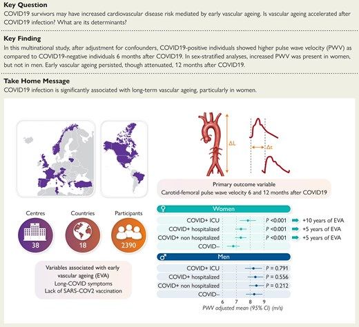
Researchers from 16 different countries recruited 2390 individuals (age 50 ± 15 years, 49.2% women) to assess the impacts of COVID-19 on vascular aging.

They found that persistent COVID-19 symptoms in women correlated with increased vascular aging.

Here is why this matters.

medpagetoday.com/cardiology/gen…
Arteries deliver oxygenated blood away from the heart.

They are surrounded by layers of cells, including smooth muscle cells (SMCs).

The diameter of the arteries expands and contracts in part due to the SMCs.

Higher O2 demand = SMC relaxation = increased artery diameter. Image
When we say higher O2 demand, you're likely thinking of exercise.

But the expansion and contraction of arteries can occur at rest.

For instance, with neurovascular coupling...
Neurovascular coupling is a fancy way of saying that increased neural activity will increase blood flow to your brain.

But not your entire brain.

Here is an example.
Reading this thread is causing neurons to fire in your temporal lobe (and elsewhere).

The firing of those neurons causes potassium ions (formerly inside the neuron) to diffuse away from the neuron and bind SMCs.

This causes SMCs on nearby arteries to relax, increasing the diameter.

Increased arterial diameter = increased blood flow to the neuron(s).

Thus, neurovascular coupling helps nourish your active neurons.
This happens rapidly - in milliseconds.

In other words, neuron firing throughout the brain conducts a symphony of highly localized vasodilation events.

This is needed for peak 🧠 performance.
Thus, vascular health is important.

But not just in the brain - your entire body.

Back to the study.
Researchers measured arterial stiffness by calculating the speed of a pressure wave traveling from the carotid artery (neck) to the femoral artery (groin).

They adjusted for factors like age and blood pressure to enhance accuracy.

This serves as a proxy for vascular health because higher values indicate stiffer arteries.
The researchers stratified the data by sex.

Males showed no significant difference in arterial stiffness.

However, women with persistent COVID-19-like symptoms had a higher pressure wave velocity (proxy for stiffer arteries).

Even mild C-19 increased stiffness.

A 12-month follow-up found a slight reduction in stiffness.

Full study:
academic.oup.com/eurheartj/adva…Image
This stiffness is likely caused by endothelial cell dysfunction and vascular inflammation.

There are hints that SARS-CoV-2 can dysregulate vascular smooth muscle cell function, but more data are needed (1).

Leave a comment below - we want to hear from you.
Literature Cited:
1. Pelisek J, et al. Clin Sci (Lond). 2022 Nov 11;136(21):1571-1590. PMID: 36367091.

• • •

Missing some Tweet in this thread? You can try to force a refresh
 

Keep Current with Brain Inflammation Collaborative

Brain Inflammation Collaborative Profile picture

Stay in touch and get notified when new unrolls are available from this author!

Read all threads

This Thread may be Removed Anytime!

PDF

Twitter may remove this content at anytime! Save it as PDF for later use!

Try unrolling a thread yourself!

how to unroll video
  1. Follow @ThreadReaderApp to mention us!

  2. From a Twitter thread mention us with a keyword "unroll"
@threadreaderapp unroll

Practice here first or read more on our help page!

More from @BrainInflCollab

Aug 28
Data suggest there is a broken ion channel on the surface of white blood cells in patients with (1):
- Long COVID
- ME/CFS

New evidence suggests it can be fixed.

Here is how...🧵 ID: Cartoon of a purple ion channel protein embedded in the plasma membrane.
Research from Dr. Sonya Marshall-Gradisnik in Brisbane, Australia, verified that Long COVID patients, like ME/CFS patients, have a dysfunctional calcium ion channel called TRPM3(1).

The function of this ion channel can be restored with low-dose naltrexone (LDN) in ME/CFS-isolated immune cells (1).

New evidence suggests the same for Long COVID patients. (below)Image
This ion channel is located on the plasma membrane of cells, where it is highly permeable to Ca2+ (ions).

This means it lets Ca2+ ions into the cell.

These calcium ions play an important role in:
- calcium-dependent signaling pathways
- calcium homeostasis
- cell differentiation
- cell adhesion
- cell division

It also plays an important role in the function of immune cells (1).
Read 7 tweets
Aug 22
New research provides further proof that ME/CFS is a biological condition.

Researchers from the University of Edinburgh found anomalies in 8 different genomic regions in more than 15,000 people diagnosed with the condition.

Here are the details...🧵 ID: Decorative heat map image of two brains with red and yellow splotches on a greenish blue backdrop.
But first, a disclaimer.

The research was published in a pre-print journal, meaning it has yet to be peer reviewed.

Thus, it could contain unverified or flawed data, methods, or interpretations.

Let's dive in.

science.org/content/articl…Image
They discovered the mutations in the following genomic regions:
- ARFGEF2/CSE1L - reduces inflammation
- RABGAP1L - viral and bacterial defense
- SUDS3 - dampens neuroinflammation
- CCPG1 - helps control viral infections
- OLFM4 - dampens inflammation
- BTN2A2 - T cell responses
- CA10 - pain signaling
- FBXL4 - metabolism

Preprint: pure.ed.ac.uk/ws/portalfiles…ID: Fig. 2: Eight genome-wide significant associations to ME/CFS. (A) Manhattan plot of the primary DecodeME genome-wide association study (GWAS-1), showing the chromosomal position of DNA variants (x-axis) against −log10p (y-axis) from genome-wide association tests accounting for sex and genetic ancestry. The upper horizontal dashed line indicates the threshold used to define genome-wide significant associations (p < 5 × 10−8); the lower horizontal dashed line indicates p < 1 x 10⁻⁵. Six loci were genome-wide significant in GWAS-1. An additional locus, OLFM4, was genome-wide significant in...
Read 7 tweets
Aug 15
45% of those with Long COVID suffer from a sleep disorder (1).

It's so prevalent that it's dubbed 'coronasomnia'.

Here is the science...🧵
\ Coronasomnia:
There is a strong association between Long COVID and the development of:
- insomnia
- sleep continuity disorders
- feeling of non-restorative sleep
- changes in the sleep-waking cycle

However, this is not unique to Long COVID.
\ Sleep and ME/CFS:
Those with Myalgic Encephalomyelitis/Chronic Fatigue Syndrome (ME/CFS) and other neuroinflammatory conditions also have similar disordered sleeping patterns (2).

This debilitating condition is symptomatically similar to Long COVID.

Like long COVID, it can also be triggered by a pathogen such as:
- Borrelia burgdorferi (Lyme disease)
- Epstein-Barr Virus
- Herpesvirus 6
- Influenza
- etc.
Read 9 tweets
Aug 3
Neuroinflammation can lead to repetitive movements similar to those observed in:
- Obsessive Compulsive Disorder (OCD)
- Autism Spectrum Disorder (ASD)

New evidence suggests it might be (partly) treatable with FDA-approved drugs...🧵 ID: A painting of a man on the far right and a women on the left - the man is pictured in grey with yellow specs on his head signifying neuropathology.
South Korean researchers found that two FDA-approved drugs can reverse these repetitive movements:
- Memantine - licensed for Alzherimers
- Interleukin-1RA (Anakinra) - licensed for rheumatoid arthritis

Here are the details... simplified.

buff.ly/WfEXTclImage
Researchers stimulated neuroinflammation in mice by activating the inflammasome (in-flam-uh-sohm) in white blood cells called macrophages.

Thus, the macrophages in the brain (i.e., the microglia) had a hyperactive inflammasome, causing neuroinflammation.

⬆️ inflammasome activity = ⬆️ inflammation.ID: Cartoon revealing how the inflammasome is primed following downstream signaling from inflammatory cytokines leading to the production and assembly of inflammasome genes. The assembled inflammasome is stimulated by factors that suggest the cell is damaged or under the threat of a pathogen. This triggers the 'cutting' of inflammatory cytokines IL-18 and IL-1b into their active forms.
Read 10 tweets
Jul 14
A new study just revealed how psychologically damaging it is to tell a patient that their symptoms are in their head.

Here is what they found...🧵 ID: ‘I still get stressed and weepy … My GP told me I was depressed when in fact I was having seizures … This has caused me so much stress and upset and anger. I’m still very angry and am crying now as I type. I have nowhere to voice this anger’ (Ppt 574, IA, England)
"More than 80% of [systemic autoimmune rheumatic disease] patients reported that having their symptoms dismissed as... psychological, along with comments like "it's in your head," harmed their self-worth. For 72%, the experience remained upsetting even decades later."

Read more here:
buff.ly/UiggOoeImage
The study included 3,396 patients later diagnosed with one or more of the following:
Lupus
Sjögren’s
Vasculitis
Systemic sclerosis
Inflammatory arthritis
Polymyalgia Rheumatica
Mixed Connective Tissue Disease
Undifferentiated Connective Tissue Disease
Other inflammatory rheumatic diseases
Read 9 tweets
Jun 28
Low-dose nicotine can help alleviate Long COVID symptoms.

Could this explain why?🧵 ID: Screenshot of a Reddit comment section of long COVID patients revealing that nicotine helped alleviate their symptoms.
To understand the nicotine hypothesis, we must start with the spike protein.

The SARS-CoV-2 spike protein contains a region similar to neurotoxic proteins found in:
- Rabies virus
- Cobra venom
- Indian krait snake venom

These neurotoxic proteins work by binding and blocking the nicotinic acetylcholine receptors (nAChR) from functioning.

Can the spike protein do the same?ID: Image of an Indian krait snake.
ID: Cartoon image revealing how ACh activates the NAChR and how some venoms do the opposite by blocking the NAChR.
When you align the amino acid sequence of the spike with the neurotoxins, you see some similarities (red letters).

In addition, functional studies suggest spike protein fragments bind and inhibit nAChR (1,2).

Also, injecting mice with the 11 amino acid spike fragment caused an inflammatory response and a decrease in memory (3).

This was reversed by injecting choline, an activator of nAChRs (4).ID: The alignment of an 11 amino acid stretch from the SARS-CoV-2 spike protein that is similar, as show by the red letters, as the Rabies venom protein and two different snake venom proteins.
Read 9 tweets

Did Thread Reader help you today?

Support us! We are indie developers!


This site is made by just two indie developers on a laptop doing marketing, support and development! Read more about the story.

Become a Premium Member ($3/month or $30/year) and get exclusive features!

Become Premium

Don't want to be a Premium member but still want to support us?

Make a small donation by buying us coffee ($5) or help with server cost ($10)

Donate via Paypal

Or Donate anonymously using crypto!

Ethereum

0xfe58350B80634f60Fa6Dc149a72b4DFbc17D341E copy

Bitcoin

3ATGMxNzCUFzxpMCHL5sWSt4DVtS8UqXpi copy

Thank you for your support!

Follow Us!

:(