🧵 Why you cannot be oedematous and hypovolaemic at steady state
“Puffy but intravascularly dry”?
⚠️The most persistent myth in IV fluid therapy
#FOAMed #physiology #MedX
“Puffy but intravascularly dry”?
⚠️The most persistent myth in IV fluid therapy
#FOAMed #physiology #MedX
2/
Capillaries filter fluid continuously.
Lymphatics return that filtered fluid continuously.
Daily:
• Filtration ≈ several litres
• Lymph return ≈ several litres
• Net plasma loss ≈ zero
No oedema. No hypovolaemia.
Capillaries filter fluid continuously.
Lymphatics return that filtered fluid continuously.
Daily:
• Filtration ≈ several litres
• Lymph return ≈ several litres
• Net plasma loss ≈ zero
No oedema. No hypovolaemia.
3/
Two states exist:
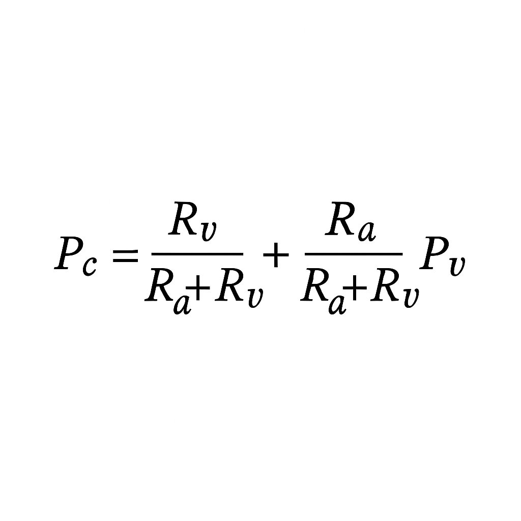
High capillary pressure (Pc) → ↑ filtration (Jv) → lymph works harder → oedema only when filtration exceeds lymph drainage.
Low Pc → filtration ↓ towards zero → lymph dominates → plasma volume ↑ → oedema resolves.
You cannot occupy both states at once.
Two states exist:
High capillary pressure (Pc) → ↑ filtration (Jv) → lymph works harder → oedema only when filtration exceeds lymph drainage.
Low Pc → filtration ↓ towards zero → lymph dominates → plasma volume ↑ → oedema resolves.
You cannot occupy both states at once.
4/
In true hypovolaemia:
• Pc is low
• Jv ≈ zero
• Lymph returns a few hundred mL/h
• Plasma volume is defended (transient reabsorption, continued lymph return)
Any oedema shrinks, not grows
In true hypovolaemia:
• Pc is low
• Jv ≈ zero
• Lymph returns a few hundred mL/h
• Plasma volume is defended (transient reabsorption, continued lymph return)
Any oedema shrinks, not grows
5/ The 1/3–2/3 myth
We were all taught: “Only 1/3 of crystalloid stays intravascular; 2/3 goes interstitial.”
This is only true in euvolaemia, when Pc lies near the J-point and filtration is brisk.
In hypovolaemia, Pc sits below the J-point.
Filtration stops.
→ 100% of infused fluid stays intravascular until Pc rises back toward the J-point.
→ No oedema forms during this phase.
Ignoring this is the source of enormous iatrogenic overload.
6/
Once euvolaemia is restored, Pc moves back toward the J-point.
Any additional fluid now raises Pc and accelerates filtration.
Lymph flow increases, but it can only recirculate fluid – not remove the excess.
Total ECF volume expands, and oedema develops once enough extra volume has been added.
Once euvolaemia is restored, Pc moves back toward the J-point.
Any additional fluid now raises Pc and accelerates filtration.
Lymph flow increases, but it can only recirculate fluid – not remove the excess.
Total ECF volume expands, and oedema develops once enough extra volume has been added.
7/
Oedema therefore reflects normal or high intravascular volume + high Pc in the context of expanded ECF volume.
Even with high lymph flow, the surplus fluid has to sit somewhere – most ends up in the interstitium.
If oedema exists at equilibrium, the intravascular space cannot be depleted.
Hypovolaemia = Pc too low to drive filtration
Oedema = Pc high with excess ECF
These states cannot coexist at steady state.
8/
Transient ≠ steady state
• Significant haemorrhage
• Sudden aggressive UF
• First hours of severe burns
• Anaphylaxis / Clarkson’s
These are transition phases, not steady-state ICU physiology.
Transient ≠ steady state
• Significant haemorrhage
• Sudden aggressive UF
• First hours of severe burns
• Anaphylaxis / Clarkson’s
These are transition phases, not steady-state ICU physiology.
9/ The sepsis misconception
Septic patients on arrival are vasodilated and hypotensive, but not oedematous – and there is no haemoconcentration.
No Hb rise = no intravascular fluid loss.
They present with high Pc, high Jv, and matched lymph return: a high-flow circuit, not hypovolaemia.
They only become oedematous after we give litres of IV fluid.
10/
What about studies showing that in early sepsis or burns, Pi becomes very negative and the lymphatic pump transiently weakens?
True – but these reflect the acute, non–steady-state phase (cytokine storm, NO surge) and are often based on modelled inferences, not direct multi-organ lymph-flow measurement.
Where lymph flow is measured directly (animal models), inflammation generally increases lymph flow, not decreases it.
These findings describe the transition, not equilibrium physiology.
11/
By the time the system reaches equilibrium, the picture is different.
Pc is high, Jv is high, and lymph flow is recirculating that filtrate – so intravascular volume is preserved unless extra fluid is added.
Oedema appears only after IV loading pushes Pc above the J-point.
Direct measurements support this; claims of sustained lymph failure are largely modelling artefacts, not steady-state human physiology.
This is why vasopressor-first is safe.
12/
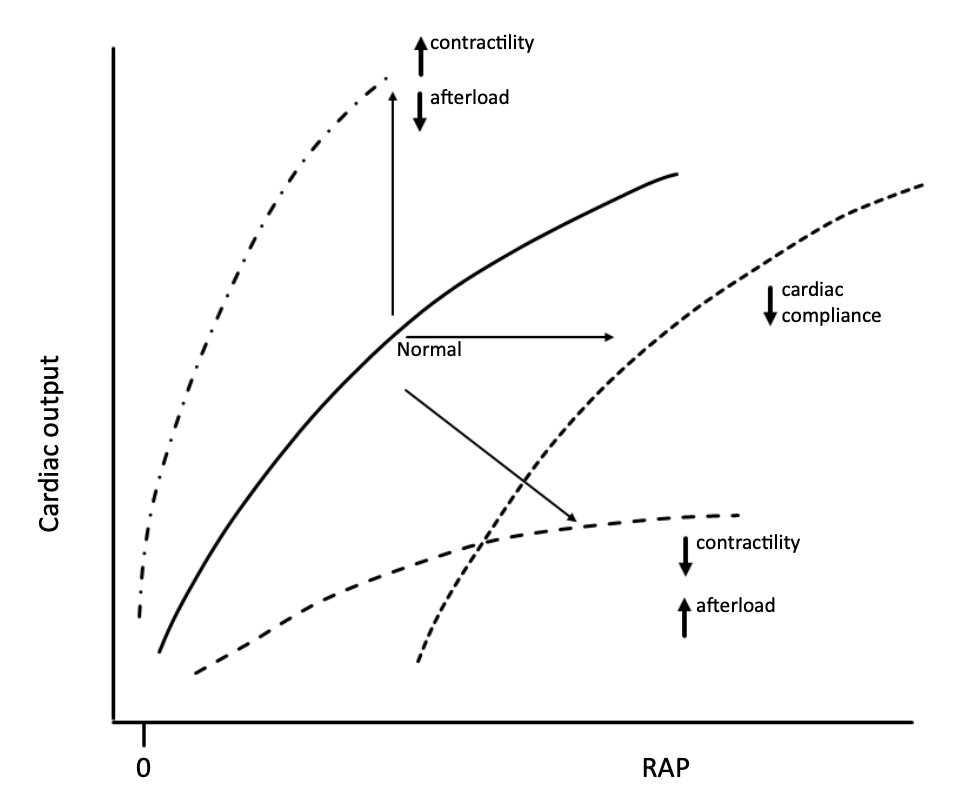
If you see oedema plus hypotension, the issue isn’t “dryness” – it’s usually:
• High RAP
• Venous congestion
• Impaired lymph return
• Low SVR
• Low Pms
A fluid bolus may nudge Pms up for a few minutes, but it quickly raises Pc and congestion again.
Net effect: adding fluid worsens all of these.
If you see oedema plus hypotension, the issue isn’t “dryness” – it’s usually:
• High RAP
• Venous congestion
• Impaired lymph return
• Low SVR
• Low Pms
A fluid bolus may nudge Pms up for a few minutes, but it quickly raises Pc and congestion again.
Net effect: adding fluid worsens all of these.
13/
Conversely, in true hypovolaemia:
Any fluid – even water – stays intravascular until Pc reaches the J-point.
You cannot generate oedema while Pc remains low.
Conversely, in true hypovolaemia:
Any fluid – even water – stays intravascular until Pc reaches the J-point.
You cannot generate oedema while Pc remains low.
14/
So the rule becomes simple:
🟦 Oedema = high-pressure, high-volume physiology (expanded ECF)
🟥 Hypovolaemia = low-pressure, low-filtration physiology
You cannot be both at steady state unless lymphatics fail catastrophically – a rare extreme, not the pattern of routine sepsis.
So the rule becomes simple:
🟦 Oedema = high-pressure, high-volume physiology (expanded ECF)
🟥 Hypovolaemia = low-pressure, low-filtration physiology
You cannot be both at steady state unless lymphatics fail catastrophically – a rare extreme, not the pattern of routine sepsis.
15/ Takeaway
Oedema requires a high Pc state.
Hypovolaemia requires a low Pc state.
These cannot coexist at steady state with functioning lymphatics.
The “oedematous but dry” idea is a relic – and has driven decades of harmful fluid overload.
Oedema requires a high Pc state.
Hypovolaemia requires a low Pc state.
These cannot coexist at steady state with functioning lymphatics.
The “oedematous but dry” idea is a relic – and has driven decades of harmful fluid overload.
16/
Next up:
Albumin — 70 years, 700 studies, zero sustained benefit.
And why almost everything people think it does is actually just lymph flow.
#MedX #ICU #FluidTherapy #Physiology
Next up:
Albumin — 70 years, 700 studies, zero sustained benefit.
And why almost everything people think it does is actually just lymph flow.
#MedX #ICU #FluidTherapy #Physiology
• • •
Missing some Tweet in this thread? You can try to
force a refresh