Ashley Miller Profile picture
#zentensivist #FUSIC Haemodynamics National Lead. Ex ICS council. BSE level 2. BJJ ⬛️⬛️🟥⬛️. No politics, no pets, just #foamed #fluids #pocus #haemodynamics
Sep 12 12 tweets 3 min read
🧵 Part 3 — Why ICU RCTs fail (beyond colliders)

1. The puzzle

Decades of critical care RCTs.
Huge effort. Tens of thousands of patients.
Very few reproducible breakthroughs.
This is Part 3 of my series on why ICU trials fail — and why physiology must guide us.Image 2. Heterogeneity (noise, even in “real” diseases)

Even when the trial is valid, patients vary hugely:
– Baseline physiology, comorbidities, genetics
– Different illness phases (early vs late, compensated vs exhausted)
– Co-interventions (ventilation, sedation, antibiotics)

That means a treatment can help some, harm others.
The “needs bigger N” argument reflects the multi-causality of critical illness.
Sep 6 8 tweets 3 min read
🧵 Part 2. Heterogeneity vs Colliders in Critical Care RCTs

1. The puzzle

Critical care RCTs keep failing.
The usual explanation?
“Patients are too heterogeneous.”
That’s partly true — but there’s a deeper problem.

Part 2 of a 3-thread series on why ICU trials fail and why physiology must guide us. 2. Heterogeneity (the usual alibi)

Heterogeneity = patients in the same trial differ in ways that matter:
– Age, comorbidities, severity
– Disease heterogeneity (e.g. pneumonia due to strep vs haemophilus)
– Different physiology

This makes treatment effect harder to see. Solution? Bigger trials, subgroups, precision medicine.
👉 RCT model still valid — just noisy.
Sep 4 12 tweets 3 min read
🧵“Thresholds, consensus & physiology”

Decades of critical care research have produced few reproducible breakthroughs.
Maybe the problem isn’t our interventions — it’s that we reduce patients to syndromes, instead of treating them as individuals with distinct physiology. 2. The RCT problem

Critical care RCTs keep failing.
Sepsis and ARDS trials are the classic examples: huge effort, massive cost… yet most results are negative, inconsistent, or impossible to replicate.
Aug 25 21 tweets 3 min read
1/21
Acid–base interpretation often feels like a maze.
But there’s a simple way to make sense of it at the bedside.
It starts with pH, strong ions, and base excess. 2/21
First: what is pH?
pH = the concentration of hydrogen ions, which come from water splitting into H⁺ and OH⁻.
Anything that changes how much water dissociates changes pH.
Aug 16 19 tweets 4 min read
1/
Shock isn’t “give fluids, then pressors, then inotropes.”
That recipe misses the physiology.
Here’s how to manage shock properly: 🧵
#MedX #haemodynamics Image 2/
First, rule out mechanical causes (tamponade, tension PTX, massive PE, acute valve rupture, etc). These need specific fixes.
Jul 30 14 tweets 3 min read
1/
In our last thread, we explored how vessels can collapse when MAP falls below a threshold — the Critical Closing Pressure (CCP) — creating a vascular choke point.
But there’s a common misunderstanding we need to clear up 🧵👇
#MedX #Physiology #Haemodynamics
2/
The concept of the vascular waterfall helps explain this:
When CCP rises above venous pressure, blood flow can cease completely — even if a pressure gradient still exists.

Why “waterfall”? Because flow is no longer influenced by the pressure downstream.
Jul 29 25 tweets 5 min read
🧵 What is Critical Closing Pressure — and why does it matter for perfusion?

A thread to clear up one of the most misused and misunderstood ideas in circulatory physiology.
👇Image 1.
We talk a lot about MAP, CVP, and perfusion pressure.
But there’s a hidden threshold that can choke flow completely — even when there’s still a gradient.
It’s called Critical Closing Pressure (CCP).
And it’s time to make sense of it.
Jul 27 17 tweets 3 min read
🧵 "What really determines tissue perfusion?"

– and why most explanations get it wrong.

Let’s sort out MAP, CVP, CCP, autoregulation, vasopressors, and the flow that actually reaches your organs.
👇 Image 1/
You’ve probably heard:
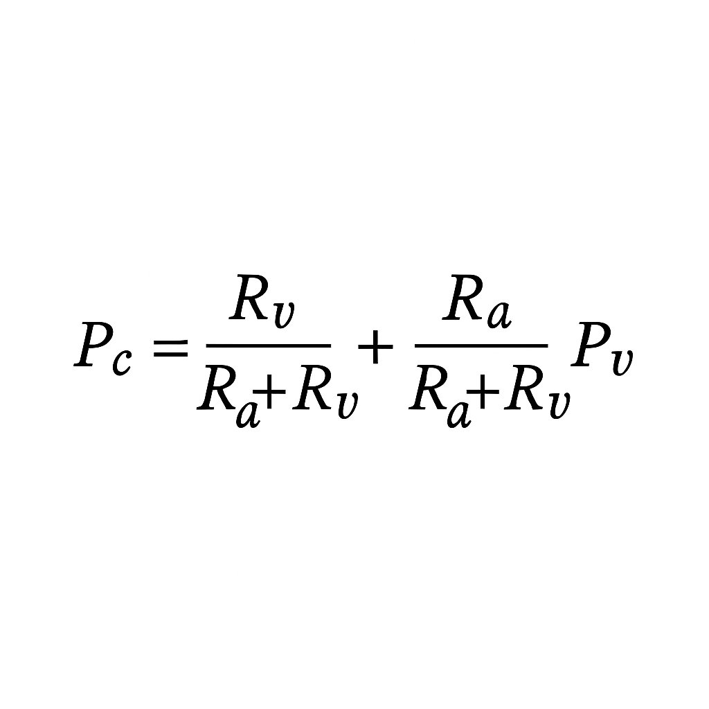
“Perfusion pressure = MAP − CVP”

Or sometimes:
“Perfusion = MAP − CCP”

But both are context-dependent.
Let’s unpack what truly drives tissue perfusion — and why it’s more dynamic than most realise.
Jul 13 13 tweets 3 min read
1/
Shock is complex. But our tools are often simplistic.
This paper proposes a new model:
🩸 Four circulatory interfaces that must stay coupled to maintain perfusion.
Uncouple any one — and shock worsens.
#Shock #MedX #FOAMcc
Here’s the framework.
🔗 doi.org/10.3390/jpm150…Image 2/
🔧 The model outlines 4 key interfaces:
1. LV to systemic arterial
2. Arteriolar to capillary
3. Capillary to venular
4. RV to pulmonary arterial

Each can be assessed. Each can fail. And each demands tailored therapy.
Jul 5 19 tweets 4 min read
🧵 Starling’s Law: Misunderstood, Misapplied, and Still Misleading
1
🚨 “Starling’s Law explains how the heart increases cardiac output.”

You’ve probably heard this a thousand times.
But it’s wrong.
Or at least - very incomplete.
Let’s fix it.
Because this matters - for heart failure, fluids, vasopressors, inotropes, afterload, and how we think about the whole system. 2
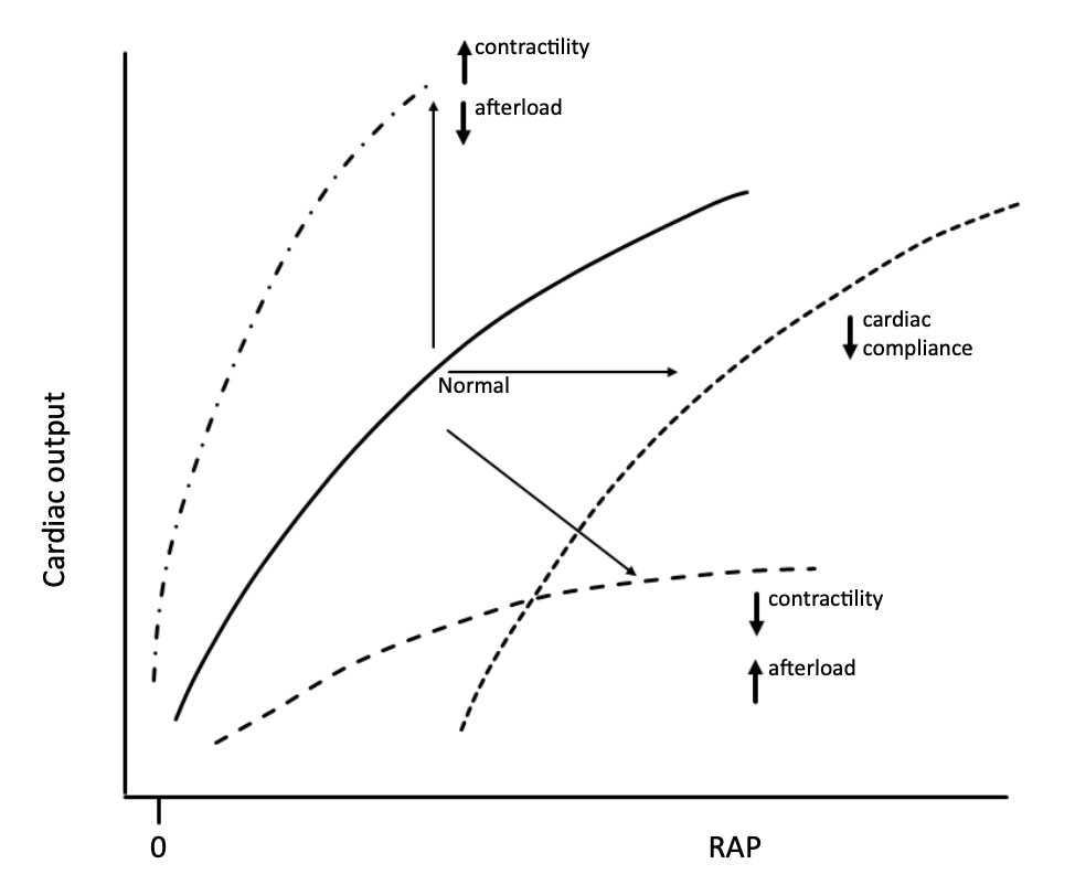
🫀 Textbook Starling curves show:

Preload (RAP or EDV) on the x-axis
Cardiac output on the y-axis
Upward shifts with “more inotropy” or “less afterload”
❌ But that’s not how the system really works.
Because in a real circulation, the system sets flow - not the heart.
Jun 27 18 tweets 3 min read
1/
Most people think the heart drives circulation.
But what if that’s backwards?
Anderson’s model flips the whole idea of cardiac output on its head — and it changes how you think about fluid, flow, and failure.
🧵👇
#physiology #FOAMed #MedTwitter #criticalCare #cardiacOutput Image 2/
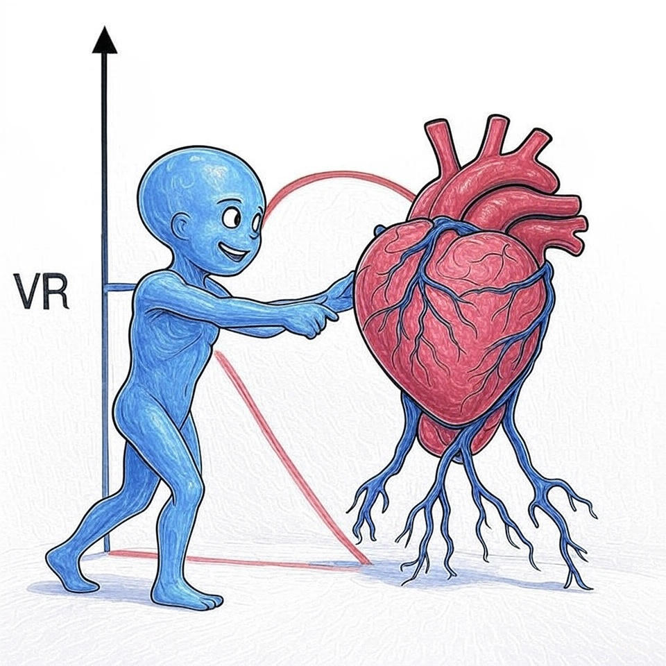
🚿The system sets the flow based on metabolic need.
It adjusts vascular tone and volume to change Pms (mean systemic pressure) and venous return.
The heart simply ejects what arrives — unless it fails.
May 12, 2024 7 tweets 2 min read
Thanks for some great comments on this thread. Some nuance and further explanation is needed here.
Being ill activates the symp NS. I’m sure you have all noticed being tachy and having a bounding pulse when you have flu or are hungover! CO goes up to meet ⬆️ metabolic demand. Sepsis of course is the same and can result in a ⬆️or ⬇️ cardiac output. The key factor is the balance between sepsis causing venoplegia (reducing Pms) and the sympathetic response trying to counter this (which increases Pms and HR).
May 10, 2024 6 tweets 2 min read
#haemodynamics #hemodynamics myths.
Myth 1.
'Sepsis causes low SVR and high stroke volume/CO' This is a common misconception. Why? Because the moment a sick patient comes anywhere near a health care provider they get fluid boluses.
Oct 18, 2021 20 tweets 8 min read
Post holiday season, @ICUltrasonica, @wilkinsonjonny & I are back to take you through the most most critical clinical questions on #haemodynamics that ultrasound can answer

We’re now on to question 3 of FUSIC HD

’Is the aorta abnormal?’

#FUSIC #echofirst #POCUS #FOAMus Aortic dissection is easily missed, carries a high mortality and should be on the differential of any patient with shock, abdo pain or chest pain. Contrary to popular belief the entire aorta can be imaged via transthoracic and abdominal ultrasound. Let’s start with some anatomy
Jun 30, 2021 25 tweets 9 min read
We’ve seen how to measure VTI and stroke volume with #ultrasound
Our next #FUSIC haemodynamic question brought to you by @icultrasonica, @wilkinsonjonny and I is:

Q2. Does SV respond to fluid, vasopressors or inotropes?

#echofirst #POCUS #haemodynamics #foamed In Q1 we saw how to measure stroke volume (SV). Q2 helps us manage someone with an inappropriately low SV. Pressors, fluids and inotropes are all treatment options. If If used correctly, they will ↑SV. If not, they won’t, and they may even be harmful.
Oct 15, 2020 7 tweets 4 min read
@iceman_ex @avkwong Really interesting study. Before I read it I assumed that 'physiological assessment' would be lots of fluid responsiveness assessment and then filling to an unresponsive (pathological) state. In fact a lot of ultrasound was used. And there was no difference in fluid administered @iceman_ex @avkwong So why did US not help? Echo doesn't tell you whether there is hypovolaemia or not. A hyperdynamic heart is a feature of low venous return which is more often from venoplegia than hypovolaemia. Echo cannot distinguish between these.
Apr 22, 2020 8 tweets 2 min read
Let's talk about fluids in COVID. @iceman_ex @Wilkinsonjonny @ThinkingCC @load_dependent
1/8
Early in the outbreak it was commonly advised to aim for a -ve fluid balance
More recently a higher than expected occurrence of AKI and RRT has been observed prompting calls for a more liberal fluid strategy.
All these miss the point about the type of fluid being administered
2/8