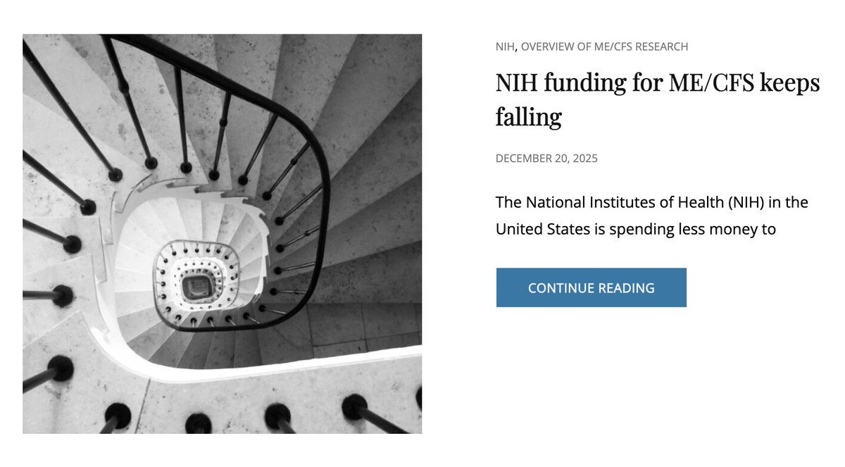
1) New blog post: NIH funding for ME/CFS keeps falling.
Last year, we calculated that the NIH funded 23 ME/CFS projects, totalling an investment of $10.1 million. In 2025, however, this amount decreased to $7.4 million for 18 projects.
2) Even if we include funding for Ian Lipkin’s team at Columbia University (which did not appear in the database), the funding still decreased by 7% to $ 9.4 million.
3) Five ME/CFS projects are planned to end in 2026, hinting that the decline will likely continue next year. The influx of new grants is currently too low to reverse the downward trend.
4) In 2025, there were only two new NIH projects on ME/CFS.
One focuses on bone marrow mesenchymal stromal cell (BMMSC)-derived exosomes. These are important in the communication between cells and play a role in modulating the immune system.
5) This exosome project is led by Vladimir Beljanski at Nova Southeastern University. His team will isolate blood cells from patients and expose them to BMMSC-exosomes. Afterwards, they will test mRNA gene expression and mitochondrial function.
They got $224,070 in 2025.
6) The other project is led by Associate Professor Ramasubramanian at San José State University and focuses on red blood cell deformability. Increased stiffness of red blood cells in ME/CFS patients might make it harder to pass through small spaces, such as tiny blood vessels.
7) Ramasubramanian has previously published about this with Ron Davis using a microfluidic device. They suspect that oxidative stress might be the reason why blood cells of ME/CFS patients are less flexible. This new grant will explore this further and received $432,431 in 2025.
8) The level of NIH funding for ME/CFS was already unacceptably low. To be commensurate with its disease burden, it would need to increase roughly 14-fold. Seeing it decrease even more, year after year, feels very unjust for the millions affected by this horrible illness.
10) There's some ambiguity about which projects to include (is it sufficiently about ME/CFS or not?), and we may have missed one, but the overall trend seems clear.
• • •
Missing some Tweet in this thread? You can try to
force a refresh
1) 🇦🇺 This new study tested the kynurenine pathway in the plasma of 61 ME/CFS patients and 61 controls.
There were no differences in tryptophan or kynurenine, but further downstream, patients had increased levels of 3HK and lower levels of picolinic acid and quinolinic acid.
2) In the white blood cells of patients, the researchers found increased AMP and a lower ATP/ADP ratio, which both hint at an issue with sufficient energy production in immune cells.
3) The authors also tested plasma cytokines (IL-1β, IL-2, IL-4, IL-6, IL-8, IL-10, IL-12p70, IL-13, IL-17A, IL-18, IL-23, IL-33, MCP-1, TNF, interferon IFN-α, IFN-γ) but found no differences.
"We did not observe any differences in pro-inflammatory cytokines between cohorts."
1) Interesting new hypothesis paper by the team of Ron Davis.
They suspect that the recently discovered glymphatic system (the 'lymph nodes' of the brain) plays a role in ME/CFS pathology.
2) The glymphatic system helps to clear waste products from the brain, similar to the lymphatic system elsewhere in the body. It assists with various clean-up processes, especially during sleep.
So ME/CFS could be due to a failed clean-up/reset problem in the brain.
3) In particular, the authors speculate that ME/CFS patients might have antibodies against the AQP4 water channels.
AQP4 proteins are expressed at the membrane of astrocytes throughout the central nervous system and facilitate the exchange of fluids in the brain.
1) This study from Cornell University tested more than 6000 proteins before and after two exercise tests.
It found altered patterns in ME/CFS patients compared to controls, particularly in proteins involved in the immune system, signal transduction, and muscle contraction.
2) The study included 79 ME/CFS patients (selected using the Canadian criteria) and 53 controls. Participants underwent 2 exercise tests, and samples were collected at 5 different time points before and between these tests.
3) At baseline, no protein was significantly different between patients and controls.
The most interesting results were found, not immediately after the first or second exercise test, but after a 24-hour recovery period following the first exercise.
1) Dr. Vanessa Velasco gave preliminary results on OMF's new neutrophil study.
She found that neutrophils of ME/CFS patients died quicker and more often than those of healthy controls, under inflammatory conditions.
2) Neutrophils are immune cells that form the first line of defence when there is an infection. The researchers were able to isolate the neutrophils from whole blood, without changing them, and then mimic inflammatory conditions in the lab.
3) The experiments showed that ME/CFS neutrophils moved slower and produced more reactive oxygen species than controls.
There was also less apoptosis among ME/CFS neutrophils: they don’t die properly when they should. This could prevent the body from resolving inflammation.
1) Impressive study from Johns Hopkins researchers.
They infected mice with SARS-CoV-2, found that female mice had a stronger immune response and more cognitive problems after 84 days than males, and that this was due to over-expression of genes such as Tlr7 on the X chromosome.
2) The study tries to answer the paradox of why males are more likely to be severely ill during infection, while females are more likely to develop Long Covid.
They confirmed this in mice: males had more severe acute illness while females had more lingering cognitive problems.
3) The working memory of the mice was tested through various behavioural tests. There was also a control group of females without infection. Yet only the infected females did worse on maze, marble burying, and novel object recognition tests.
1) The most interesting presentation during the 2025 Stanford symposium was on PET scans of the entire body.
Dr. Michelle James explained that they found a striking pattern with more TSPO signal in various muscle groups of ME/CFS patients, such as the thigh and shoulders.
2) PET scans work by injecting a radioactive molecule into the veins. The scan records annihilation events where a positron leaves the cell and collides with an electron, creating two photons. That signal tells us where in the body the radioactive molecule is binding.
3) Ideally, you want a radioactive tracer that is highly specific to a certain cell type or process so that you know exactly what the signal means.
In this ME/CFS study, they used the popular TSPO tracer, which binds to microglia, astrocytes, myeloid cells, etc.