1/How low can you go??
All the hype nowadays is about high field MRI, but what about low field??
Read on for this month’s @theAJNR SCANtastic for what to know about what may be the next biggest thing in MRI!
ajnr.org/content/47/3/7…
All the hype nowadays is about high field MRI, but what about low field??
Read on for this month’s @theAJNR SCANtastic for what to know about what may be the next biggest thing in MRI!
ajnr.org/content/47/3/7…

2/The growing strength is for larger & larger field strengths for higher & higher resolution
So why would we possible go backwards to lower field strength?
Turns out there are some advantages.
So why would we possible go backwards to lower field strength?
Turns out there are some advantages.

3/Low field strength magnets are much for flexible
They can be put in non-traditional settings (clinics) & can also possibly be moved to the bedside
It is truly POC MRI!
But how does it perform?
They can be put in non-traditional settings (clinics) & can also possibly be moved to the bedside
It is truly POC MRI!
But how does it perform?

4/For infarcts, it tends to see the forest instead of the trees.
It is very good at detecting large infarcts & performs equivalently to normal field strengths
It is very good at detecting large infarcts & performs equivalently to normal field strengths

5/However, it can miss smaller infarcts or they can be much more difficult to see
But, with the advent of deep learning AI algorithms, the image quality can be enhanced to lower the risk of missing small findings
But, with the advent of deep learning AI algorithms, the image quality can be enhanced to lower the risk of missing small findings

6/The question is: how good does the quality HAVE to be?
Yes, we may love amazing 3T images, but often, we don’t need that degree of image quality or detail to make the correct diagnosis.
Lower field strengths can suffice.
Yes, we may love amazing 3T images, but often, we don’t need that degree of image quality or detail to make the correct diagnosis.
Lower field strengths can suffice.

7/It’s like food.
Would you like a Michelin starred meal? Yes.
Do you really need it? No. Regular food is good enough to suffice.
Would you like a Michelin starred meal? Yes.
Do you really need it? No. Regular food is good enough to suffice.

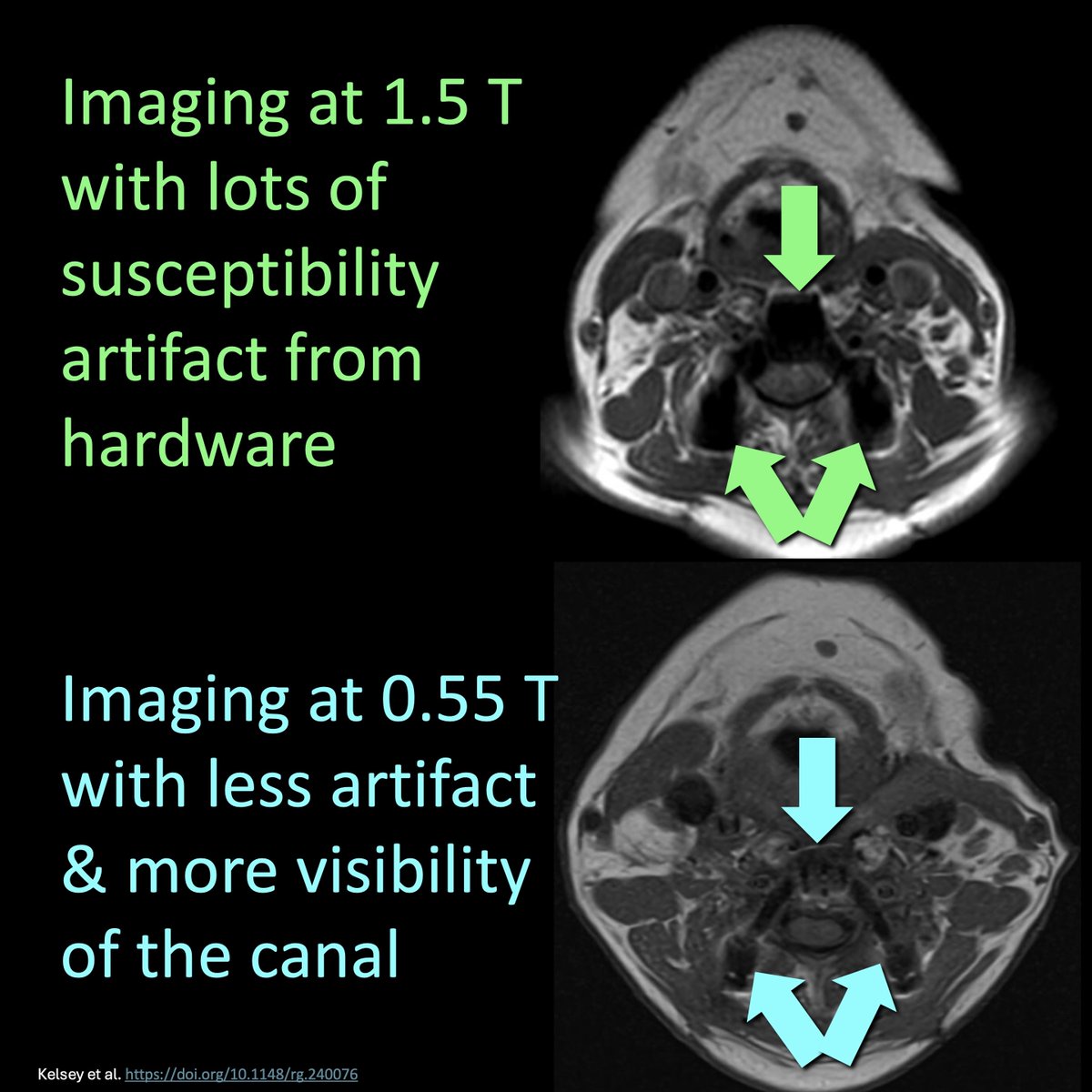
8/And low field MRIs have a big advantage when it comes to susceptibility artifact
Larger field strengths are more sensitive to susceptibility—which can obscure pathology.
It’s just like how larger ears are more sensitive to noise—it’s not always a good thing!
Larger field strengths are more sensitive to susceptibility—which can obscure pathology.
It’s just like how larger ears are more sensitive to noise—it’s not always a good thing!

9/You can see here how lower field strengths actually make the canal MORE visible than on higher field strength bc there is less susceptibility artifact from the patient’s hardware.
10/In this months @theAJNR, Rao et al. found low-field MRI was 92% sensitive & 86% specific for detecting infarcts in ventilated ICU patients
And it changed management in nearly 40%
And it changed management in nearly 40%

11/Hopefully now you know that good things can often come in small field strengths!!
But this only scratches the surface!
Follow @theAJNR & check out the article for yourself:
ajnr.org/content/47/3/7…
But this only scratches the surface!
Follow @theAJNR & check out the article for yourself:
ajnr.org/content/47/3/7…

• • •
Missing some Tweet in this thread? You can try to
force a refresh