Today in @Nature, we report MouseMapper: foundation-model AI to map disease perturbations across the entire mouse body cell-by-cell.
In obesity, it revealed body-wide inflammation & unexpected facial nerve damage. 🧵👇🔉
led by @Dorie00 & @yingchen733 nature.com/articles/s4158…
In obesity, it revealed body-wide inflammation & unexpected facial nerve damage. 🧵👇🔉
led by @Dorie00 & @yingchen733 nature.com/articles/s4158…
Many diseases, including obesity, affect multiple organs and body systems simultaneously. But studying these systemic effects at cellular resolution across the whole body has remained a major challenge.
Using DISCO tissue clearing and light-sheet microscopy, we are able to image intact mouse bodies in 3D at single-cell resolution. The bottleneck has been analyzing these massive datasets in an unbiased and scalable way.
MouseMapper solves this challenge: an AI framework combining deep-learning-based segmentation of peripheral nerves, immune cells and organs/tissues across the entire mouse body. #AI #DeepLearning #BiomedicalResearch 
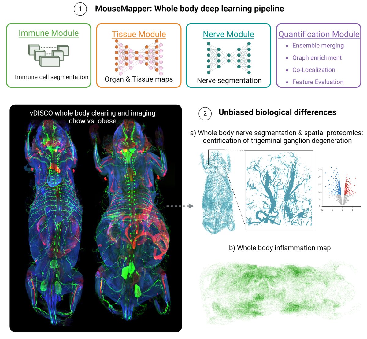
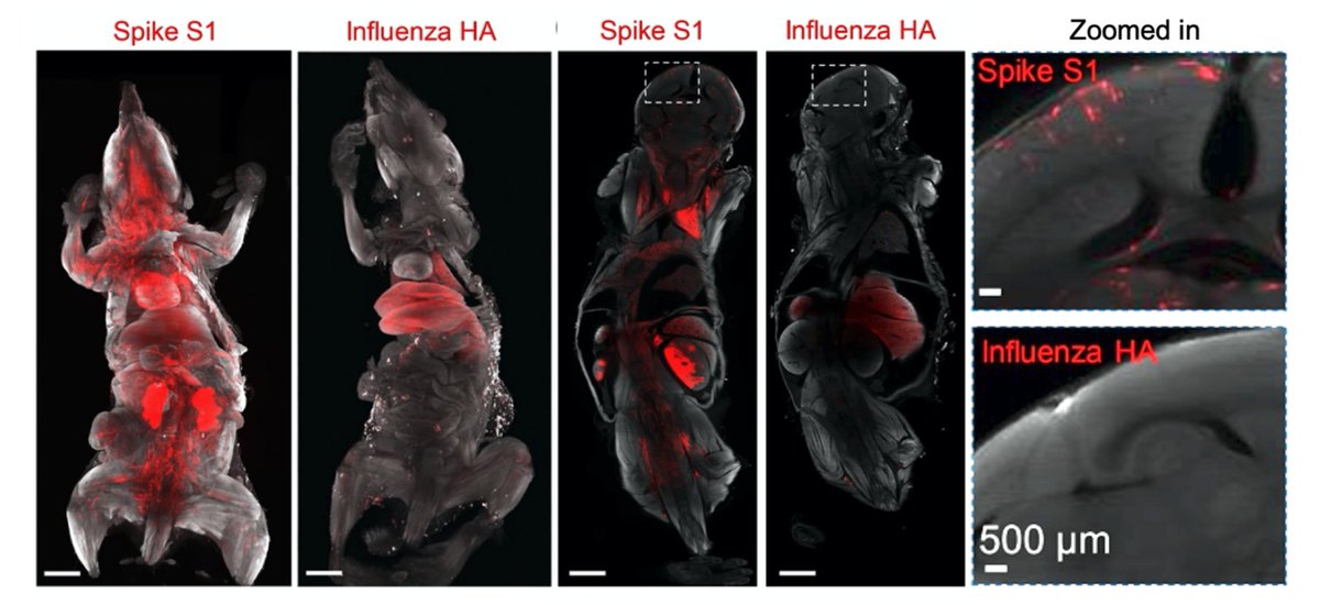
To study obesity-induced changes, we used vDISCO clearing and light-sheet imaging to visualize the whole peripheral nervous system (Uchl1-eEGFP) in lean and obese mice.
We also visualized obesity-associated inflammation (CD68-eGFP+ immune cells) throughout intact mouse bodies, revealing widespread systemic inflammation.
To enable comprehensive whole-body analysis of disease, we developed MouseMapper, which consists of three integrated AI modules:
• Nerve-Module → segments peripheral nerves
• Immune-Module → detects immune cells and clusters
• Tissue-Module → maps 31 organs and tissues
• Nerve-Module → segments peripheral nerves
• Immune-Module → detects immune cells and clusters
• Tissue-Module → maps 31 organs and tissues

The AI models were trained using uniquely curated 3D datasets annotated in virtual reality, enabling accurate segmentation of nerves, immune cells, organs and tissues across terabyte-scale whole-body datasets.

Using MouseMapper, we generated the first whole-body peripheral nerve maps in obesity and discovered that overall nerve density is reduced in obese mice.

We also converted the segmented nerve networks into whole-body nerve graphs, enabling extraction of structural features such as local nerve radii. This revealed a shift toward smaller nerve calibers.

Graph analysis also revealed uncovered structural alterations in the infraorbital branch of the trigeminal nerve, which is essential for facial sensory perception. Obese mice showed a reduction in nerve endings, accompanied by impaired whisker sensation.

To understand the molecular basis of these changes, we performed spatial proteomics on trigeminal ganglia and identified dysregulated pathways linked to axon guidance, cytoskeletal remodeling and complement signaling.

Importantly, we found that several obesity-associated molecular signatures identified in mice were also conserved in post-mortem human trigeminal ganglia from individuals with obesity.

This provides a translational bridge between whole-body imaging in mice and human disease biology, linking obesity to structural and molecular changes in peripheral sensory nerves.
MouseMapper also generated whole-body inflammation maps by quantifying CD68+ immune-cell clusters across tissues and categorizing them by cluster size.

Beyond obesity, MouseMapper provides a scalable blueprint for studying systemic diseases at whole-body resolution, including neurodegeneration, cancer, cardiovascular disease and immune disorders.
Feel free to explore our data! The online atlas allows interactive exploration of whole-body nerve and immune-cell alterations in obesity:
discotechnologies.org/MouseMapper/
discotechnologies.org/MouseMapper/
Huge thanks to all co-authors and collaborators who contributed to this interdisciplinary effort spanning AI, tissue clearing, imaging, neuroscience, metabolism and spatial proteomics.
We hope MouseMapper helps uncover previously inaccessible system-wide disease mechanisms and accelerates discovery of new therapeutic targets.
and huge congrats to other co-first authors @Rami96614090
• • •
Missing some Tweet in this thread? You can try to
force a refresh