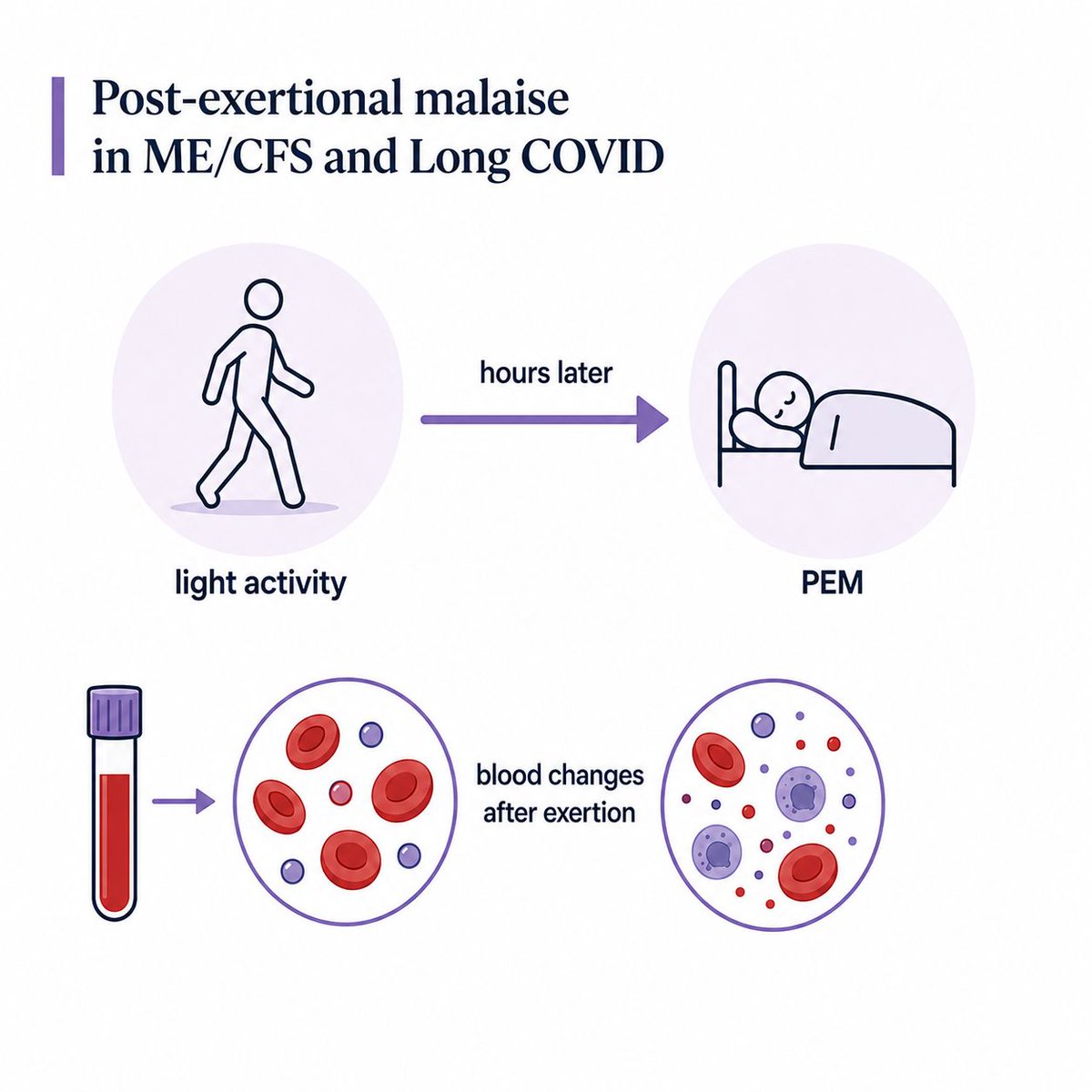
Mitochondrial findings in ME/CFS and Long COVID include altered fuel use, lower
energy efficiency, immune-cell energy stress, muscle-cell abnormalities and worse
muscle findings after PEM.
This thread breaks down what that means in simple language.
energy efficiency, immune-cell energy stress, muscle-cell abnormalities and worse
muscle findings after PEM.
This thread breaks down what that means in simple language.

The main idea is not "mitochondria are broken".
The research points more towards cells struggling to meet energy demand cleanly, especially during immune stress, muscle contraction or PEM.
The research points more towards cells struggling to meet energy demand cleanly, especially during immune stress, muscle contraction or PEM.
Mitochondria help cells turn fuel and oxygen into usable energy.
They also help control immune signalling, cell stress and inflammation.
So mitochondrial findings can reflect energy strain, immune activation or both.
They also help control immune signalling, cell stress and inflammation.
So mitochondrial findings can reflect energy strain, immune activation or both.
Naviaux et al. found broad chemical differences in ME/CFS blood samples.
Most of the altered chemicals were lower than normal.
In simple terms: the blood chemistry looked like the body was running on a
lower-output setting.
Most of the altered chemicals were lower than normal.
In simple terms: the blood chemistry looked like the body was running on a
lower-output setting.
Fluge et al. found a pattern suggesting impaired pyruvate dehydrogenase function.
This enzyme helps move sugar-derived fuel into the main energy-making route inside mitochondria.
This enzyme helps move sugar-derived fuel into the main energy-making route inside mitochondria.
In simple language: cells may start breaking down sugar, but struggle to feed that sugar-derived fuel into the strongest energy-making system.
That could push cells to rely more on backup fuel routes.
That could push cells to rely more on backup fuel routes.
The same study exposed healthy muscle cells to blood serum from severe ME/CFS patients.
Those cells increased respiration and released excess lactate.
That suggests blood-borne factors may push muscle cells into abnormal energy
behaviour.
Those cells increased respiration and released excess lactate.
That suggests blood-borne factors may push muscle cells into abnormal energy
behaviour.
Tomas et al. found impaired mitochondrial respiration in immune cells from ME/CFS patients.
A later study found severe ME/CFS showed both mitochondrial and glycolytic
impairment.
So severity may involve different energy changes, not only stronger symptoms.
A later study found severe ME/CFS showed both mitochondrial and glycolytic
impairment.
So severity may involve different energy changes, not only stronger symptoms.
Mandarano et al. studied T cells directly.
They found altered metabolism in CD4 and CD8 T cells from ME/CFS patients, including reduced mitochondrial membrane potential in CD8 T cells.
They found altered metabolism in CD4 and CD8 T cells from ME/CFS patients, including reduced mitochondrial membrane potential in CD8 T cells.
Fernandez-Guerra et al. found lower mitochondrial coupling efficiency in immune
cells.
In other words:
the cells may be using oxygen, but turning less of that effort into usable energy
cells.
In other words:
the cells may be using oxygen, but turning less of that effort into usable energy
Their protein findings pointed towards pyruvate dehydrogenase and coenzyme A
metabolism.
That overlaps with other ME/CFS findings.
Different methods keep pointing towards altered fuel use
metabolism.
That overlaps with other ME/CFS findings.
Different methods keep pointing towards altered fuel use
Missailidis et al. found that ME/CFS lymphoblasts may rely more on alternative fuels to feed mitochondria.
Put simply:
the cells may be changing which fuels they use to keep energy production going.
Put simply:
the cells may be changing which fuels they use to keep energy production going.
Another Missailidis study found Complex V inefficiency, increased proton leak and lower mitochondrial membrane potential.
In simpler language:
the final energy-making machinery looked less efficient.
In simpler language:
the final energy-making machinery looked less efficient.
Sweetman et al. found altered immune-cell proteins linked to oxidative
phosphorylation, electron transport and ATP production.
This points towards immune cells being under energy strain.
phosphorylation, electron transport and ATP production.
This points towards immune cells being under energy strain.
A 2025 multi-modal ME/CFS study found higher AMP and ADP with a lower ATP/ADP ratio in immune cells.
In simple terms:
the cells showed signs of low usable energy or energy stress
In simple terms:
the cells showed signs of low usable energy or energy stress
Muscle-cell studies add another layer.
When muscle cells from ME/CFS patients were stimulated in the lab, they did not
increase AMPK activation and glucose uptake normally.
AMPK helps cells respond when energy demand rises.
When muscle cells from ME/CFS patients were stimulated in the lab, they did not
increase AMPK activation and glucose uptake normally.
AMPK helps cells respond when energy demand rises.
A later muscle-cell study found ME/CFS cells were worse at using glucose for
mitochondrial energy.
But use of some other fuels was more preserved.
So the problem may involve how sugar-derived fuel is used, not total mitochondrial failure.
mitochondrial energy.
But use of some other fuels was more preserved.
So the problem may involve how sugar-derived fuel is used, not total mitochondrial failure.
The WASF3 study found that a stress-linked protein can disrupt mitochondrial
respiratory supercomplexes.
In simple language:
cell stress may interfere with how mitochondria organise their energy-making
machinery.
respiratory supercomplexes.
In simple language:
cell stress may interfere with how mitochondria organise their energy-making
machinery.
The same study found higher WASF3 and ER-stress signals in ME/CFS muscle biopsy samples.
This does not prove WASF3 explains ME/CFS.
But it gives one possible route from cell stress to poor energy production.
This does not prove WASF3 explains ME/CFS.
But it gives one possible route from cell stress to poor energy production.
Long COVID has overlapping evidence.
A small immune-cell proteomics study found 162 proteins differed from controls.
Twenty-one were linked to mitochondrial metabolism, translation, dynamics or oxidative phosphorylation.
A small immune-cell proteomics study found 162 proteins differed from controls.
Twenty-one were linked to mitochondrial metabolism, translation, dynamics or oxidative phosphorylation.
Appelman et al. took muscle biopsies before and after PEM in Long COVID.
They found lower exercise capacity, lower mitochondrial respiration, lower TCA-cycle metabolites, more fast-fatigable fibres and worse muscle abnormalities after exertion.
They found lower exercise capacity, lower mitochondrial respiration, lower TCA-cycle metabolites, more fast-fatigable fibres and worse muscle abnormalities after exertion.
Consistent mechanism: cells may be able to make energy, but not scale it properly when demand rises.
That could mean poor reserve during infection, immune activation, standing, exertion or PEM.
That could mean poor reserve during infection, immune activation, standing, exertion or PEM.
Less-replicated signals include WASF3, blood-serum effects on healthy muscle cells, and newer ATP/ADP findings.
These are interesting because they suggest possible routes into the energy problem, but they need replication.
These are interesting because they suggest possible routes into the energy problem, but they need replication.
Study links: pubmed.ncbi.nlm.nih.gov/?term=27573827…
At Amatica Health, we're working to build one of the largest linked biological and
patient datasets in this space, to support research into the biology of conditions like ME/CFS and Long COVID.
Our mission and methods: amaticahealth.com
patient datasets in this space, to support research into the biology of conditions like ME/CFS and Long COVID.
Our mission and methods: amaticahealth.com
• • •
Missing some Tweet in this thread? You can try to
force a refresh