Mourad H Senussi, MD, MS Profile picture
Dec 12, 2017 3 tweets 5 min read Read on X
What is the underlying rhythm? See reply for the answer @amalmattu @MasriAhmadMD @MusaSharkawi @jelevenson @ @mbelshazly #FOAMed #EKG Image
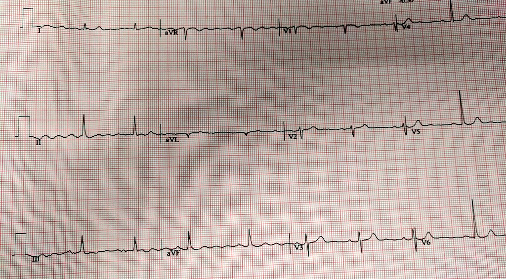
@amalmattu @MasriAhmadMD @MusaSharkawi @jelevenson @mbelshazly A cursory glance may fool one to think this is atrial flutter. On closer inspection clear preceding p-waves are seen. This gentleman has Parkinson's disease and the undulating baseline coincides with his resting tremor frequency. Image

• • •

Missing some Tweet in this thread? You can try to force a refresh
 

Keep Current with Mourad H Senussi, MD, MS

Mourad H Senussi, MD, MS Profile picture

Stay in touch and get notified when new unrolls are available from this author!

Read all threads

This Thread may be Removed Anytime!

PDF

Twitter may remove this content at anytime! Save it as PDF for later use!

Try unrolling a thread yourself!

how to unroll video
  1. Follow @ThreadReaderApp to mention us!

  2. From a Twitter thread mention us with a keyword "unroll"
@threadreaderapp unroll

Practice here first or read more on our help page!

More from @msenussiMD

Feb 8, 2023
**Hepatic Veins : A window to the heart**
What do you see here?

Clue: look at the rhythm
Hepatic veins can provide a plethora of information on the "right heart apparatus".
1. Tricuspid valve
2. RV function /PA pressures
3. Volume status

Often overlooked but can also glean some info on electrical activity of the atria i.e. atrial dysrhythmias.
Lets go over circuitous methods of doing so :)

Note good antegrade flow.

But one can clearly see the f-waves from the atrial flutter!
Read 7 tweets
Feb 7, 2023
***Bloodless central venous cannulation***
1. Preload the needle with your guidewire
2. Use wrap around technique to control the wire
3. Impeccable DYNAMIC ultrasound guidance
4. Rejoice - not a single drop of blood in your field
Alternatively can use the back end of specially designed needles that obviate the need to remove your syringe from the needle. I find it more cumbersome but allows for aspiration of blood!
Important caveats

Must use impeccable dynamic dynamic ultrasound technique and localize the needle tip intraluminally!

Most times when needle tip position is lost is during the syringe removal process. So try out this technique!
Read 4 tweets
Oct 25, 2021
** Bubbles of Truth **

We can gain a tremendous amount of clinical information by observing the flow of bubbles within our vascular system.

Here are a few illustrative examples!
Exhibit A:
Below is a subxiphoid view highlighting markedly distended hepatic veins and IVC. Note the reflux of bubbles into the HV/IVC consistent with high right sided filling pressures.
The bubbles in the above case are arising from rapidly infusing IV fluids through an upper extremity central venous catheter.
Read 14 tweets
Oct 16, 2021
*Hyperdynamic LV: Harbinger of Doom*

This clincial scenario has played out before my eyes on multiple occasions

Echo performed on a pt with undifferentiated shock

"EF > 70%...hyperdynamic.. all good."

A hyperdynamic LV is more sinister than you may think. Lets find out why..
Consider why the LV would be hypercontractile and avoid the knee jerk reflex to assume that it is ONLY due to hypovolemia/hemorrhage i.e decreased RV preload/low mean systemic filling pressure .
Start thinking in terms of LV preload.
1. RV failure
2. Mitral regurgitation
3. VSD
4. Vasodilated state

You need to actively rule out these conditions in the right clinical context.
Read 8 tweets
Sep 22, 2020
(1/)I've had several people ask me about details on how to use bedside ultrasound to confirm ET tube placement, detect esophageal and mainstem intubations, and adjust positioning.

So here is a step by step guide!

Ref: journals.lww.com/ccejournal/Ful…
(2/)**Overview** this protocol utilizes sequential tracheal and bilateral thoracic lung ultrasound.
1. Tracheal ultrasound to r/o esophageal intubations
2. Right Lung US
3. Left Lung US
4. Adjustment to achieve bilateral lung sliding
(3/)**Tracheal Ultrasound**
This is simple. Place your transducer transversely at the level of the suprasternal notch. Below is an example of the appearance of the trachea with a hyperechoic anterior wall and acoustic shadowing. Transverse ultrasonographic appearance of the trachea. The t
Read 17 tweets
Jul 7, 2020
(1/24)*De-mystifying Hepatic waveforms*
Welcome to Part 2. Now that we have gone through the basics:(). Time for a deep dive where we can start to decipher the important clinical information we need to apply at the bedside #showmethewaveforms #shocksquad
(2/24)We must acquaint ourselves with the different guises that HV waveforms may appear, they can be
- biphasic and tetrainflectional
- tetraphasic and tetrainflectional
- Increased antegrade flow (S and D magnitude)
- Increased retrograde flow (A-reversal, SR, DR)
See⬇️
(3/24)The fun doesn't stop there! We all know how the atrium augments flow to the ventricle. With atrial relaxation (analogous to x-descent on JVP) that downward deflection helps increase forward flow during systole. With TV closure, you can see a notched S-wave (S1, S2).
Read 26 tweets

Did Thread Reader help you today?

Support us! We are indie developers!


This site is made by just two indie developers on a laptop doing marketing, support and development! Read more about the story.

Become a Premium Member ($3/month or $30/year) and get exclusive features!

Become Premium

Don't want to be a Premium member but still want to support us?

Make a small donation by buying us coffee ($5) or help with server cost ($10)

Donate via Paypal

Or Donate anonymously using crypto!

Ethereum

0xfe58350B80634f60Fa6Dc149a72b4DFbc17D341E copy

Bitcoin

3ATGMxNzCUFzxpMCHL5sWSt4DVtS8UqXpi copy

Thank you for your support!

Follow Us!

:(