Mourad H Senussi, MD, MS Profile picture
Intensivist. Cardiologist. Lifelong martial artist. Hemodynamics, Shock & Resuscitation.
Feb 8, 2023 7 tweets 2 min read
**Hepatic Veins : A window to the heart**
What do you see here?

Clue: look at the rhythm 
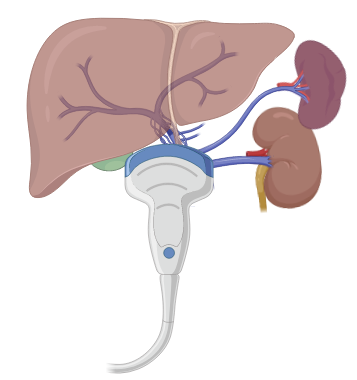
Hepatic veins can provide a plethora of information on the "right heart apparatus".
1. Tricuspid valve
2. RV function /PA pressures
3. Volume status

Often overlooked but can also glean some info on electrical activity of the atria i.e. atrial dysrhythmias.
Feb 7, 2023 4 tweets 2 min read
***Bloodless central venous cannulation***
1. Preload the needle with your guidewire
2. Use wrap around technique to control the wire
3. Impeccable DYNAMIC ultrasound guidance
4. Rejoice - not a single drop of blood in your field Alternatively can use the back end of specially designed needles that obviate the need to remove your syringe from the needle. I find it more cumbersome but allows for aspiration of blood!
Oct 25, 2021 14 tweets 4 min read
** Bubbles of Truth **

We can gain a tremendous amount of clinical information by observing the flow of bubbles within our vascular system.

Here are a few illustrative examples! Exhibit A:
Below is a subxiphoid view highlighting markedly distended hepatic veins and IVC. Note the reflux of bubbles into the HV/IVC consistent with high right sided filling pressures.
Oct 16, 2021 8 tweets 4 min read
*Hyperdynamic LV: Harbinger of Doom*

This clincial scenario has played out before my eyes on multiple occasions

Echo performed on a pt with undifferentiated shock

"EF > 70%...hyperdynamic.. all good."

A hyperdynamic LV is more sinister than you may think. Lets find out why.. Consider why the LV would be hypercontractile and avoid the knee jerk reflex to assume that it is ONLY due to hypovolemia/hemorrhage i.e decreased RV preload/low mean systemic filling pressure .
Sep 22, 2020 17 tweets 14 min read
(1/)I've had several people ask me about details on how to use bedside ultrasound to confirm ET tube placement, detect esophageal and mainstem intubations, and adjust positioning.

So here is a step by step guide!

Ref: journals.lww.com/ccejournal/Ful… (2/)**Overview** this protocol utilizes sequential tracheal and bilateral thoracic lung ultrasound.
1. Tracheal ultrasound to r/o esophageal intubations
2. Right Lung US
3. Left Lung US
4. Adjustment to achieve bilateral lung sliding
Jul 7, 2020 26 tweets 15 min read
(1/24)*De-mystifying Hepatic waveforms*
Welcome to Part 2. Now that we have gone through the basics:(). Time for a deep dive where we can start to decipher the important clinical information we need to apply at the bedside #showmethewaveforms #shocksquad (2/24)We must acquaint ourselves with the different guises that HV waveforms may appear, they can be
- biphasic and tetrainflectional
- tetraphasic and tetrainflectional
- Increased antegrade flow (S and D magnitude)
- Increased retrograde flow (A-reversal, SR, DR)
See⬇️
Jul 2, 2020 16 tweets 12 min read
(1/ )There has been an overwhelming interest in the use of hepatic waveforms as a congestive parameter. With social media, somewhat obscure concepts like these are quickly brought to the forefront and implemented in clinical practice. Many are eager to incorporate #VEXUS but... 
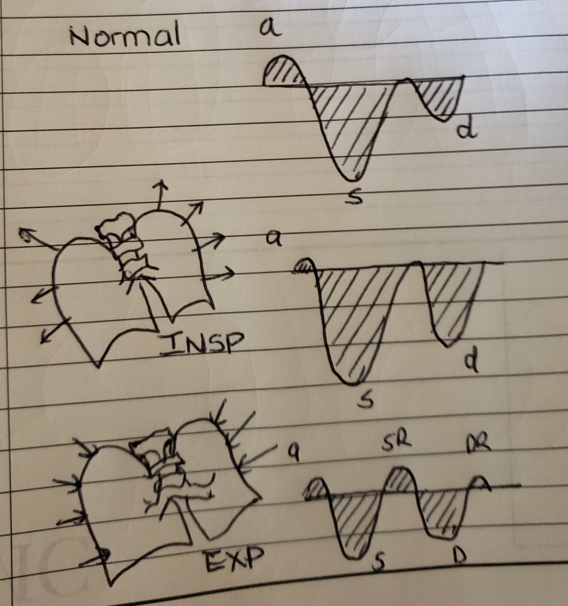
...a thorough understanding of these waveforms, hemodynamic correlates, and dynamic changes that occur in physiology and disease, is paramount to incorporating this into practice. #shocksquad #tweetorial #VEXUS #showmethewaveform. Here are rules of interpreting hepatic waveforms
Mar 31, 2018 9 tweets 5 min read
Tips & tricks for axillary/subclavian central line
1. Start at midclavicular line -> the deltopectoral groove.
2. Collapsible, thin-walled
3. Continuous Doppler flow!
4. Presence of valves denotes a venous structure
5. BONUS inject fluid via ipsilateral peripheral IV! Longitudinal view note the alignment of the rib underneath to utilize the PART method