drshukla14 Profile picture
Emergency Medicine Attending (Consultant) PGY23. History, Physical, #POCUS!
Dr RAMY Profile picture HYOUNES2 Profile picture 2 subscribed
Sep 15, 2023 4 tweets 2 min read
1/n Acute right hip pain on the background of subacute migratory polyarthritis, probably reactive. #POCUS helped diagnosis of right hip effusion, consistent with synovitis. Normal left hip joint image for comparison, showing no effusion.

Image
Image
Image
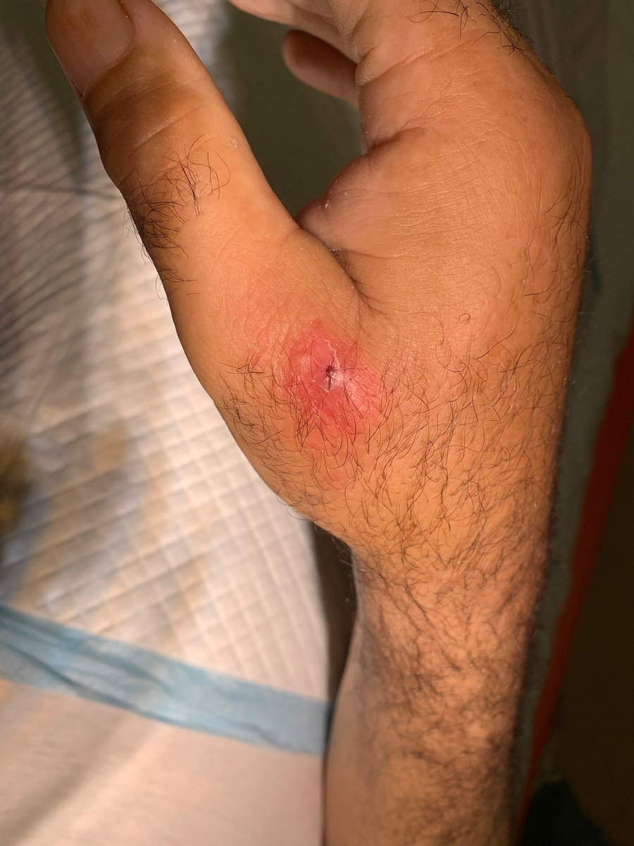
2/n Joint aspirated. Low clinical suspicion of septic arthritis, thus simultaneously injected with triamcinolone+lidocaine (rheumatology colleagues supported the decision).
May 26, 2023 6 tweets 3 min read
1/n: Thread of interesting ED #POCUS case. F/70+, known epigastric hernia (relapsed post repair years ago). Repeated vomiting since 1 week. Labs and plain x-rays - ImageImage 2/n Distended stomach noted on #POCUS -
Mar 11, 2023 7 tweets 3 min read
1/n #POCUS Acute right scrotal pain. Performed awaiting scrotal exploration. "Saddle view" from inferior poles to assess gross difference between echotexture 
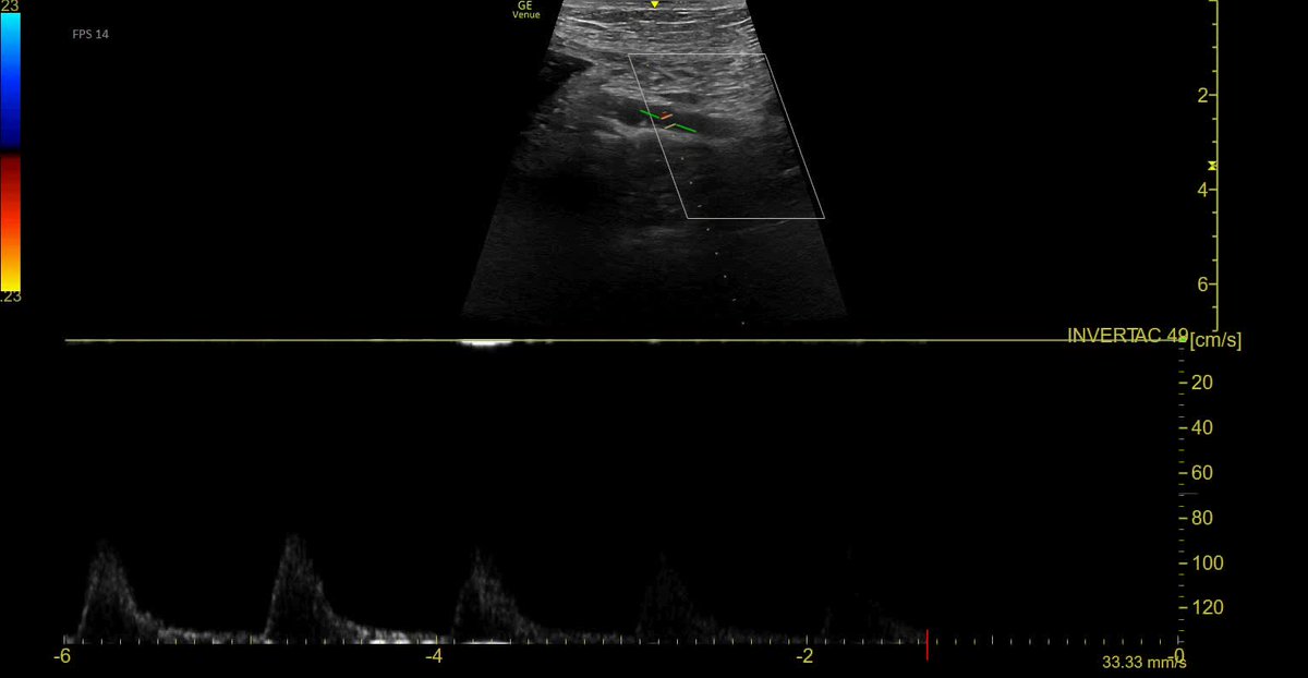
2/n The normal (Left) testis - shows normal internal septal architecture with pulsatile blood flow on color power doppler, with normal overlying skin thickness.
Nov 19, 2022 5 tweets 2 min read
#POCUS case 1/n - Chest pain since 2 hours. Is it posterior STEMI ? 2/n #POCUS Apical Long Axis (A3C)
Apr 21, 2022 5 tweets 2 min read
1/4 #POCUS EM case: M/50, anginal pain since 2 hours. ECG: lateral STEMI. Decision for fibrinolytic therapy (non-PCI site) 2/4 #POCUS appearance of lateral hypokinesia on Short Axis LV:
Apr 9, 2022 4 tweets 2 min read
1/n #POCUS case - F/28. Left wrist pain since 2 weeks, severe since 48 h -> ED visit. Unable to grip due to painful thumb movements. Tender over lateral radial styloid area, Finkelstein test +. Dx: De Quervain tendinopathy. 2/n Comparison with normal - * marks fluid pockets around thickened tendons APL and EPB.
Mar 14, 2022 7 tweets 4 min read
1/n #POCUS case - Pneumothorax. M/20, abrupt left pleuritic chest pain. Absent breath sound left side, hyper-resonant percussion. Absent Pleural sliding on #POCUS . No lung point found, consistent with clinical impression of large pneumothorax. 2/n Normal pleural sliding on #POCUS right anterior thorax
Mar 13, 2022 6 tweets 3 min read
Thread 1/n #POCUS as a mystery solving tool. M/50, SYNCOPE. Seemed benign orthostatic, and I was upset a Head CT was done. Was expecting it normal, but surprise - reported as left parietal WATERSHED territory infarct. MRI later shows infarct better - 2/n Re-examined carefully, and indeed patient had a homonymous right inferior quadrant-anopia. But WHY does he have watershed infarct unilaterally, from what is a global hypo-perfusion event? Left carotid looks ok...
Mar 10, 2022 6 tweets 3 min read
Thread 1/n #POCUS case - (?) COVID - cured by metoprolol. Acute dyspnea with cough, referral as ? COVID-19 with CXR. Sinus rhythm 120/min, RR 40/min, Sats 68% RA, 93% with O2 15 lpm by NRBM. 
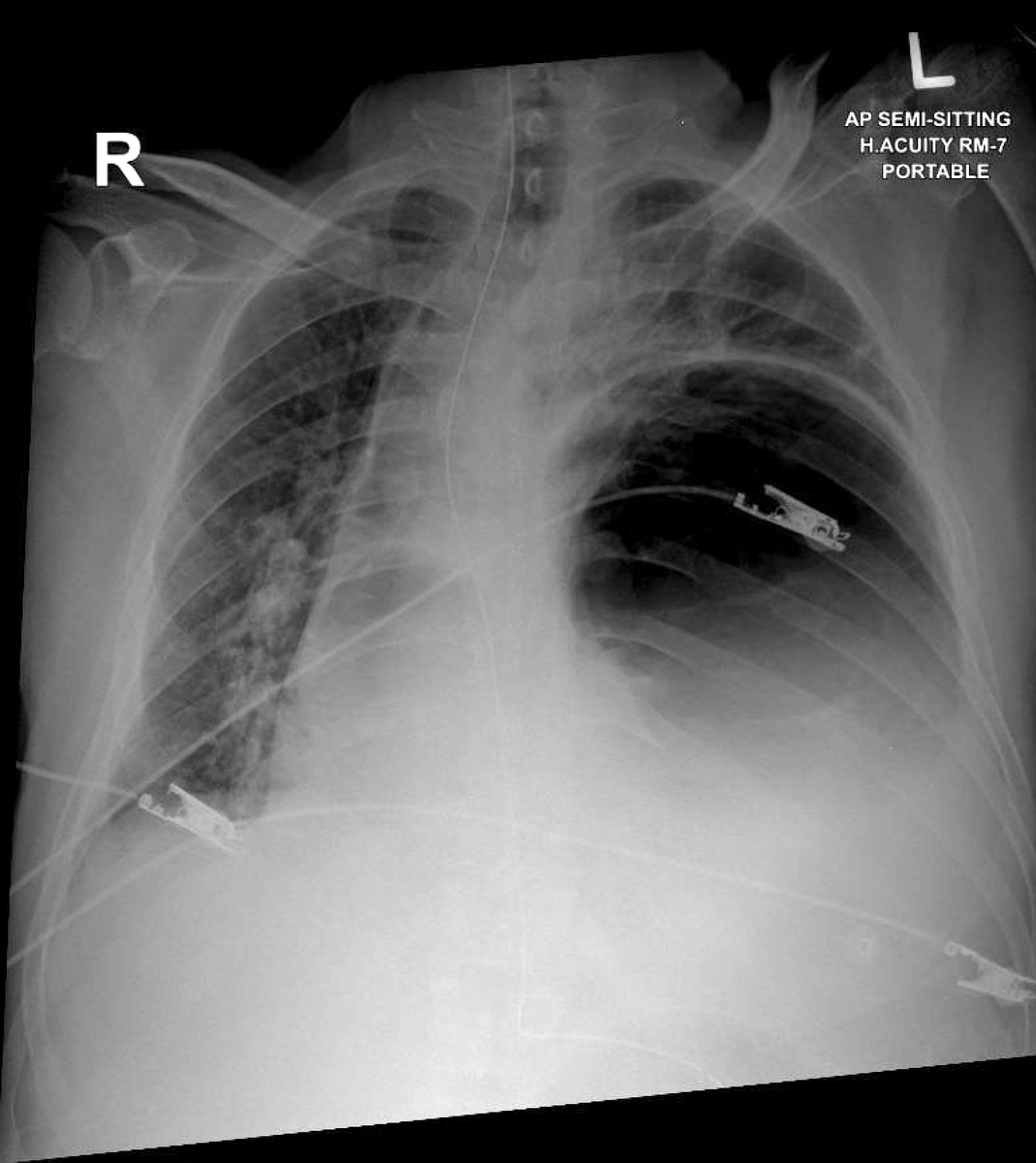
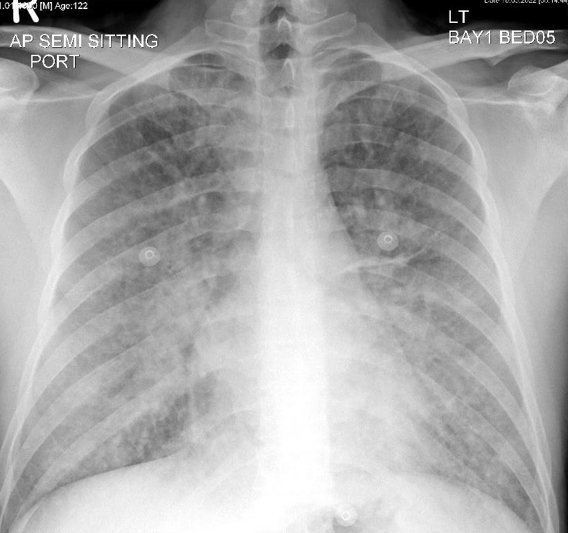
2/n #POCUS all lung zones show B-lines as expected, but minimal left pleural effusion which is NOT common with COVID-19.
Feb 20, 2022 4 tweets 2 min read
Thread: 1/3 Clinically anterior glenohumeral dislocation, confirmed by #POCUS within 5 min of arrival 
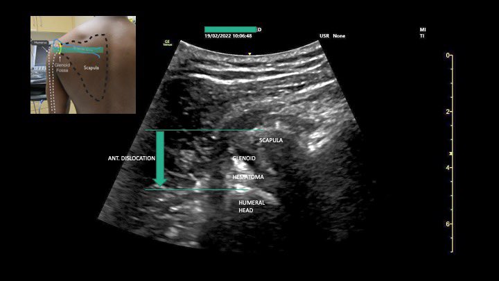
2/3 Pain 10/10 to 0/10 within 5 min of interscalene block
Nov 25, 2021 6 tweets 3 min read
Thread, 1/n - Male 75. Acute dyspnea since 1 hour, can't talk, wheezy. SPO2 75%. No previous record. "All that wheezes is not asthma". #POCUS leaves no doubt about pulmonary edema, Dx while patient is being attached to monitor.BP 180/110 mmHg. 2/n CPAP 10 mmHg, IV infusion of nitrate being prepared. Pectus excavatum, parasternal long axis impossible from usual 3/4 ICS. But Apical 4 chamber suggests mild LV systolic dysfunction
Dec 11, 2020 6 tweets 3 min read
1/n #POCUS is more sensitive than CXR for subtle consolidation detection. M/40, fever, right sided chest pain. Had cough 3 d ago, now resolved.WCC 13k, CRP 230. Normal physical exam. 2/n #POCUS showed bilateral small basal consolidations, B-lines at right base with sub pleural consolidations. Right posterior basal region sub pleural consolidation -
Nov 8, 2020 9 tweets 3 min read
1/n #POCUS findings of effusive constrictive pericarditis. Patient presented with 2 months progressive pedal edema and ascites. Pericardial effusion, funny bounce of IVS 2/n A4C septal bounce
Oct 26, 2020 6 tweets 2 min read
1/n #POCUS as a part of ED evaluation for first episode of atrial fibrillation (this demographic has significant prevalence of rheumatic heart disease) - appears grossly normal - moving on to other views... 2/n Apical 4C also grossly normal....
Oct 5, 2020 9 tweets 3 min read
1/n #POCUS M/68, vomiting since 6 hours, back pain since 1 h. Says no to chest pain /abd pain / dyspnea. Diaphoretic, vomiting, HR 90, RR 24, BP 128/70, SpO2 91% (air). Reduced BS left side, Abd distended, nontender. ECG normal. Is it acute aortic? Tried to get PLAX heart - 2/n - scanning right parasternal area - found the heart - hyperdynamic, but nothing grossly wrong there.
Sep 30, 2020 5 tweets 2 min read
Thread 1/4 - #POCUS Case - MPA+bilateral PE. HR 110, BP 94/68, Sats 98% room air. Subjectively, patient feels well, no dyspnea/pain. Warm skin. Shifted to EM Resus. Lytic or not? Young pt, no known co-morbids / provocation. 2/4 #POCUS - RV does not seem dilated
Jul 4, 2020 6 tweets 3 min read
#POCUS case - F/80. Known mesenteric stenosis (celiac origin, and SMA). Has upper abdominal pain off and on - ? mesenteric angina. Now presents with severe upper abdominal pain. Tender+++ epigastrium, no peritonism. Normal GB ultrasound and lipase. Lactate 0.4. Previous CT: Epigastric tenderness and normal lactate argue against acute mesenteric occlusion. #POCUS - SMA origin
Jun 27, 2020 8 tweets 2 min read
Thread 1/n. #POCUS findings in a M/31, leg swelling and abdomen distention progressive since 1/12. Starting with right IJV (high gain setting) Abdomen RUQ Image
Sep 22, 2019 6 tweets 3 min read
Thread - (1/n) #POCUS findings in Left Main coronary occlusion.. this is not a STEMI - its WORSE ! M/50, 12 h intermittent chest pain. 2 hours of constant chest pain. Normal BP, no rales... but.. ECG horrible. Thread - (2/n) #POCUS findings in Left Main coronary occlusion
Sep 16, 2019 5 tweets 3 min read
#POCUS guided above elbow radial nerve block thread (1/n) Identification or radial nerve #POCUS guided radial nerve block thread (2/n): Actual block administration
Jul 16, 2019 5 tweets 2 min read
Patient referred for arthrocentesis, suspected septic arthritis. #POCUS shows triceps abscess, NO effusion. Aspiration attempt could have contaminated the joint & CAUSED septic arthritis. Consider scanning before sticking any needles. Image