#IMPOCUS educational account sharing cases (fictionalized), ideas, EBM, and clinical integration. Not medical advice. Formerly PittIMPOCUS. #FOAMus.
2 subscribers
Jan 26, 2022 • 25 tweets • 12 min read
#POCUS for post-renal AKI: a basic approach and a review of key images.
Overview here, more details in thread. 1/
Sample case: 60s m with HTN, DM2, presents with fatigue, Cr 6.0 from b/l 0.5. No flank pain or fever. UA no blood, protein, WBCs, LE, nit. No nephrotoxic meds. Empiric 2 L IVF given. Foley placed - some blood, 10 cc UOP over 8 hours. Renal USG ordered. Nephrology consulted.
2/
Jan 9, 2022 • 23 tweets • 11 min read
Classic conundrum... PNA vs cardiogenic pulmonary edema
60 y/o no PMH presents with dyspnea, hypoxemia. SpO2 92% 6L NC HR 120 RR 30 BP 110/70. B/l crackles. No JVP or LE edema. Lactate 4.0. BNP 3x ULN (nco baseline). Tn 2xULN Procal 0.2. Pre-COVID.
How can we apply #POCUS 1/
For the sake of argument, let's assume for this case 1) pre-test probability of PNA is 50% 2) pre-test probability of cardiogenic pulmonary edema is 50% 3) one or both of the above must be present to explain the presentation
2/
Dec 20, 2021 • 14 tweets • 7 min read
Humbling pleural procedure case to share.
70 y/o admitted with fever, hypoxia, R flank pain, loculated pleural effusion (right lower) on CXR. Concern for empyema prompting abx, chest US and possible intervention.
A tweetorial on optimal machine settings for B line image acquisition, with a focus on: focal position at pleura, harmonics off, and increased far gain.
1/18
Let's start with some polls
We will stick to low frequency probes here (i.e. curvilinear and phased array - linear has its own purpose for detailed pleural assessment)
Which preset do you prefer for B line assessment?
2/
Dec 21, 2020 • 31 tweets • 7 min read
Hello everyone! We're here with another #IMPOCUS case for those of you as excited about pocus as you are about the holidays.
65 yo F w hx HFpEF, COPD on 3L O2 at home who presents after a recent CHF admission (diuresed and improved) with fevers, SOB, and diarrhea. COVID+.
1/2/ She is HDS without significant respiratory distress, requiring 4L O2.
Before we start, some polls for you all: 1. How often do you find lung POCUS useful in COVID19?
Nov 22, 2020 • 25 tweets • 18 min read
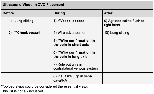
1/ Thread: Ultrasound for CVC placement - more than just follow the needle tip and confirm the wire.
There are many potential uses of ultrasound during CVC placement, shown here. Not all are essential.
#POCUS#IMPOCUS#FOAMED#FOAMUS2/ Pre-procedure
Lung sliding on ipsilateral side - for a comparison to post-procedure
-use linear probe
-apical and lateral/anterior
Nov 15, 2020 • 31 tweets • 21 min read
Let's talk about placement of small bore nasoenteric feeding tubes and use of ultrasound for this (A thread)
Scenario: Pt with gastroparesis and COVID/ARDS, planning to prone, want post-pyloric tube. No dedicated team for this. Can we place safely w #POCUS guidance?
1/
How is small bore feeding tube (SBFT) different from standard NG?
Narrower (6-8 Fr vs 14-18 Fr), more flexible (but w rigid stylet for placement), longer. Thus: more comfortable, gastric or post-pyloric, ineffective for suction, prone to clogging, prone to PTX if placed in lung.
Mar 9, 2020 • 36 tweets • 21 min read
1/ Happy Monday everyone! We have a special case for you today written by UPMC PGY2 #POCUS enthusiast @MikeTao15.
A 52 yo M with hx of EtOH/HCV cirrhosis, VTE on Eliquis, HFrEF presents w worsening abd distention and pain. He is disoriented and unable to give much history.
2/ Cirrhosis previously complicated by esophageal varices, hepatic encephalopathy, and ascites. No follow up since last hospital admission at outside facility ~1 year ago. Has not been taking meds.
Vitals in the ED: Temp 38.1, HR 90, BP 98/70, Pulse Ox 96% on 2L
Feb 28, 2020 • 39 tweets • 25 min read
Ever found DVT POCUS challenging? We do.
Here is a #Tweetorial on #POCUS for Lower Extremity DVT assessment. Let’s walk through a case and discuss common questions that arise.
50s f no PMH presenting to the ED on a Friday evening with right leg swelling and redness...
1/
History/exam/labs:
No travel, surgeries, immobilization. No dyspnea, CP. No meds. No FH of VTE. no tobacco.
HR 88 SpO2 98% RA BP 160/90 T 37.
Exam unremarkable other than 2+ LE edema erythema and tenderness from right ankle to knee.
CBC/BMP/coags normal
2/
Jan 11, 2020 • 30 tweets • 21 min read
1/ After a break for the new year, we are back with a case of #POCUS and abdominal distension.
59 y/o m with no medical history presenting with increasing abdominal girth and now altered mental status.
#IMPOCUS2/ HPI:
well until 3 months ago. Since then, progressive fatigue, nausea, mild diffuse itching without rash. Few weeks of early satiety, poor PO intake, and now increasing abdominal girth. Last BM 3 days ago. No vomiting. Weight unchanged over 3 months. No fevers/chills.
Sep 27, 2019 • 19 tweets • 21 min read
A lines, B lines, Consolidation… the ABCs of Lung Ultrasound
But what do they really mean?
Here we attempt a #tweetorial presenting a way of thinking about common patterns seen on lung ultrasound.
See table here for a summary, and thread below for details
1/
We will start with a hypothetical case for illustration
65 y/o f w HFpEF, COPD admitted 4 days ago with acute pancreatitis (now resolved). You are prepping her for discharge but she is now short of breath/hypoxic requiring 4 L O2.
2/
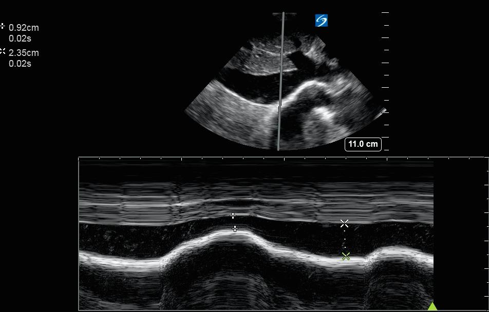
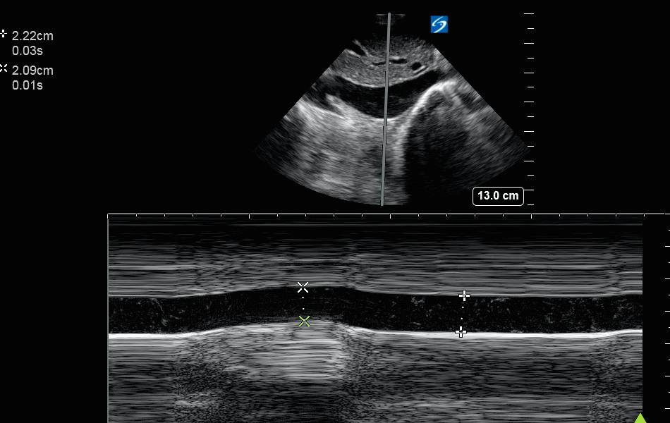
These 2 IVC M-mode images were obtained from the same patient 10 seconds apart. How could they be so different?
1/
60 m with hx of COPD, HFpEF, admitted w LLL PNA s/p abx and 1 L LR. HR 118 BP 110/70. IVC US was performed as a piece of info in determining whether to administer more IVF.