I help high-performers reclaim 2 hrs of deep sleep in 30 days using sunlight & quantum biology. DM “SUN” for free Light Audit. #CircadianCEO #QuantumSleep”
 2 of 3 📸🔆💫👑🎆🎇🌅
 2/5
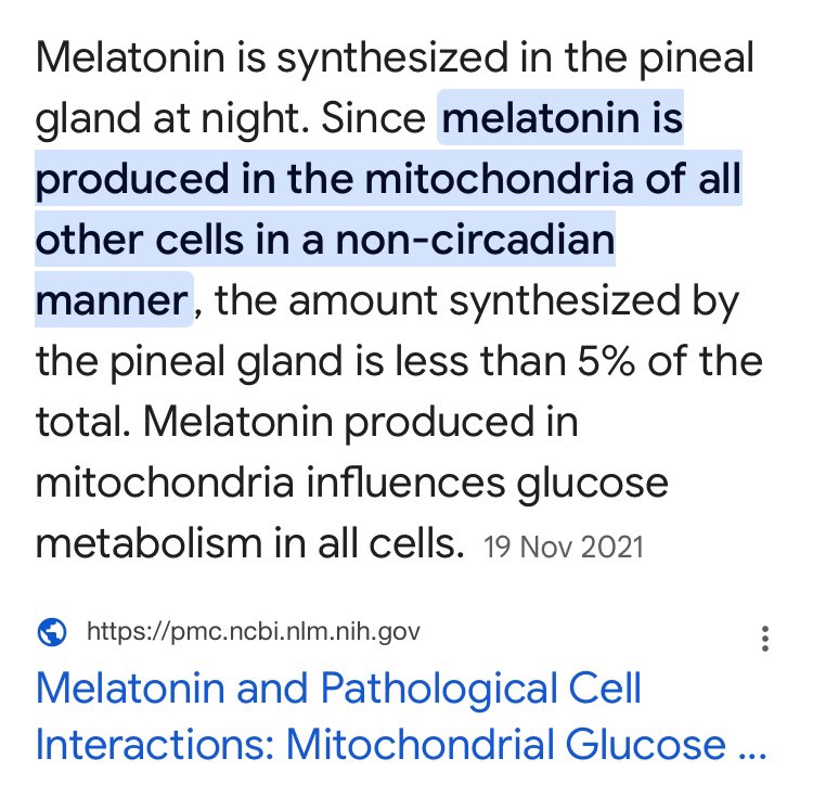
2/5 2 of 3 The Mitochondrial Faucet
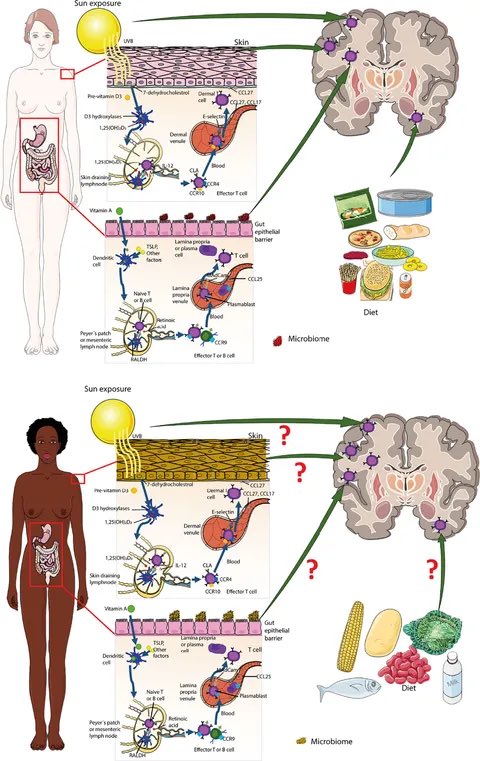
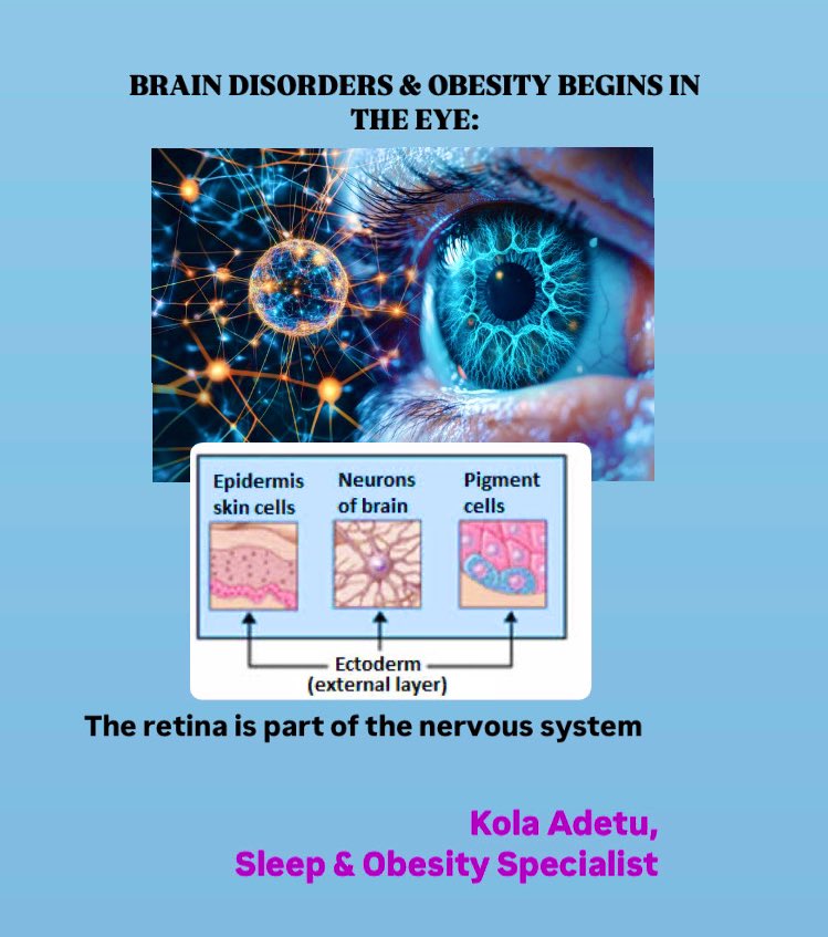
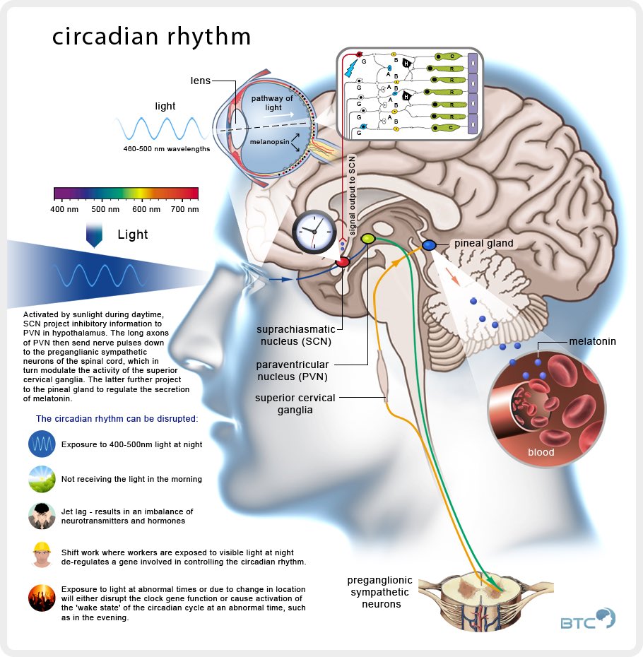
 MS prevention: Never Miss a sunrise, omega 3 fatty fish maxxing
 Part 1 instagram.com/p/DIdYe7zImfE/…