Official Neuroscience News Twitter. Brain research news articles on neuroscience, psychology, AI, neurology, brain cancer, robotics, mental health & science.
2 subscribers
Jan 11 • 4 tweets • 2 min read
Red Light Therapy May Reduce Deadly Blood Clots
A new study shows that exposure to long-wavelength red light reduces the formation of blood clots, which are a leading cause of heart attacks, strokes, and other serious health conditions.
Researchers found that mice exposed to red light had five times fewer clots compared to those exposed to blue or white light.
The study linked red light exposure to reduced inflammation and lower platelet activation, which are key drivers of clot formation.
Analysis of human data revealed that cancer patients using blue light-filtering lenses had fewer blood clots, suggesting light exposure could influence clot risks in people as well.
The research highlights the potential of red light therapy as an inexpensive, non-invasive way to lower clot risks in high-risk populations.
Red Light Therapy May Reduce Deadly Blood Clots
Red light exposure may reduce blood clot risks, according to groundbreaking research. By lowering inflammation and platelet activity, it could prevent strokes, heart attacks, and more. Clinical trials are next.
Sleep Loss Weakens Brain’s Ability to Block Unwanted Memories
Sleep deprivation impairs the brain's ability to suppress unwanted memories, a function critical for maintaining mental health.
In a study, well-rested participants could engage the prefrontal cortex to block intrusive thoughts, while sleep-deprived individuals struggled.
The hippocampus, responsible for memory retrieval, showed reduced activity during suppression in rested participants but remained overactive in those deprived of sleep.
REM sleep played a key role in restoring the brain's memory suppression mechanisms.
These findings help explain why poor sleep is linked to conditions like anxiety, depression, and PTSD.
Researchers suggest targeting sleep improvement as a potential therapy for enhancing mental resilience.
Sleep Loss Weakens Brain’s Ability to Block Unwanted Memories
Sleep deprivation disrupts the brain’s ability to suppress intrusive memories, vital for mental health. REM sleep restores this function, offering new insights into conditions like PTSD.
Early Substance Use Linked to Brain Structure Differences in Teens
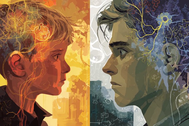
A large-scale study of nearly 10,000 adolescents revealed that those who used substances like alcohol, nicotine, or cannabis before age 15 had distinct brain structural differences compared to non-users.
Many of these differences existed before substance use began, suggesting underlying factors like genetics and environment may predispose individuals to early substance initiation.
The researchers found global differences, such as greater brain volume, as well as regional variations in cortical thickness and surface area.
Some structural differences were unique to the type of substance used, emphasizing the complexity of brain development.
While these findings could help guide prevention strategies, the researchers stress that brain structure alone cannot predict substance use.
Further studies are needed to explore how these differences interact with behavior, environment, and genetics over time.
Early Substance Use Linked to Brain Structure Differences in Teens
Early substance use is linked to brain structure differences in adolescents, many of which exist before use. This highlights the role of genetics, environment, and brain biology in addiction risk. Findings may inform prevention strategies and improve models of addiction.
Serotonin, GABA, and Dopamine Drive Hunger and Feeding
Researchers have identified how brain circuits and neurotransmitters regulate meal initiation, shedding light on the mechanisms of hunger and satiety.
Serotonin, a neurotransmitter that suppresses appetite, is inhibited by GABA and dopamine when hunger signals dominate, enabling meal initiation.
As feeding progresses and satiety sets in, these inhibitory signals decrease, allowing serotonin levels to rise, which suppresses further food intake.
The study highlights the synergistic role of GABA and dopamine in controlling serotonin-producing neurons in the dorsal Raphe nucleus, which connect to the hypothalamus.
These findings improve our understanding of how the brain manages feeding behavior and body weight.
The insights could inform the development of more effective treatments for obesity and related disorders.
Serotonin, GABA, and Dopamine Drive Hunger and Feeding
How does the brain decide when it's time to eat? New research reveals that GABA, dopamine, and serotonin collaborate to regulate hunger and satiety—insights that could revolutionize obesity treatments.
Brain imaging of fetuses and newborns reveals a rapid surge in functional brain connectivity during the transition at birth, suggesting neural reorganization to adapt to the external world.
Researchers analyzed data from 126 fetal and 58 infant scans, showing region-specific growth in brain networks.
Subcortical, sensorimotor, and superior frontal regions experienced significant changes, with the subcortical network showing increased communication efficiency.
These findings highlight the dynamic nature of early brain development, with some areas reorganizing while others remain stable.
The study offers insights into how prenatal factors, sex, and prematurity might influence brain network growth patterns.
This research lays a foundation for understanding how early-life brain adaptations impact long-term cognitive and emotional outcomes.
Birth Spurs a Surge in Brain Connectivity
New research shows birth sparks a rapid surge in brain connectivity, especially in subcortical and sensorimotor networks. These findings illuminate how the brain adapts to external stimuli early in life and sets the stage for future neural development.
Home-Based Brain Stimulation Shows Promise for Treating Depression
A new study reveals that home-based transcranial direct current stimulation (tDCS) is a safe and effective treatment for moderate to severe depression.
The research shows that patients using active tDCS at home experienced significant improvements in their depressive symptoms compared to those in a control group.
The noninvasive treatment involves applying a mild electrical current to the scalp, which has been used in clinical settings for other mental health conditions.
In this study, participants underwent 10 weeks of treatment, with tDCS sessions initially five times a week and then reduced to three.
Results showed that the active tDCS group had three times higher rates of remission compared to the placebo group.
This innovative approach could make depression treatment more accessible for millions of people who struggle with traditional therapies.
Home-Based Brain Stimulation Shows Promise for Treating Depression
A new study shows home-based transcranial direct current stimulation (tDCS) effectively reduces depressive symptoms, offering a safe, accessible alternative to traditional therapies for moderate to severe depression.
Fear memories are first stored as broad associations, making it hard to forget but difficult to recall specific details.
Over time, these memories become tied to specific timelines and events, a process that involves a shift from the hippocampus to the dorsolateral prefrontal cortex.
This integration occurs after the fear-inducing event, helping the brain to limit the scope of fear and focus on the event sequence.
People with high anxiety show weaker integration of these episodic memories, which may lead to prolonged fear and a higher risk of developing PTSD.
By tracking brain activity with fMRI and using machine learning, researchers identified how memory processing changes from general fear associations to time-based recall.
These findings offer new insights into why some individuals develop PTSD while others do not and open up potential new avenues for trauma treatment.
Why PTSD Develops in Some but Not Others
New research uncovers how fear memories shift from broad associations to specific timelines. This time-based integration is weaker in those with high anxiety, offering fresh insights into PTSD risk and potential treatment strategies.
Researchers have discovered how morphine alleviates pain by targeting a specific group of neurons in the brain, termed the 'morphine ensemble,' located in the rostral ventromedial medulla (RVM).
These neurons play a crucial role in inhibiting pain signals by connecting to the spinal cord, where they interact with inhibitory neurons to block pain transmission.
By inactivating this group of neurons, scientists found that morphine's pain-relieving effects were completely abolished, and reactivating them restored pain relief.
This breakthrough provides a clearer understanding of how morphine works in the body, potentially paving the way for safer pain management strategies that reduce the risks of addiction and overdose.
The findings also offer insights into the neural pathways that differentiate pain relief from the euphoric effects of morphine.
Future research aims to explore why morphine's effectiveness diminishes with long-term use.
Morphine’s Pain Relief Mechanism Unveiled
New research uncovers how morphine relieves pain by activating a 'morphine ensemble' of neurons. This discovery could lead to safer pain relief options by reducing the risk of addiction and overdose.
New Imaging Technique Identifies Autism Markers with 95% Accuracy
Researchers have developed a novel imaging technique that identifies genetic markers of autism in brain images with 89-95% accuracy.
This breakthrough method uses transport-based morphometry (TBM) to detect specific brain structure patterns linked to autism-related genetic variations.
Unlike traditional methods that rely on behavioral cues for diagnosis, this approach focuses on the genetic basis of autism, allowing for earlier and more personalized interventions.
The technique could revolutionize autism care by enabling doctors to diagnose and treat the condition based on genetic information.
By distinguishing between normal and autism-linked genetic variations, TBM offers a deeper understanding of the biological roots of autism.
This research marks a significant step forward in the quest for more effective and targeted autism treatments.
New Imaging Technique Identifies Autism Markers with 95% Accuracy
New research identifies autism markers in brain images with up to 95% accuracy, paving the way for earlier diagnosis and personalized treatment.
New research reveals the surprising role of the anterior precuneus (aPCu) in our brain, a key player in forming our physical sense of self or "I". A disruption in its activity can trigger feelings of unreality.
#neuroscience
1/4
Sense of Self: The Brain Structure That Holds Key to “I”
The findings further our understanding of self-awareness and consciousness.
A new study breaks ground on the intricate connections between cognition and personality traits. With data from over 2 million participants, it's a giant leap in understanding human individuality. Cognitive prowess? Linked to activity levels. Open-mindedness? Tied to cognitive… twitter.com/i/web/status/1…
Untangling the Human Mind: The Interplay Between Cognition and Personality
This study revolutionizes our understanding of human individuality, shedding light on the diverse tapestry of the human mind.
New research reveals that daily mindfulness meditation can counteract cognitive bias, making us more receptive to negative information. Better decision-making could be just a meditation away.