
Cardiologist 🇧🇪 | ExerciseEchocardiography | HeartFailure | ValvularHeartDisease | @JessaZiekenhuis | @hartcentrumh
 1/8
1/8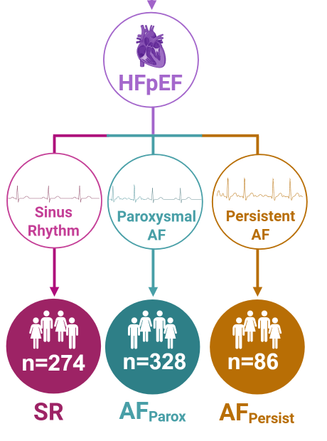
 2/14
2/14
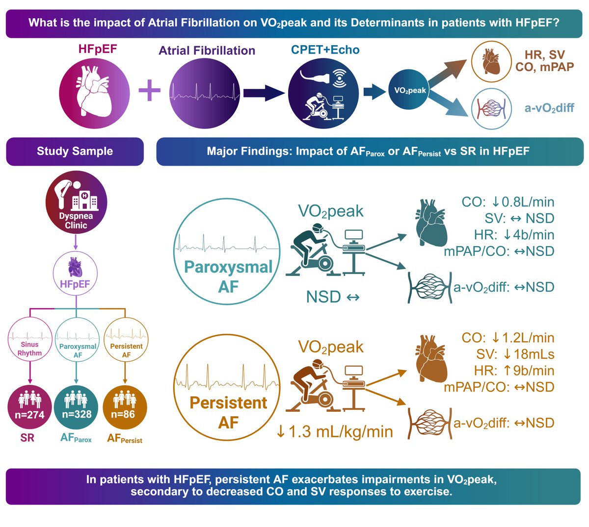
 22 pts with simultaneous #iCPET and #ExerciseEchocardiography: Elevated