Neurophilia Profile picture
Neuro-Interventionist & Neurointensivist, Autodidact, Philomath, Iconoclast, Bibliophile, Technophile, ‘Normie’, Sempiternal scholar (Nerd) & Teacher @ Kochi
2 subscribers
Mar 2 13 tweets 8 min read
Here are my slides on Nodo-paranodopathies : these disorders are neither Inflammatory nor demyelinating.
They are ‘Suis generis’ : a category unto themselves, even though they mimic AIDP and CIDP ( inflammatory demyelinating disorders) Image
Image
Image
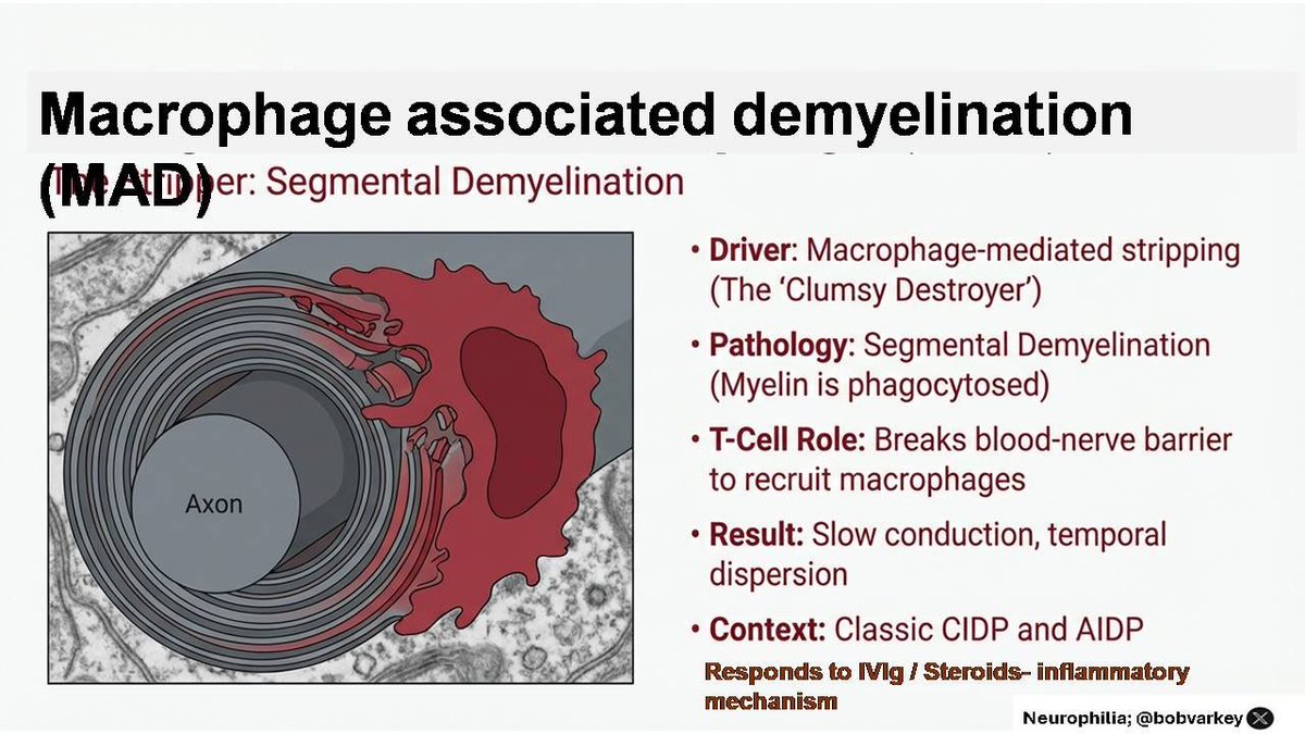
Florid macrophage infiltration in between myelin lamellae and T cell recruitment lead to segmental demyelination. Due to antigenic mimicry, the Schwann cells themselves undergo differentiation into an inflammatory demyelinating subtype (iDSc) which invites macrophages and T cells and unravels the myelin lamellae for them to jump in and chew away
Jun 5, 2025 6 tweets 2 min read
A sweet syndrome
30 yr old 👩‍🦳 came with progressive memory loss of 2 months duration. 10 days earlier she had a seizure and was admitted to
MRI shows this ⬇️ Image
Image
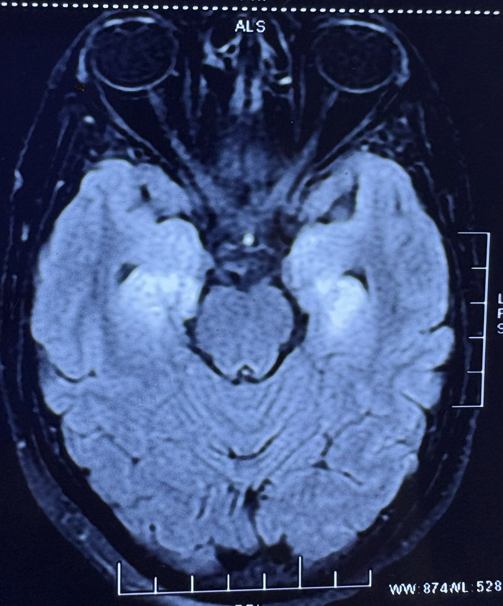
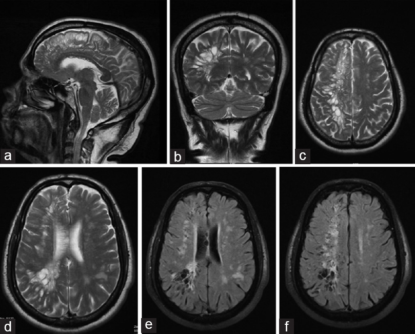
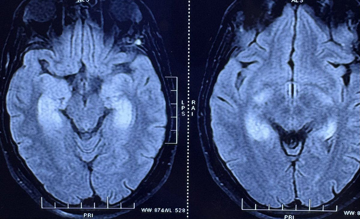
A diagnosis of limbic encephalitis was made. She was started on IVIG x 5 days. HbA1c was 6.4%. An autoimmune encephalitis panel and PET CT turn up negative. She comes for a second opinion. After admission her blood sugar is noted to be 500 mg 🍩🍩🍭
Feb 22, 2025 6 tweets 3 min read
This young girl's quest for a diagnosis ended today. She came to us after a 2.5 year ordeal in different places.
🌟This 23 year old 👩‍🚒bariatric Surgery (Sleeve Gastrectomy) in October 2022 after which her weight ⬇️⬇️ from 120 kg to 72 kg in 8 months.
🌟Experienced GERD and was bedbound for 6 months. She developed emotional disturbances requiring Qutipin.
🌟2 months later, she developed progressive weakness in hands (2 months).
🌟o/e weakness in distal hand muscles (extensors > flexors). Brisk DTRs, flexor plantar response, normal sensory function.
🌟NCS showed a generalized motor CMAP reduction, predominantly affecting radial nerves.
🌟She was diagnosed with a unclear myeloneuropathy vs. Progressive Motor Neuropathy post-bariatric surgery.
🌟MRI Spine: normal. Holo vitamin panel normal, except high Pantothenic Acid.
🌟Creatinine 1.6 (Medical Renal Disease - ? etiology). 🌟In january 2025, for severe menstrual pain, & given leuprolide & Midazolam for severe pain
🌟The next day, she developed quadriparesis.
🌟NCV showed conduction block (Possibly Guillain-Barré Syndrome - GBS).
🌟When she worsened , she was administered 2 gm/kg IVIg. After 3 weeks of improvement, and ambulation, she worsened to grade 2/5 power in the limbs and severe sensory loss over the proximal limbs.
🌟ANA: Negative, MRI Whole Spine: Normal, Ganglioside antibody: Negative.
🌟NCV B/L UL and LL: Mixed axonal > demyelinating motor neuropathy in all four limbs.
🌟At which point she came to us.Image
Image
Image
Image
Nov 30, 2024 10 tweets 3 min read
MRI for the non-radiologists. Do the terms FFE, GRE, T2*, SWI, SWAN & Phase imaging confuse you? Here is a handy reference sheet for that. Without going into any of the physics of MRI, the 1st point to understand is;
1. T2* (pronounced "T2-star") is the basis of other sequences, such as SWAN, GRE, and FFEImage 
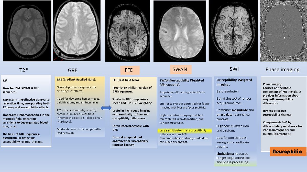
SWAN or GRE may be enough for the majority of MRI aficionados [as they require shorter acquisition times]
But.....SWI provides the best resolution for detailed susceptibility contrast (e.g., microbleeds).
Phase Imaging is useful to differentiate between iron and calcium.
Just like the German Iron cross, Iron remains black in both GRE/SWAN/SWI and Phase imaging.

But Calcium (and Mg, comparitively less) is like Yin and Yang......Black on GRE/SWAN/SWI and White on Phase imaging
Of course it is the opposite in Left handed MRI scanners.........I won't get into the differences, but ask your radiologist if you have a right handed MRI scanner.Image
Image
Nov 9, 2024 4 tweets 2 min read
A young 👩‍🦱 w/ 6 year H/o weakness starting in the hands and now in the legs - slowly progressing. Initially Dx as b/ L Carpal tunnel syndrome. O/e has wasted thenar muscles (spared hypothenar) and b/l foot drop. No sensory. Reflexes absent Image
NCV ↘️ Striking conduction blocks in Ulnar and tibial nerves , axonal motor median and perineal nerves and absent Surals - suggestive of a MADSAM neuropathy. Have offered IVig Image
Image
Nov 2, 2024 7 tweets 3 min read
Is it hard to remember the branches of the Middle cerebral artery? With Stroke, forming a large part of emergency neurology services and the advent of mechanical thrombectomy, it is important to know the branches of MCA. Fortunately, simple tricks will help you remember these branchesImage The MCA can be conveniently divided into 4 segments, each of which moves in a different direction.

[Ref Harrigan, M. R., & Deveikis, J. P. (2018). Essential Neurovascular Anatomy] Image
Jul 3, 2023 5 tweets 2 min read
A confusing presentation- elderly lady with sudden onset unsteadiness after a colonoscopy. On exam - had bilateral limb ataxia and gait ataxia Initially had only ataxia. Over the next few days developed chorea. Paraneoplastic and autoimmune work up negative
Feb 10, 2023 5 tweets 2 min read
Things that make you go hmm..... A tweetorial on the Symptomatic Nonstenotic Carotid Disease. This ⬇️ is what Ugly looks like. Sometimes, it is not significant enough on CT or MRi (< 50% stenosis) but could still be a troublemaker that requires a stent or an endarterectomy. 
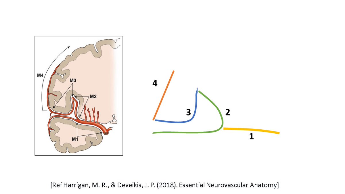
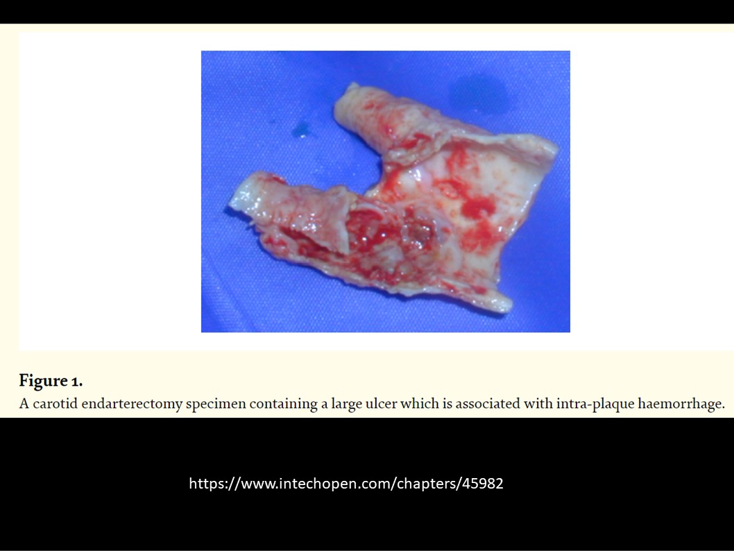
This is how the normal artery progresses from a fatty streak to total occlusion
Feb 7, 2023 4 tweets 2 min read
Hypometric saccades to the right in this pt with a right dorsal cerebellar stroke Localisation- Dorsal Oculomotor vermis (OMV) on the right. Why? ⬇️ The OMV is an accelerator for Saccades, whereas the Fastigial region (FOR) is a brake for termination of saccades.
Feb 7, 2023 6 tweets 2 min read
New back pain in a pt with Leukemia! Reported as normal, but pt comes back with worsening pain. MRI shows dark vertebral bodies with intense enhancement s/o marrow infiltration 
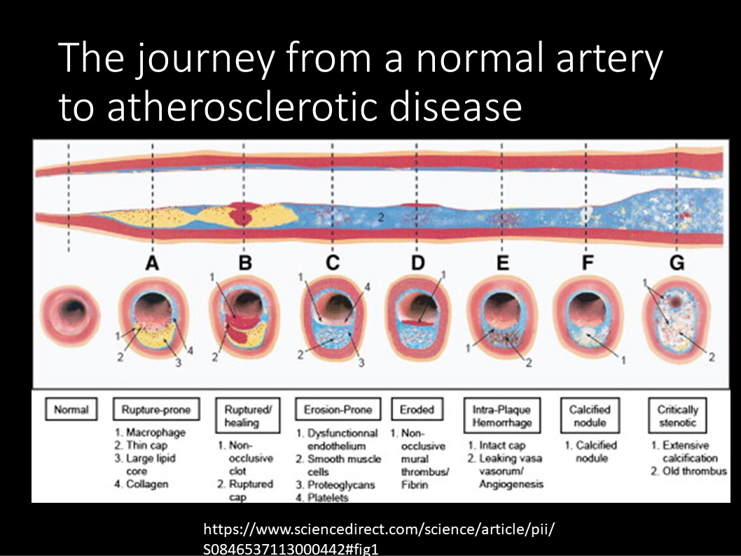
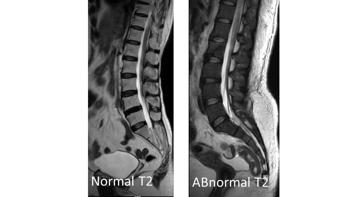
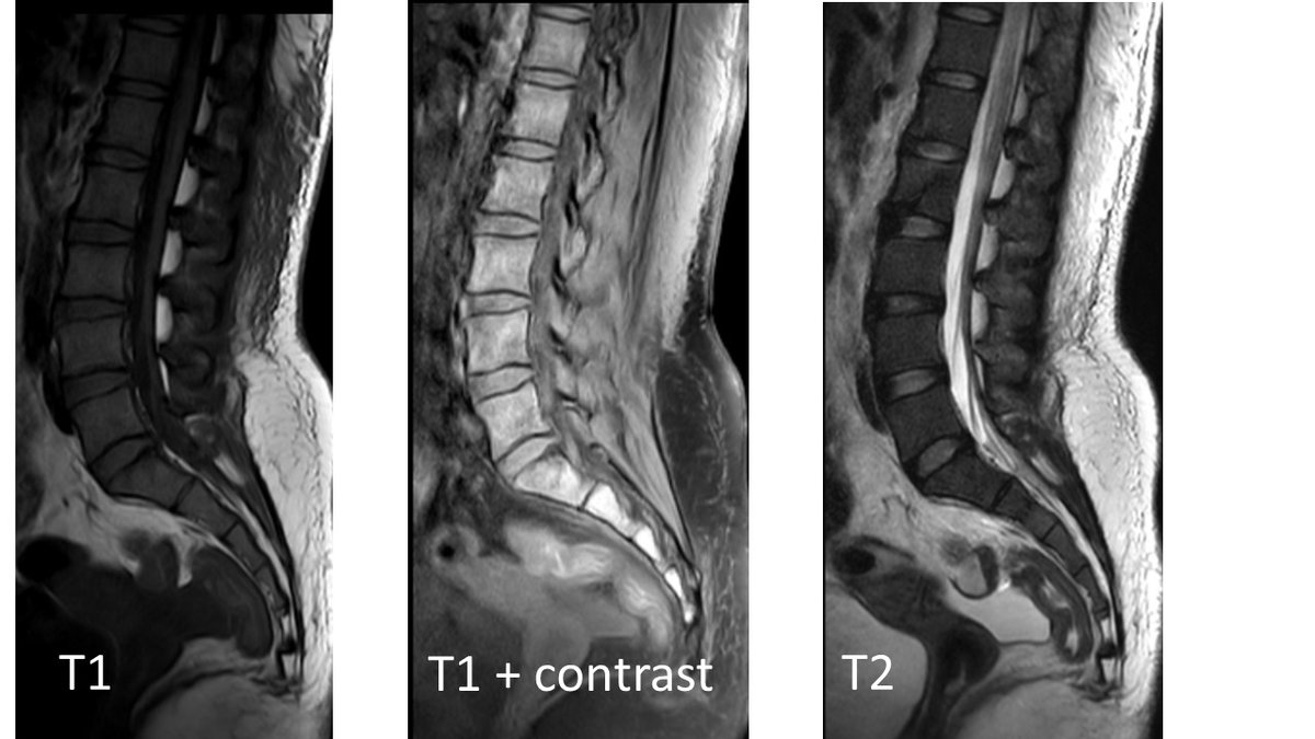
MRI in leukemia-often;
T1: diffuse low signal
T2: increased signal
T1 C+ (Gd): diffuse enhancement
Aug 29, 2022 7 tweets 2 min read
Clinical pearl- 70 yr old 👩 with Urosepsis. c/o severe neck pain and restriction x 1 day. O/e restricted neck movements, severe paraspinal spasm. No other deficits ESR 31mm/hr. Blood counts normal after Abx for urosepsis. r/w head CT shows small calcific lesion in front of the odontoid
Aug 19, 2022 5 tweets 2 min read
Clinical pearl: Orofacial and limb dyskinesias in NMDAR encephalitis. Note the lip mutilation.( pt consent for video without identification obtained)Pt is tracheostomised & off Ventilator.
Aug 13, 2022 8 tweets 4 min read
No pain, no gain! Young man with severe headache, disorientation of 2 days duration. MRI outside-left temporal hematoma. O/e agitated, wernicke's aphasia, right hemiparesis 
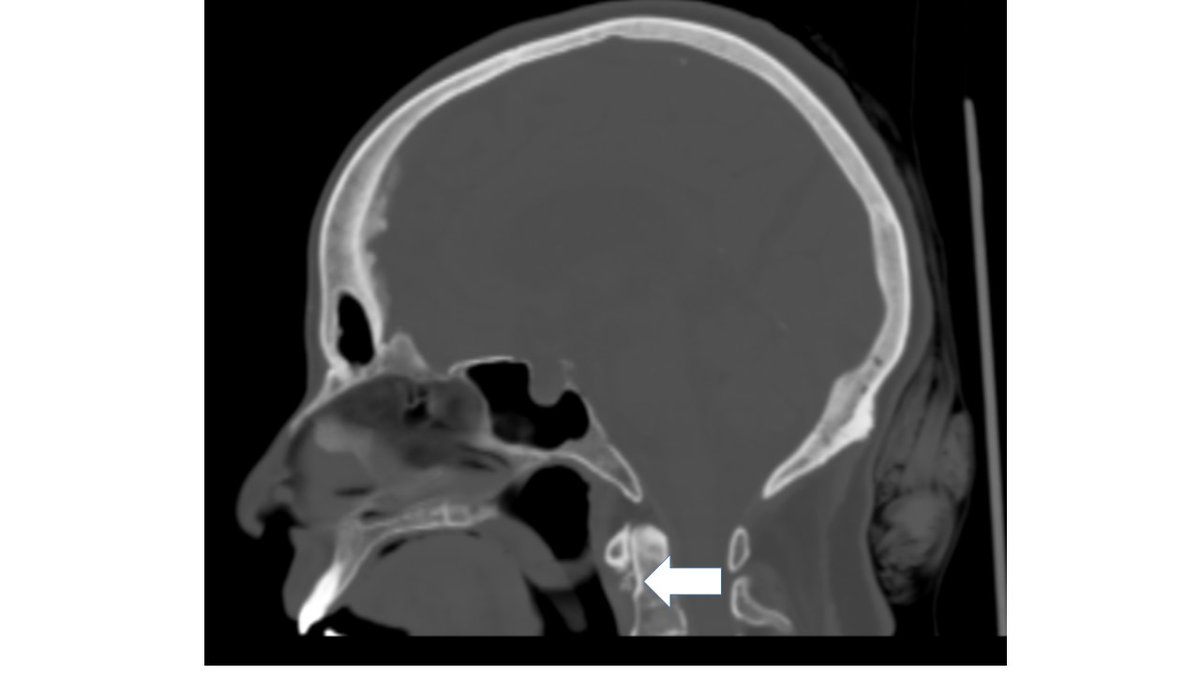
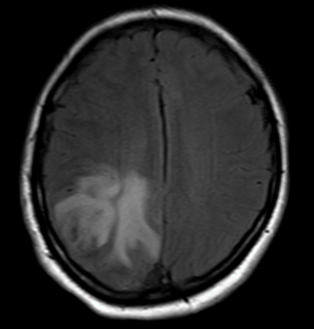
MRV shows left Transverse and sigmoid sinus thrombosis. No improvement after 2 days of IV heparin. Pt requires heavy sedation, ventilation for headache/agitation.
Aug 12, 2022 8 tweets 4 min read
Clinical pearl; ' A Hot Carotid'. A 74 year old man with acute onset weakness and tremors of the right hand. 
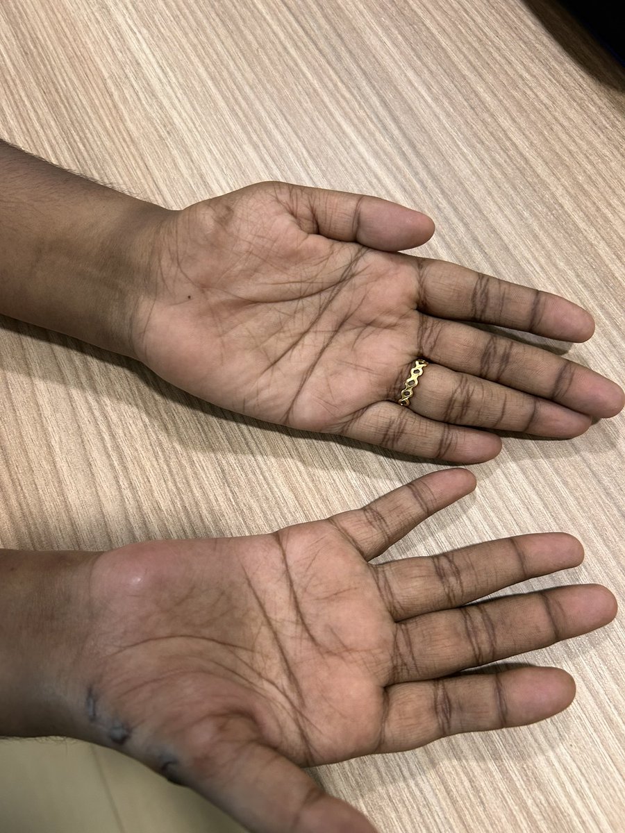
Acute dystonic tremor is an exceedingly rare manifestation of a stroke. This patient had ulnar deviation, finger flexion at MCP jts and postural tremors with mild right hand weakness.
Aug 10, 2022 5 tweets 4 min read
Clinical oddity; A mountain of a clot! A young post partum 👩‍🦰 20 days after LSCS with worsening headache and quadriparesis. MRI- right > left hemispheric edema. MRA- shows complete occlusion of the SSS and TS ImageImage 
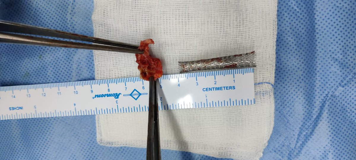
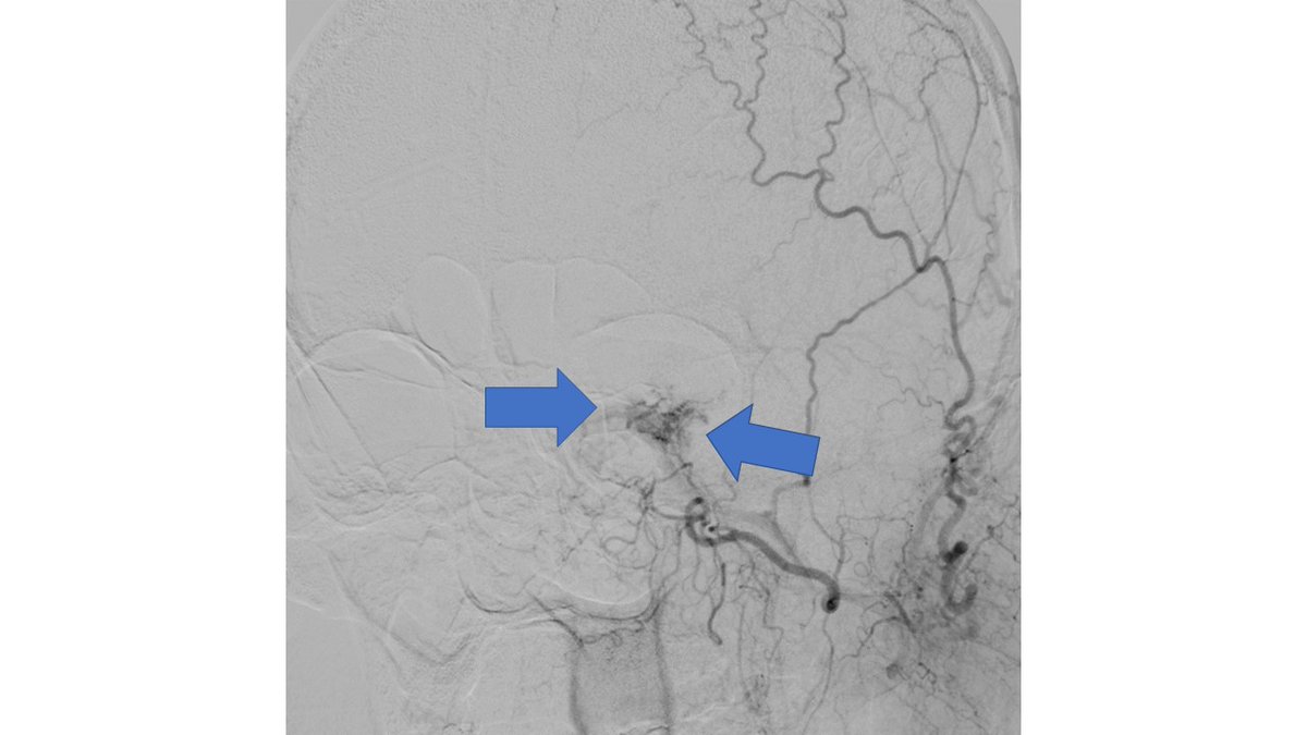
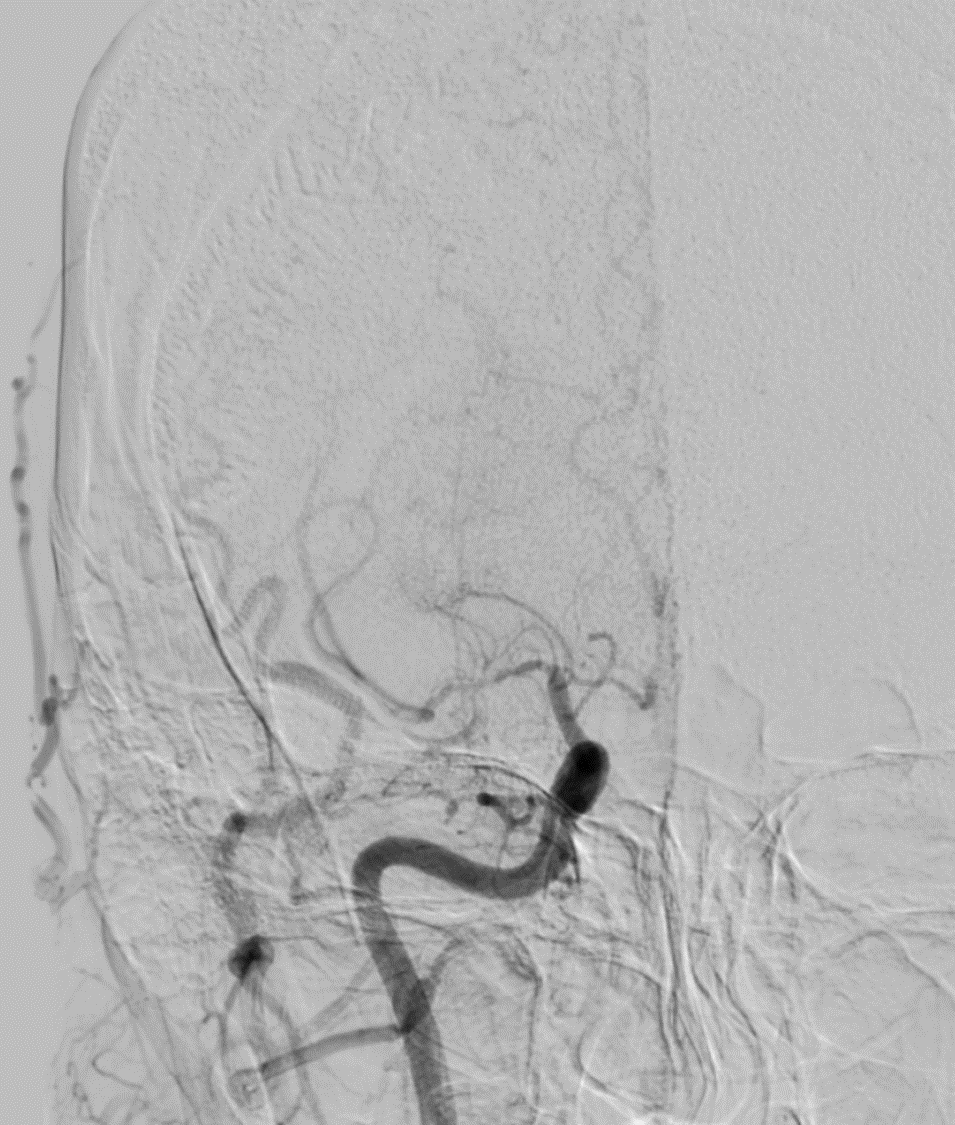
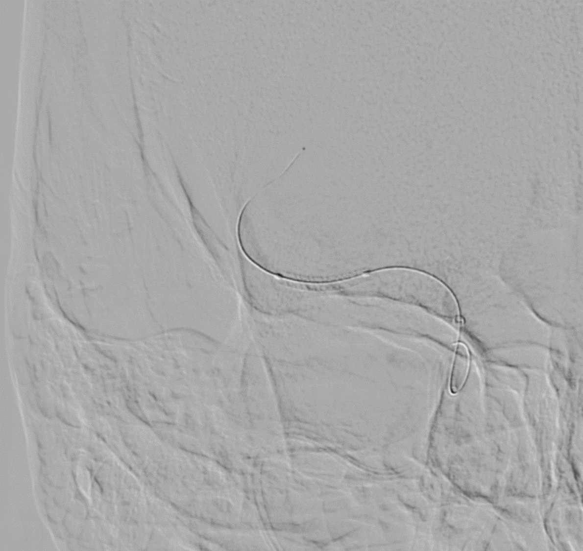
DSA also confirmed the extensive clot. In view of her recent LSCS 20 days ago, we were hesitant about giving IV lytics (urokinase infusion). Hence went in with an ACE 5 Max catheter and sucked out a mountain of clot Image
Aug 8, 2022 11 tweets 3 min read
Clinical pearl; 35 yr old with severe left sided headache & diplopia x 4 days. O/E- Left 6th nerve palsy. CTA, MR, MRA normal. PMH- h/o head injury with closed multiple skull # 1.5 years ago. D/d- Meningitis, Raised ICP, dural A-V fistula. 1/🧵 DSA shows
Aug 4, 2022 5 tweets 2 min read
Clinical conundrum- Young man with progressive stroke over 6 hrs. NIHSS -8. DSA shows a tight MCA M1 right sided stenosis, persisting after repeated angiplasty. Finally opened up with a Coronary stent 2.25 x 18 mm. 🤞 Waiting for tomorrow's CT 💓 ImageImage 
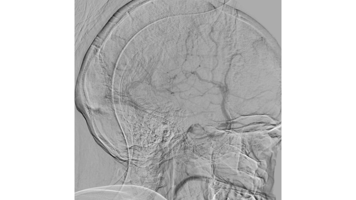
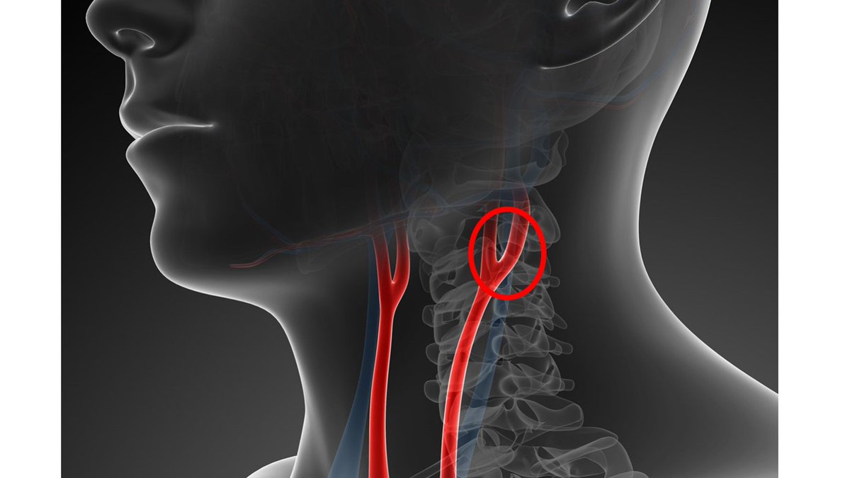
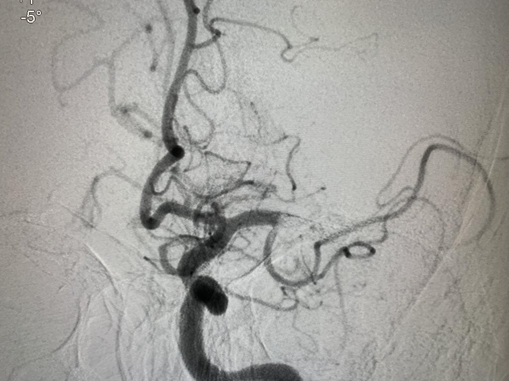
Tight stenosis opened up with a Balloon mounted stent and final result ImageImage
Aug 1, 2022 7 tweets 2 min read
Clinical pearl; Looks Can't be deceiving! Two axillary folds hide a whole story. Young man with left sided wasting. O/e has ⬇️ appearance. 
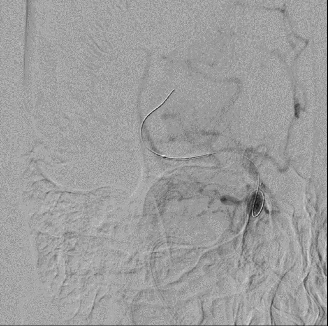
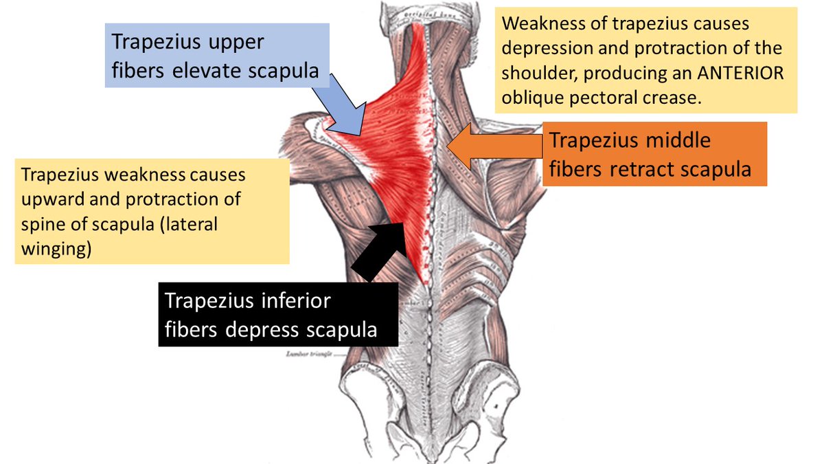
Quick recap of various scapular movements and the role of the Trapezius in stabilising the scapula
Jul 30, 2022 5 tweets 2 min read
Clinical pearl: Rescue Stenting in failed mechanical thrombectomy. A 45 year old 👩‍🔬2 hrs of aphasia, Rt Hemiplegia , NIHSS 18. CTA left MCA stump occlusion DSA⬇️ Image 5 attempts at aspiration + stent retriever -only some red and white clots come out ➡️mTICI score still Zero Image
Jun 13, 2022 7 tweets 3 min read
Hole'y Smokes. A 🧵 about a rare case I've never seen again! Why a neurological history is absolutely essential to the diagnosis of these "holes in the brain' A 71-year-old man presented to us with right-sided headache after a flight. Immediately after ascent, he developed severe right eye pain and headache and lost consciousness.
Jun 12, 2022 5 tweets 3 min read
Carotid stent explantation after stenting restenosis occurs. When a vascular surgeon friend in need is a friend in deed! He goes a step further and does a carotid endarterectomy also!