
Neuroradiologist @UMiamiHealth l Former Neurorad fellow @PennRadiology l SGU ’18 l Educational Cases I Posts are my own #RadRes #Neurorad #RadEd
8 subscribers
How to get URL link on X (Twitter) App

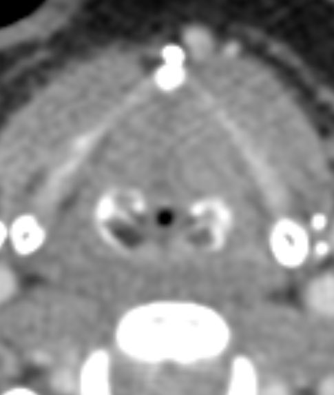

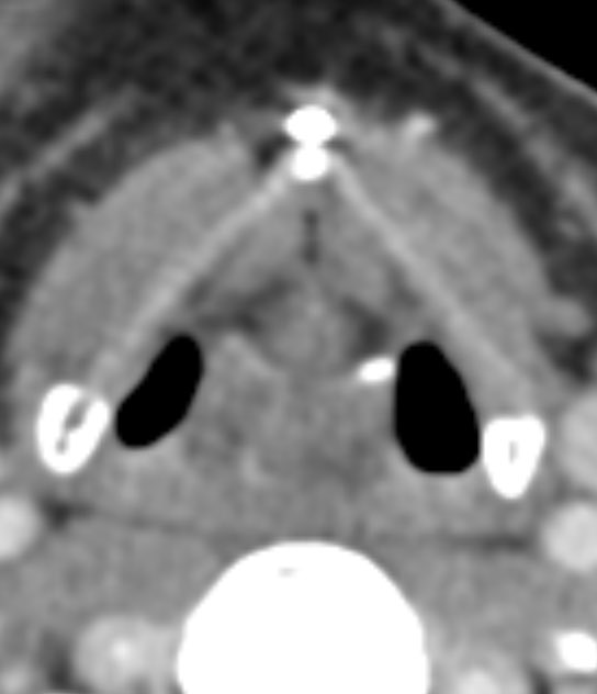
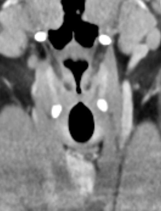
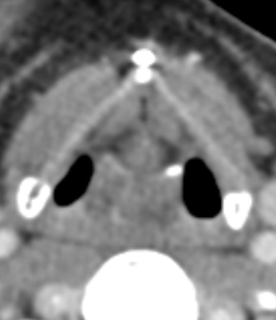
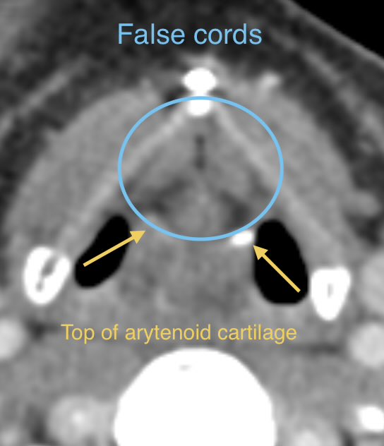
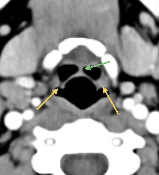 🔷False cords (vestibular folds): Basically an inferior reflection of the aryepiglottic fold around the vestibular ligament
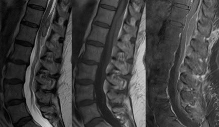

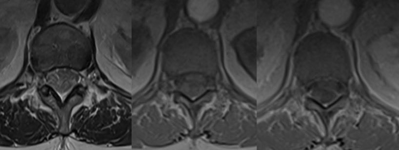
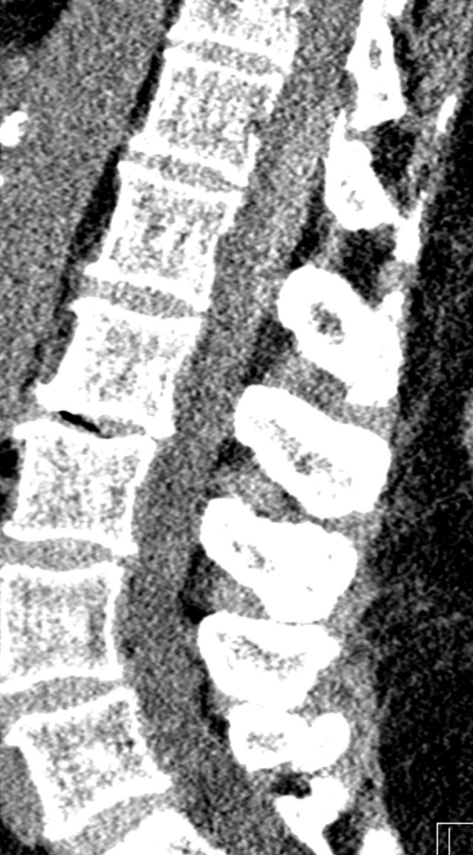 ⭐️ Answer: Spontaneous spinal epidural hematoma (no clear risk factor in this case)


 🔷For glioblastoma we need to rely on many clinical and imaging features to distinguish (no one feature is specific enough to diagnose so we need to take the whole clinical and radiographic picture into account)
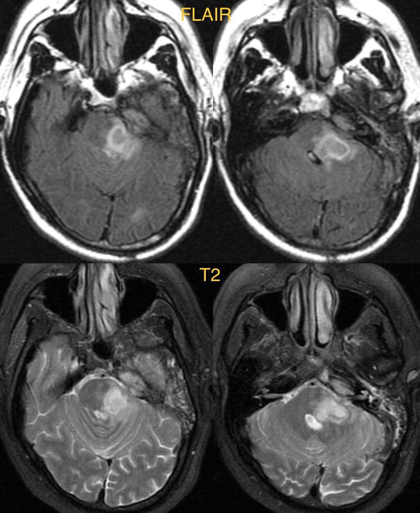

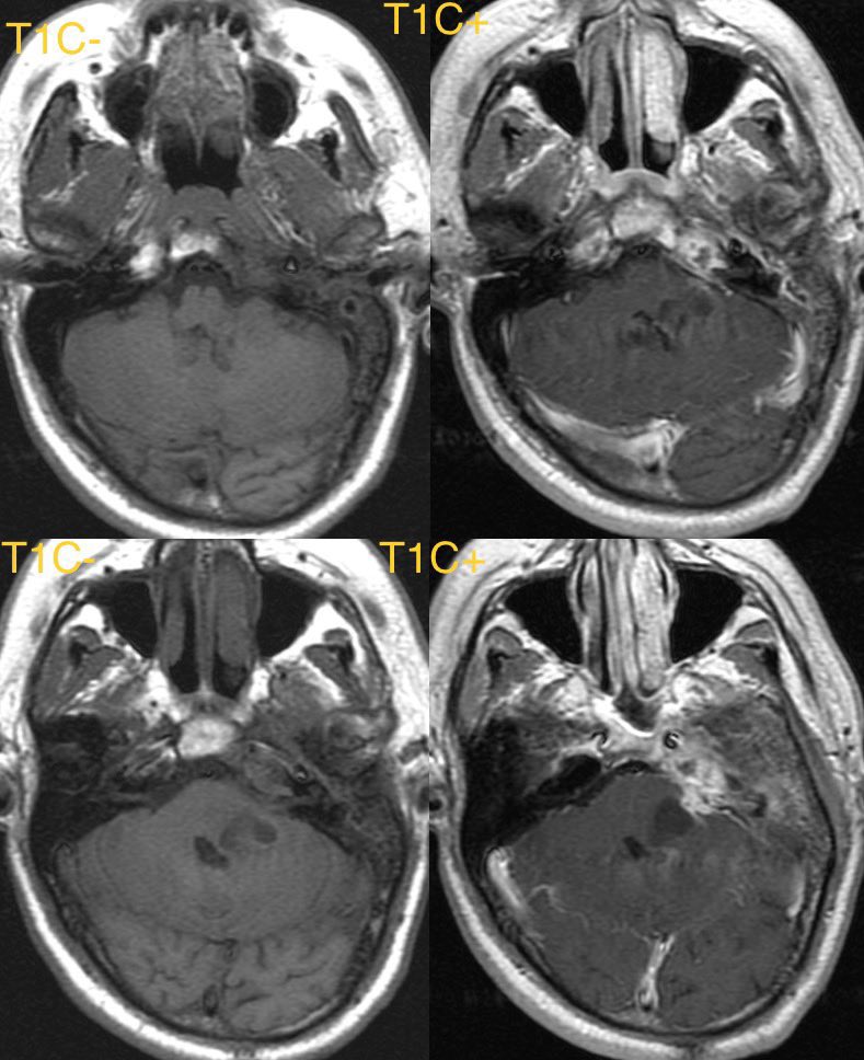

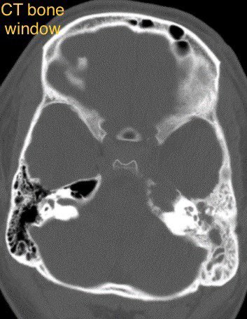 ⭐️ Answer: petrous apicitis complicated by brainstem abscess


 ⭐️ Answer: Cortical vein thrombosis (CVT)
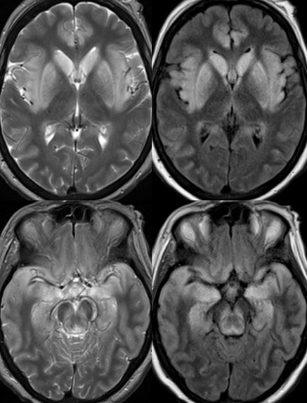

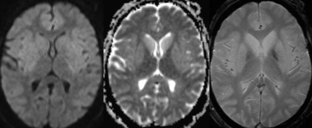

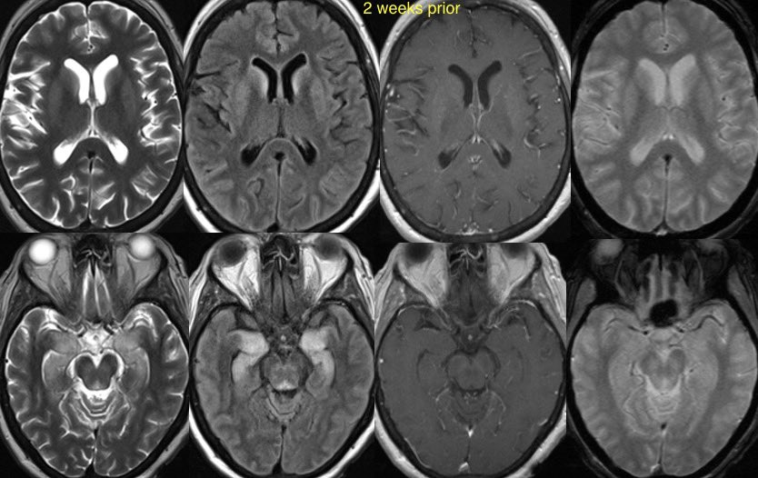 ⭐️ Answer: Viral encephalitis (Specifically Rabies)

 ⭐️ Answer: Tumefactive demyelination (MS in this case)
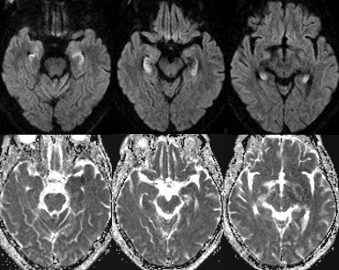
 ⭐️ Answer: Opioid-associated amnestic syndrome


 🔷Comparison from ~4 months ago 👇


 🔷Ddx for venous causes of PT:


 Answer: Linear Scleroderma aka Scleroderma en coup de sabre


 Answer: Benign-appearing notochordal lesion (formally ecchordosis physaliphora, EP)


 ⭐️ Answer: Transient Perivascular Inflammation of the Carotid Artery (Carotidynia or Fay syndrome)


 Additional image 👇


 Answer: Glomus Tympanicum Paraganglioma

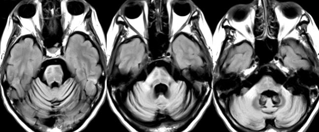

 Answer: Multiple System Atrophy Cerebellar Type (MSA-C)


 🔷First thing we notice is hemorrhage in the left basal ganglia


 Answer: Hypothalamic adhesion
