
Author & Educator @EKGDX — 75K+ learners mastering ECGs | Published in Circulation - JACC - Electrocardiology | Speaker | Innovator of an advanced ECG Simulator
 2/ In the beginning, as novices, many doctors, nurses, and students struggle with accurately interpreting an electrocardiogram. This challenge is often due, among other factors, to a lack of organization in the interpretation process. When I was a medical student, I faced the same issue until one day I decided to create an organized approach. That's when the idea of using my last name, ROIG, as an acronym was born, to help me streamline the process. Everyone eventually develops their own way of reading an EKG, but if you're just starting out, this basic method may help you.
  2/ A 61 year-old male c/o chest pain radiating to the back and left arm weakness for the past 3 hrs. The EKG shows inverted T waves in III and aVF with high levels of troponin.
 2/ The first EKG was obtained in triage.
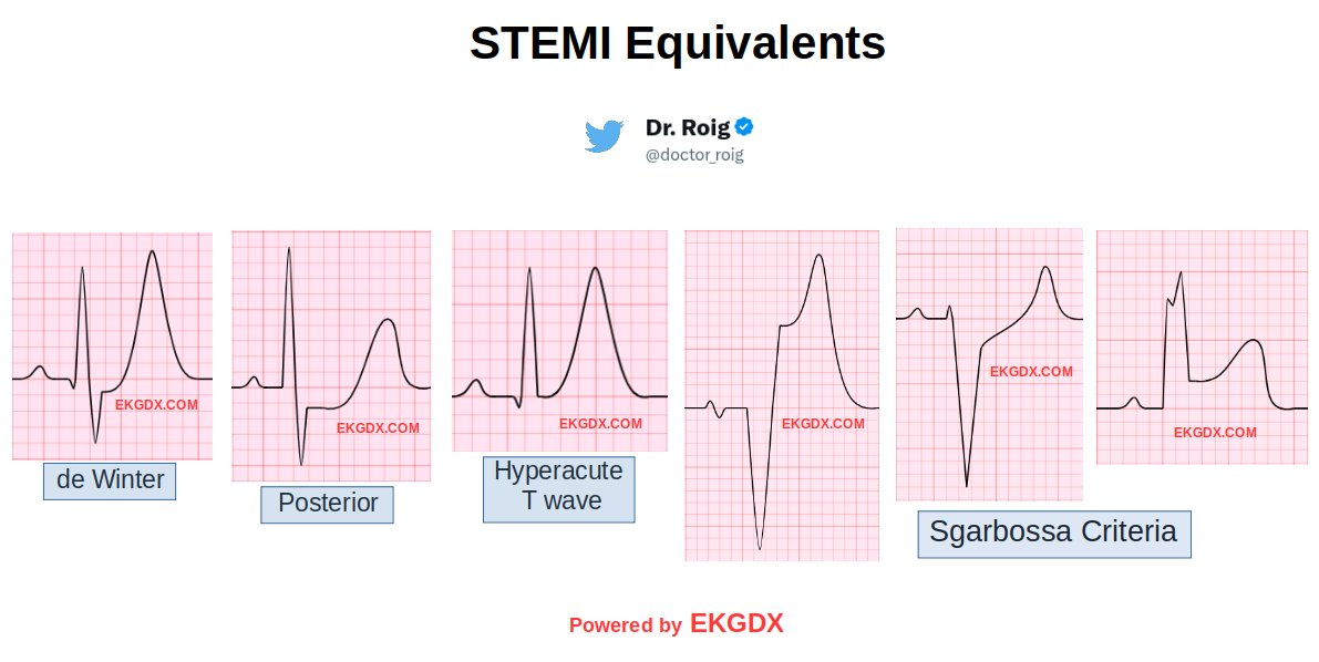 2/ For the last twenty years, studies have demonstrated that acute coronary occlusion (ACO) does not always produce the classic ST-segment elevation in contiguous leads (Koyama et al Am J Cardiol 2002). In fact, the following study has suggested that >25% of patients with ACO will not demonstrate expected ST-segment elevation (Wang, T. @CMichaelGibson et al Am Heart J 2009).
 2/ PAC occurs when an ectopic focus within the atria, generates an action potential before the next scheduled sinus beat.
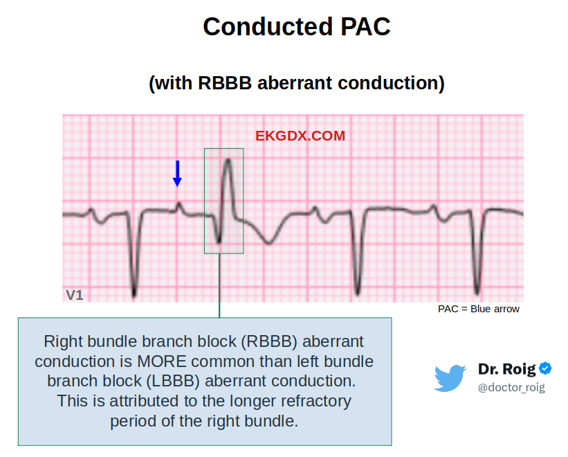
 2/ History
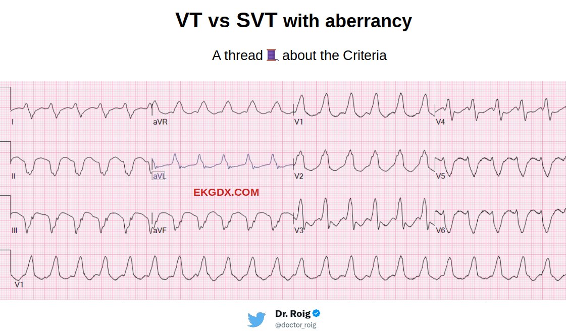
 2/ Criteria
 2/ The de Winter pattern holds significance as it is linked to the occlusion of the proximal left anterior descending coronary artery (LAD) when identified in the electrocardiogram (ECG) of individuals experiencing chest pain or displaying a history suggestive of acute coronary syndrome. In fact, this pattern is present in approximately 2% of patients diagnosed with proximal occlusion of the LAD.
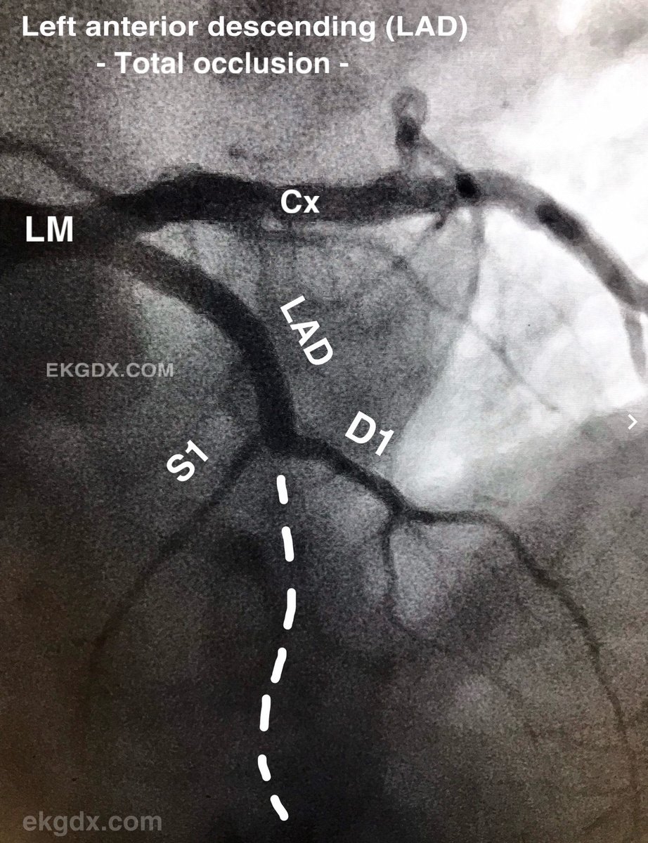
 2/ Step 1: Identify the chamber(s) paced.
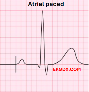
 2/ The section of EKG Challenges from @ekgdx is located at:
  2/ The Sgarbossa criteria were initially introduced over two decades ago to enhance the diagnostic precision for MI in the setting of LBBB. This criteria is widely accepted as one of the most valuable tools to assist in the diagnosis of MI when LBBB is present.
 2/x
2/x