
Emergency Medicine doc, forever POCUS fellow. Interested in Trauma & Resus. obsessed with Echocardiography. love Regional Anaesthesia. ex-Gamer.#FOAMed #POCUS
 He’s called to see a gentleman in his 40s.
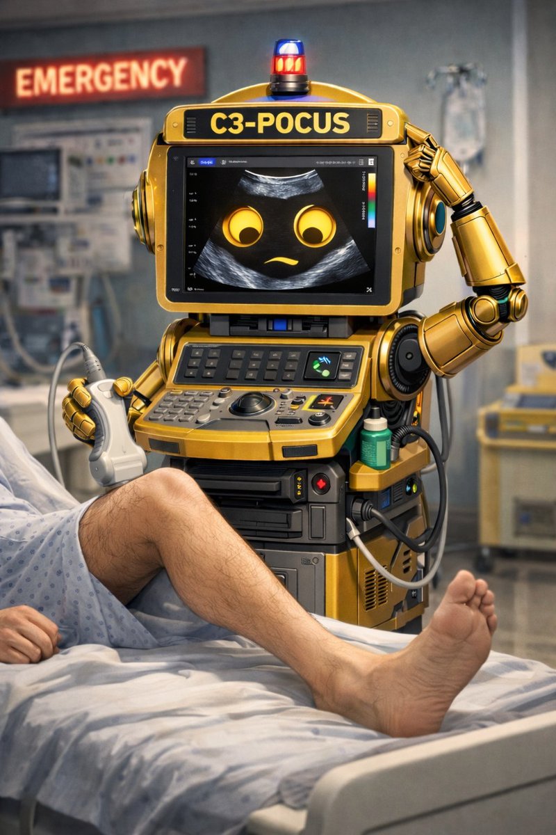 It’s 1pm and the department is busy. Ambulances are pouring in and the triage queue is getting longer by the minute.
 A quick scan of the right lower quadrant reveals:
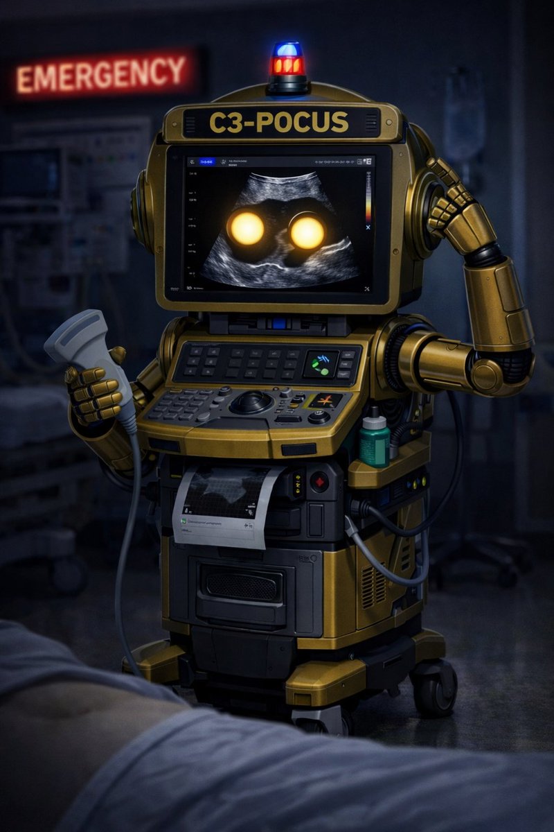
 Hi, my name is C-3POcus, your Emergency Department ultrasound machine.


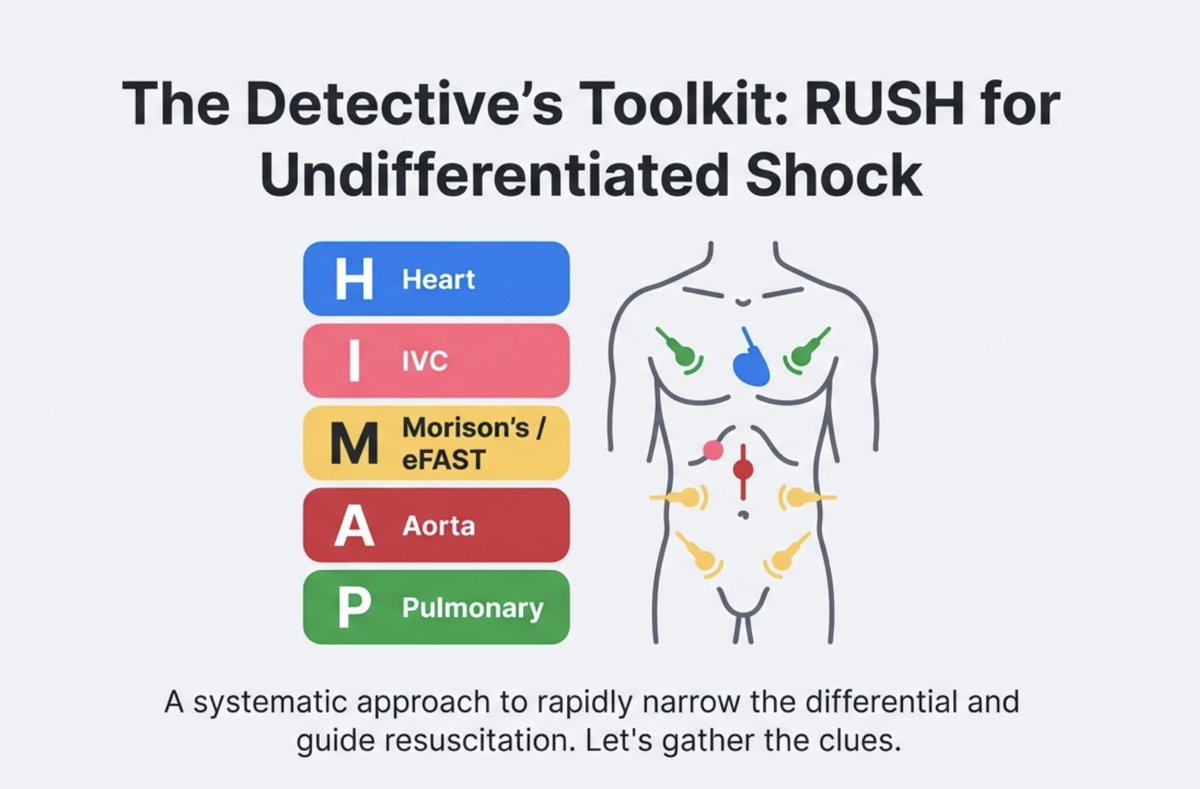
 Lets start with some basics.