Hippocampal Subfield Group Profile picture
We are working toward the formation of a harmonized hippocampal subfield segmentation method for MRI
Jun 16, 2021 10 tweets 7 min read
Guest post by @DrAnaDaugherty on hippocampal (Hc) vascularization.

Brought to you by “anastomosis” (noun): a cross-connection between adjacent blood vessels; plural “anastomoses”.

As in, “Wow! 54 years of mapping anastomoses”

#SubfieldWednesday 🧵 1/ Figure 3 from Muller & Shaw...Figure 6 from Spallazzi et ... The unique features of its arteries make Hc vulnerable to anoxia. Superficial arteries travel long tangential routes, and intrahippocampal arteries have few anastomoses and travel with the rolling CA and dentate gyrus tissue. 2/

#SubfieldWednesday
May 12, 2021 9 tweets 7 min read
Last week we talked about hippocampal variability in the hippocampal head.

The hippocampal tail is another area of the hippocampus that can vary anatomically across individuals

#SubfieldWednesday (1/n) But first, what do we mean by the hippocampal tail? Definitions vary, but one way to define the tail is the part of the hippocampus located posterior to the corpora quadrigemina (i.e. superior and inferior colliculi).

#SubfieldWednesday (2/n) Hippocampal tail is outlined in purple on T1 and T2 weightedHand drawn depiction of the hippocampus with the head, body,
May 5, 2021 7 tweets 4 min read
Today we continue our thread series discussing anatomical variability in the medial temporal lobe (MTL). Today’s topic is anatomical variability in the hippocampal head with a focus on the hippocampal digitations. 🍤🤓📢

#SubfieldWednesday (1/n) But first, what do we mean by the hippocampal head? We are talking about the anterior part of the hippocampus that contains or is adjacent to the uncus.

#SubfieldWednesday (2/n) Figure 1 from https://doi.org/10.1038/nrn.2015.24 a| A schem
Apr 14, 2021 10 tweets 6 min read
Happy #SubfieldWednesday! Today we will be sharing a 🧵 on anatomical variability in the medial temporal lobe (MTL). MTL subregions vary in size and shape across individuals & these variations can make landmark identification and segmentation difficult

#SubfieldWednesday (1/n) Today we will focus on the *most* consistent structure in the MTL: the hippocampal body. Most of our followers will be familiar with this canonical hippocampal body shape shown below in the coronal plane.

#SubfieldWednesday (2/n) Coronal plane from a T2 MRI depicting the canonical C shape
Feb 10, 2021 10 tweets 5 min read
We are pleased to announce that today we have a guest #SubfieldWednesdsay 🧵 from @MarkCembrowski !

Check it out below!

(1/n) When you look at a textbook diagram hippocampus, one sees a series of subfields - DG, CA3, and CA1. All of these regions have specialized properties relative to one another. But it raises the question: within each region, are the cell types uniform?

#SubfieldWednesdsay (2/n) Image courtesy of the Cembrowski lab: https://www.cembrowski
Dec 17, 2020 10 tweets 7 min read
Is anyone planning to do some reading about hippocampal neuroanatomy over the holidays?

If you answered, "yes", this week's #SubfieldWednesday is for you! We will give you a list of "must read" atlas references about our favorite brain structure. 🍤❣️

#SubfieldWednesday (1/n) In our 2015 paper (Yushkevich et al., NeuroImage, 2015), we provided a list of common atlases used for hippocampal subfield definition across labs.

#SubfieldWednesday (2/n) Table 2 from http://dx.doi.org/10.1016/j.neuroimage.2015.01.
Dec 9, 2020 8 tweets 4 min read
This week I thought I would share some terminology that you may or may not know!

#SubfieldWednesday (1/n) Most of our followers probably already know how that the name "hippocampus" means "seahorse" in Greek.

#SubfieldWednesday (2/n) https://www.wbur.org/npr/505754756/the-seahorse-in-your-brai
Nov 25, 2020 12 tweets 5 min read
Hello and happy #SubfieldWednesday! Today we are going to get a bit more familiar with how the hippocampal subfields differ in their composition of different cell types, cell sizes, and layer thickness. 🍤🔬

#SubfieldWednesday (1/n) Here are some images taken from five different hippocampal subfields (CA1, CA2, CA3, dentate gyrus, and subiculum). Can you tell which number corresponds to which subfield? 🤔

#SubfieldWednesday (2/n) Five different screenshots of histology slices of five diffe
Nov 11, 2020 11 tweets 3 min read
Happy #SubfieldWednesday! After a two-week hiatus we are returning to our quiz about the mysterious transentorhinal cortex!

This quiz even stumped some of us at @hipposubfields headquarters! We had to contact a neuroanatomist to confirm which answer is correct! (or at least "the most correct")
Oct 21, 2020 9 tweets 4 min read
Last week we received over 100 (!) responses on our quiz about which hippocampal subfield accumulates tau pathology earliest in the disease.

#SubfieldWednesday (1/n) This week we will explain that the correct answer for Alzheimer's disease (but not all types of dementia) is CA1!

#SubfieldWednesday (2/n)
Oct 8, 2020 12 tweets 6 min read
Hello subfield-fans! Last week's #SubfieldWednesday topic was the layered composition of the hippocampal subfields. We learned that the subfields contain three major cellular layers which makes them a part of the allocortex.

#SubfieldWednesday (1/n) Table 1 from: Insausti, Ric... 
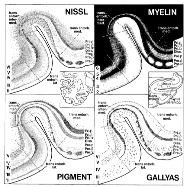
@thomcat992 replied that the hippocampus is archicortex, which is also correct! Archicortex is a type of allocortex.

#SubfieldWednesday (2/n)
Oct 1, 2020 7 tweets 4 min read
Better late than never! (It's still Wednesday in some timezones!) This week's #SubfieldWednesday will be a bit briefer than normal.

#SubfieldWednesday (1/n) Some of you might have been surprised last week when we described the hippocampal subfields as layered structures.

#SubfieldWednesday (2/n)
Sep 23, 2020 18 tweets 10 min read
It's Wednesday...so you know what that means! It's time for #SubfieldWednesday! 🥳📢

(1/n) Two weeks ago we posted about whether you can visualize subfields on in vivo MRI. Here is a link if you missed it!


#SubfieldWednesday (2/n)
Sep 9, 2020 15 tweets 5 min read
This question for this week's #SubfieldWednesday is:

Can you visualize subfields using in vivo magnetic resonance imaging (MRI)? (1/n) Answer:

Yes, there are specific anatomical features in MRI that we can use to identify different hippocampal subfields (2/n)

#SubfieldWednesday
Sep 2, 2020 10 tweets 4 min read
Drum roll please...it's time for our very first #SubfieldWednesday! One thing you might be wondering is: How many subfields are there in the human hippocampus and what are they called??

The answer actually depends on who you ask! Let me explain more!

#SubfieldWednesday