korbinhaycockmd Profile picture
POCUS, NBE CCE diplomat, Echo, Resuscitation, VExUS, Emergency Medicine, 🏴yes I’m an anarchist.
SrivatsaNagachandan Profile picture 1 subscribed
Dec 20, 2023 14 tweets 3 min read
@ross_prager @msiuba @NephroP @ArgaizR @iceman_ex @katiewiskar @ThinkingCC @rudyunni 1/ It is useful, but there’s loads more to look at besides the septum. I’ll say that amd move on to your question. Regarding pressure v volume overload I think there is a bit of inaccurate and misleading nomenclature. @ross_prager @msiuba @NephroP @ArgaizR @iceman_ex @katiewiskar @ThinkingCC @rudyunni 2/ Strictly speaking, the septum will shift to the lower pressure chamber at any time during the cardiac cycle.
Feb 25, 2023 7 tweets 25 min read
@IM_Crit_ @MH_EMultrasound @RJonesSonoEM @katiewiskar @TomJelic @msiuba @thepocusatlas @NephroP @MynephCC @HeyDrNik @ICUltrasonica @NickJohnsonMD @NephroGuy Excellent case. Loved it @IM_Crit_ @MH_EMultrasound @RJonesSonoEM @katiewiskar @TomJelic @msiuba @thepocusatlas @NephroP @MynephCC @HeyDrNik @ICUltrasonica @NickJohnsonMD @NephroGuy 1/ Here is a recent case where I did a bedside echo in a cardiogenic shock patient, then started nicardipine and repeated the echo: here is before with CO 2.8 L/min
May 31, 2022 16 tweets 6 min read
@EM_phile 1/ If this isn’t clear, let me know and I will try to do better. The core issue is if the arterial system and heart are working together at maximum efficiency. @EM_phile 2/ As an aside, the same issues apply whether we are talking LV and the system or RV and the pulmonary circuit, but the missing conditions are different for either system. But let’s stick to the LV for now.
Nov 21, 2021 12 tweets 8 min read
1/ A case: I was asked to assess a pt that just arrived with hypotension and HR in the 20s by my co-attending in the ED. ECG was with no P waves and RBBB w/ LAFB & severe bradycardia. POC K was ordered, and echo done as epinephrine gtt started at 5 mcg/min. 2/ POC echo done immediately. IVC plethoric. Here is LVOT VTI. Note SV 78 cc, HR 30, CO 2.3, calc SVR is 1,160 dynes*sec/cm^5. I didn’t trust diastolic parameters due to the bradycardia so I won’t include but E/e’ was 12.7 & I caught a small A wave not associated with the QRS. Image
Aug 22, 2021 12 tweets 4 min read
1/ Massive PE patient with high clot burden and saddle embolism. RVOT VTI before milrinone/epoprostenol medneb: Image 
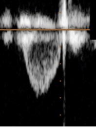
RVOT VTI after milrinone/epoprostenol medneb: Image
Aug 20, 2021 9 tweets 3 min read
1/ TCAV as a ventilator strategy in the Emergency Department? Here’s a case: A patient with COVID intubated for hypoxemic respiratory failure. O2 sats were mid 70% on 100% CPAP prior to intubation with persistent & significant increased WOB. Here’s the CXR after intubation: 
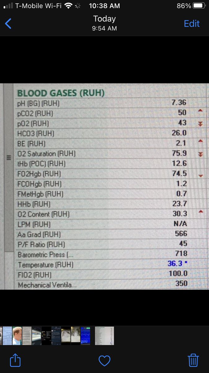
2/ Initial vent settings were with 6cc/kg TV. Initial ABG on FiO2 100% is seen below.
Jun 8, 2020 15 tweets 18 min read
@deanoburns @ThinkingCC @EMNerd_ @Thind888 @iceman_ex 1/ Quick summary: RVOT VTI can be obtained from either PSAX, modified PLAX (with the probe slightly rotated counter clockwise and angled superiority), or modified subcostal (angle the beam in a direction that’s almost parrellel to the sternum). @deanoburns @ThinkingCC @EMNerd_ @Thind888 @iceman_ex 2/ Normally, the RVOT VTI is pretty parabolic in morphology—just a little bit of a triangular shape
May 1, 2020 19 tweets 42 min read
@ArgaizR @ThinkingCC @Thind888 @msiuba @ICUnephroSuarez @VelezNephHepato @NephroGuy @NephroP @load_dependent @iceman_ex 1/ The approach to hypotensive sepsis management is complex, and there are a multitude of strategies based on this or that variable, but I think the best approach is based on having as much information available. @ArgaizR @ThinkingCC @Thind888 @msiuba @ICUnephroSuarez @VelezNephHepato @NephroGuy @NephroP @load_dependent @iceman_ex 2/ I congratulate the authors on this paper. I think one of the the starting points to consider a give volume strategy must be volume responsiveness. If no responsiveness, don’t give fluids for hypotension.
Apr 28, 2019 26 tweets 15 min read
@kyliebaker888 @LMSaxhaug 1/Sorry if this is common knowledge (I know I wasn't asked) but if anyone is confused by diastology, perhaps this will help (what follows is the most basic explaination and reality is more complicated). Ventricular filling is mostly governed by 3 factors. @kyliebaker888 @LMSaxhaug 2/ the factors are: 1)active relaxation or the detachment of the actin/myosin/troponin complex. The more Ca2+ in the cytosol the more linkage and the less ability of the fibers to "let go" thus less ability to relax. ATP is required to pump the Ca2+ out of the cytosol.