POCUS Med Ed Profile picture
Learn point-of-care ultrasound (POCUS) and become a clinician of the modern era: https://t.co/z62WHlXJRW
May 13 7 tweets 3 min read
Lung auscultation has been the standard tool for bedside pulmonary diagnosis for 200 years.

What's rarely discussed in clinical training is how poorly it actually performs when tested empirically.

A thread on the evidence. Image In a study of dialysis patients with and without severe pulmonary edema, lung crackles on auscultation were only 9% sensitive, detecting 9 out of every 100 cases.

Lung auscultation, the authors wrote, has "an unsuspectedly low sensitivity for detecting interstitial edema."

doi.org/10.2215/CJN.03…
May 4 8 tweets 3 min read
The classic jugular venous pressure exam has a hidden flaw that has been embedded in textbooks for 75 years.

It assumes the right atrium sits exactly 5 cm below the sternum in every patient.

CT data says otherwise. A thread on what POCUS changes. The 5 cm assumption traces back to a 1946 study that chose the number for "the sake of simplicity."

CT measurements show the actual right atrial depth ranges from 5 to 15 cm — and the 5 cm rule is correct only 29% of the time.

doi.org/10.1183/090319…
Dec 4, 2025 5 tweets 3 min read
This is jugular venous distension (JVD) seen with #POCUS. But how can we quantify it at the bedside?

Using the Istrail Method described in The POCUS Textbook, we can estimate the actual right atrial pressure to within about 3 mmHg.

#foamed #meded #Medtwitter #cardiotwitter
@nephrop @IM_Crit_ @pocuspeek Once JVD is discovered, the next step is to slide the probe up the neck to find the collapse point. The vertical distance from the base of the neck to the collapse point is measured. This can best be done at the bedside using your outstretched fingers.

Measure and memorize the height of your outstretched fingers, and this can act as your bedside ruler.

This will give you the pressure in centimeters of water of the visible portion of the jugular vein.Image
Mar 1, 2024 6 tweets 2 min read
Chest x-rays miss up to 60% of lung consolidations. Especially if they are retro-cardiac.

#pocus misses about 5% if you do a full exam.

Here is a good example with a corresponding left lower posterior lobe ultrasound taken with @ButterflyNetInc IQ3
Image
Chest X-ray was clear but pocus shows DENSE left lower lobe consolidation with static air bronchograms!

Here is the same consolidation 3 days later after antibiotics via the @ButterflyNetInc IQ3 again:
Mar 9, 2023 10 tweets 6 min read
Reason # 76587 why lung #pocus is important: it can often give more information than CXR or CT Chest can. Case in point:

Elderly female on methotrexate and prednisone presenting with fever and cough.

Initial CXR: 
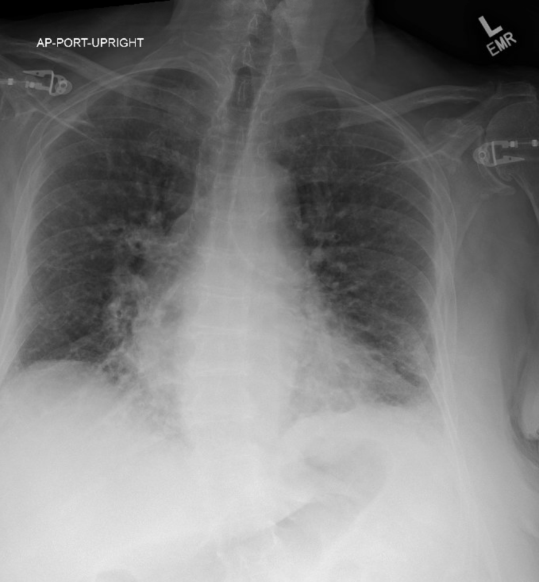
Read as "Bibasilar atelectasis." She was started on antibiotics. The following morning had abdominal pain and CT chest abdomen and pelvis ordered. CT chest seen here:
Dec 17, 2022 9 tweets 5 min read
Reason # 67543 why #pocus is important: sometimes mild symptoms are a sign of impending disaster.

41 year old thin athletic male presented to the hospital with a few weeks of mild shortness of breath with exertion.

BP 170/70, HR 100. CXR with cardiomegaly, tortuous aorta. Bedside #pocus exam shown below:
Jan 12, 2022 11 tweets 7 min read
Why we #pocus: unexpected complications and daily changes. A thread.

70 year old male presented with hypoxic respiratory failure. Initially diagnosed with bilateral pneumonia and started on zosyn. Very hypoxic requiring 15L. The following morning #pocus revealed: Large bilateral pleural effusion a with compression atelectasis. Clinically was never pneumonia. Was upgraded to ICU and intubated. R sided thoracentesis drained 1.8L of fluid and was stepped out of ICU
Sep 2, 2021 10 tweets 5 min read
The JVP exam has been with us since 1930 when Sir Thomas Lewis first proposed this exceptionally clever technique. A #pocus thread. Image 
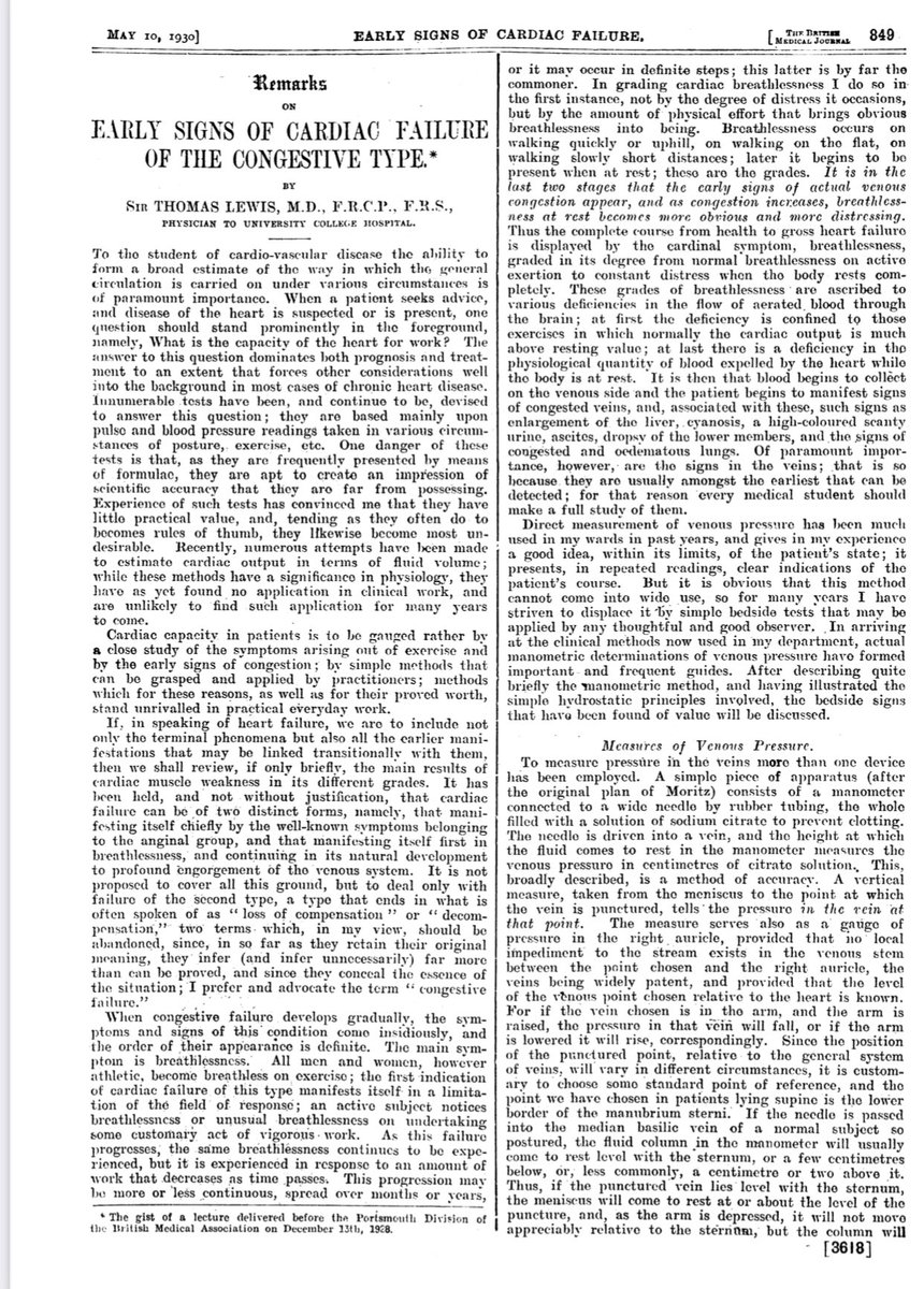
He knew that in heart failure there in “congestion of the veins” and when they become engorged can be detected on the neck.