On behalf of @American_Heart CLCD/CVRI Cardiac Imaging Committee, I would like to invite you to join us for this great & unique educational symposium at #AHA19:
⚡️ Diagnosis & Characterization of #CMD: Role of Imaging ⚡️
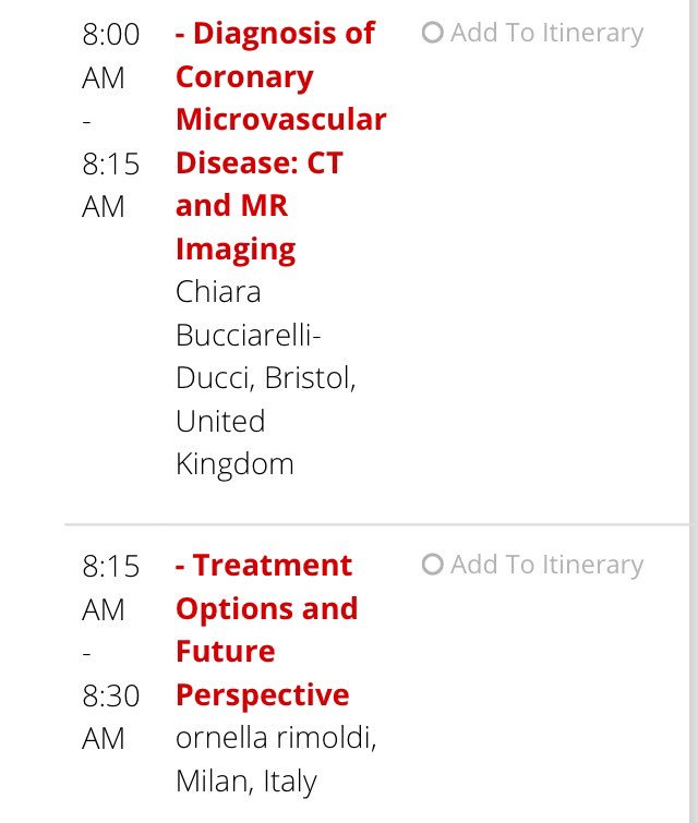
We will have a great & diverse group of national & international experts (60% ♀+ cardiologists + radiologists) covering all imaging modalities: @VTaqMD@DorbalaSharmila@chiarabd Carl Pepine & Ornella Rimoldi. This is a unique opportunity for an imaging focused session on #CMD
Dr Pepine is teaching us about invasive imaging role in Dx of #CMD #AHA19
• • •
Missing some Tweet in this thread? You can try to
force a refresh
#CMD shares similar RFs w epicardial atherosclerosis (traditional ASCVD RFs) & is associated with several biomarkers of inflammation, oxidative stress & coagulation
In our IVUS study:
✅ Most patients with #CMD had significant plaque burden
✅ Patients with #CMD had higher plaque burden & more diffuse atherosclerosis as compared to patients w/o CMD
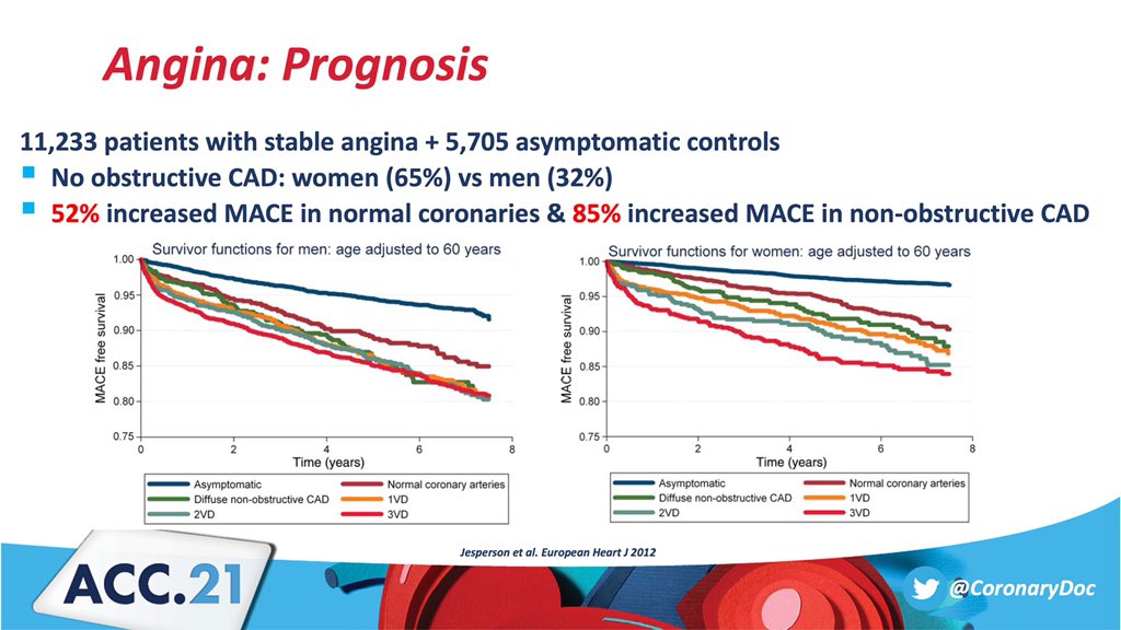
✅ Angina ➡ poor prognosis
✅ Angina with no obstructive CAD = ⬆ risks of CV events compared to no angina, even after adjusting for traditional RFs & co-morbidities
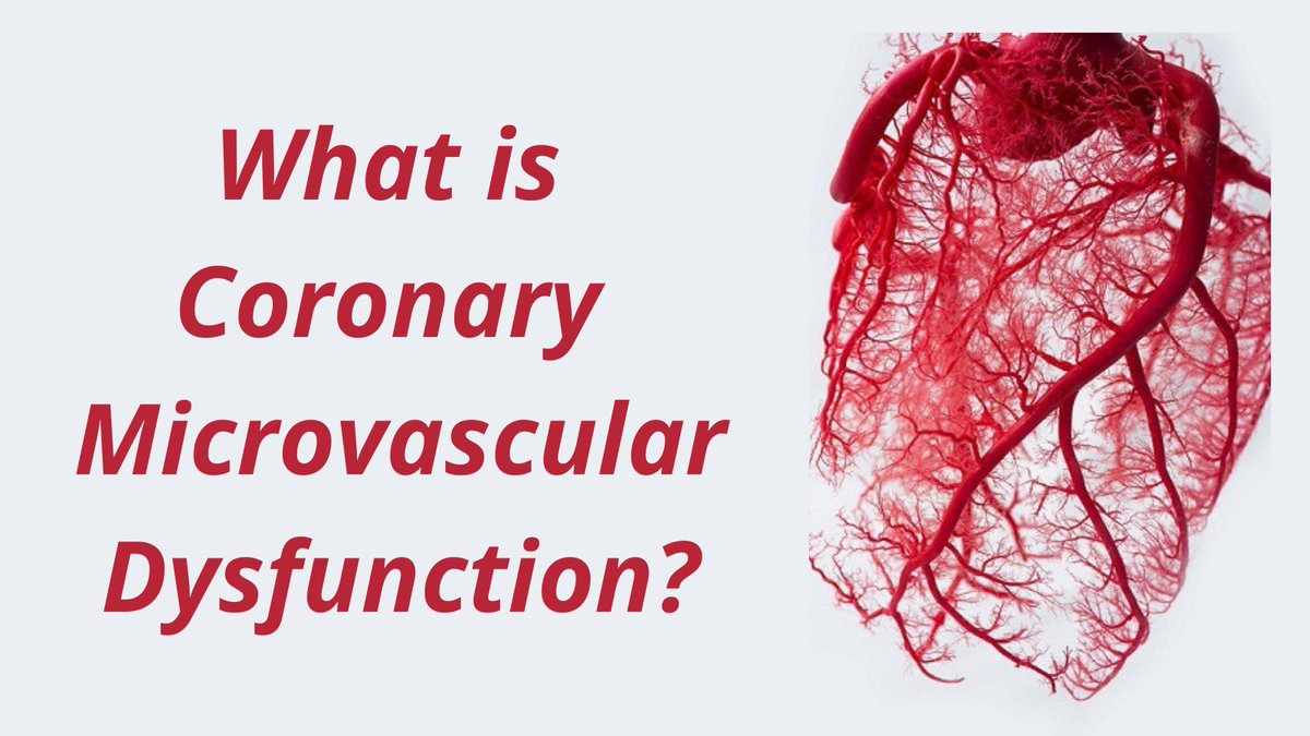
✅ What Is #CMD & Why Is It Important?
✅ Dx & Risk Stratification of #CMD: Stress Testing & Noninvasive Imaging
✅ Catheter-Based Techniques in Dx of #CMD
✅ Emerging Link Between #CMD & #HFpEF
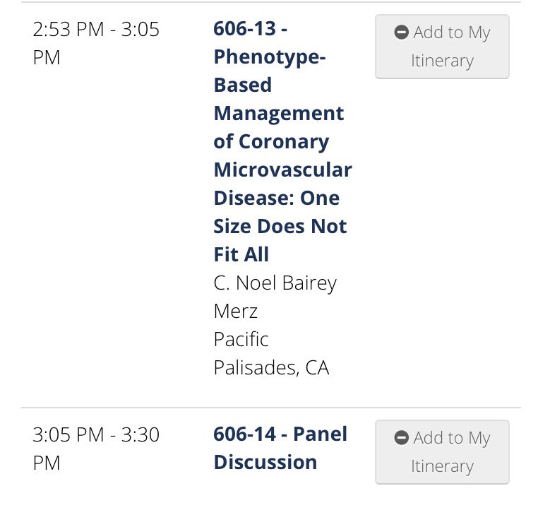
✅ Phenotype-Based Mngmt of #CMD
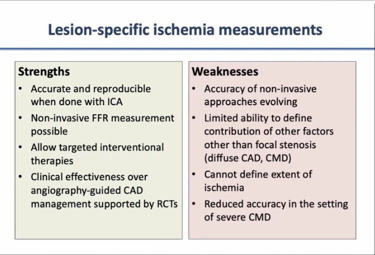
🔘 Strengths/Limits of Lesion Specific vs Myocardial Ischemia
🔘 Comparing Dx Accuracy of Tests
🔘 Ischemia Testing in #INOCA
🔘 How to Select Best Noninvasive Test
☢ Issues with proposed #FFR threshold 0.8
☢ Exercise MPI correlates well with FFR but not at 0.8
☢ Benefit of FFR-guided revasc dominantly occurs w/ thresholds <0.8
☢ Quantification of lesion-specific #ischemia insufficient for patient mgmt