Vidya Monappa Profile picture
Jan 18, 2020 β€’ 7 tweets β€’ 10 min read β€’ Read on X
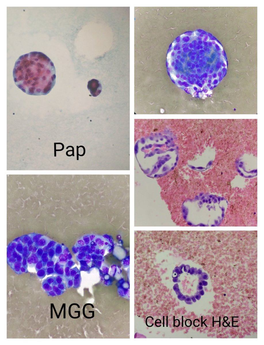
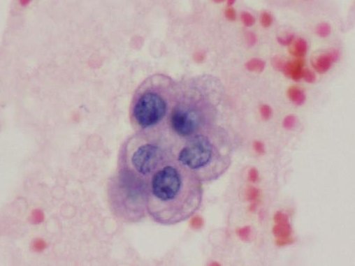
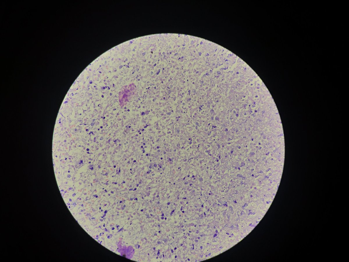
#pathology #crittersontwitter #Cytopathology Hmm.. Is this a πŸ„ or an inverted umbrella? I'm hooked onto itπŸ˜‰ @kriyer68 @smlungpathguy @ParasiteGal @VijayPatho @pembeoltulu @DraEosina @kis_lorand Image
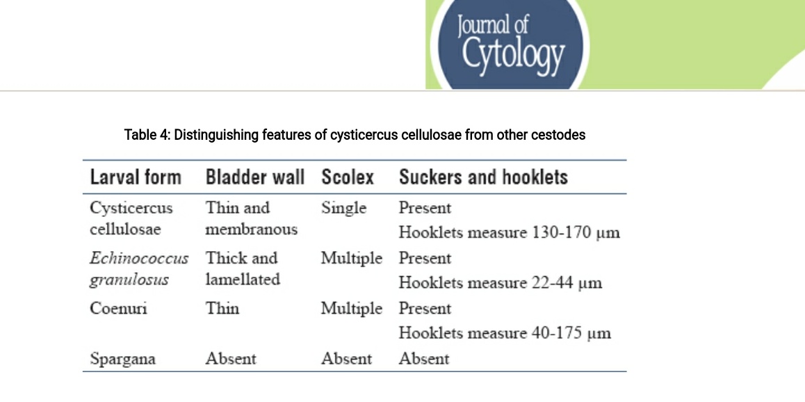
jcytol.org/text.asp?2019/… To differentiate from cysticercosis: 1. Scolices are usually not seen in Cysticercosis on Cyto, 2. As in table below, the hooklets are smaller in Hydatid. ImageImage
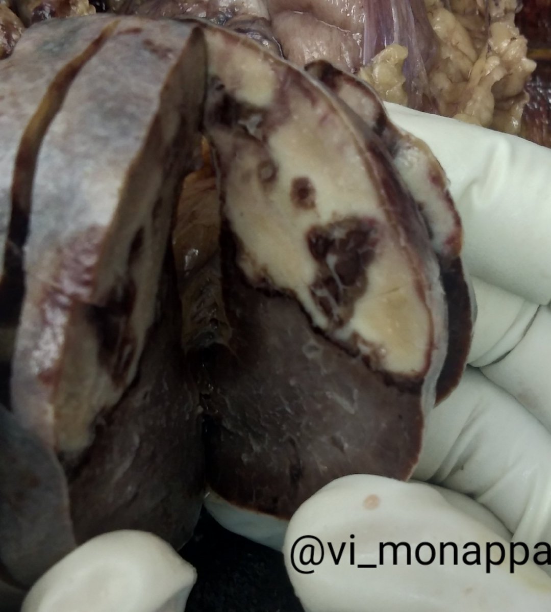
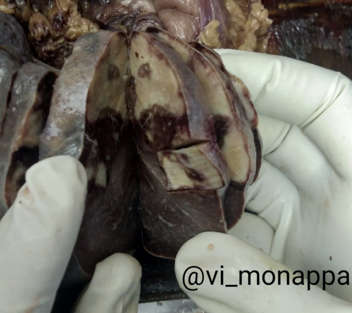
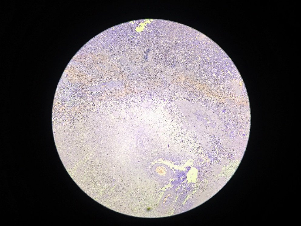
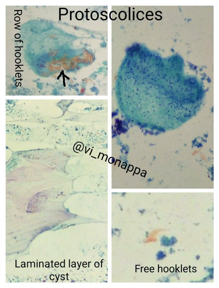
4/2020 #crittersontwitter #pathbugs As promised @VijayPatho @aanchalkakkar posting pics of the laminated cyst wall. Hydatid shows multiple protoscolices and free hooklets in a dirty background. Image

β€’ β€’ β€’

Missing some Tweet in this thread? You can try to force a refresh
γ€€

Keep Current with Vidya Monappa

Vidya Monappa Profile picture

Stay in touch and get notified when new unrolls are available from this author!

Read all threads

This Thread may be Removed Anytime!

PDF

Twitter may remove this content at anytime! Save it as PDF for later use!

Try unrolling a thread yourself!

how to unroll video
  1. Follow @ThreadReaderApp to mention us!

  2. From a Twitter thread mention us with a keyword "unroll"
@threadreaderapp unroll

Practice here first or read more on our help page!

More from @vi_monappa

Jul 30, 2020
#gipath #granulomas #pathology
Ruptured crypt granuloma in UC.
1. LP - ruptured crypt; giant cell +
2. Deeper- histiocyte aggregate with only part of crypt seen (crypt loss)
@kriyer68 @smlungpathguy @pembeoltulu @RunjanChetty @ariella8 @DraEosina @DrMarkOng
TB granulomas are large and confluent, may show necrosis.
AFB + confirms the diagnosis
@vhnguyenmd @anueru432 @anugnya_ran
Here is a foreign body (food particle) granuloma in peritoneal aspect secondary to rupture.
Read 5 tweets
Jan 4, 2020
1/2020 Massive pericardial effusion in an elderly male. Metastatic lung adenocarcinoma. #cytology #CytoPath #cellblock #lungca Pap stain, MGG, cell block and IHC.
1. Multilayered cell balls, smooth outlines, psammoma body 2. Acini in CB @kriyer68 @smlungpathguy @VijayPatho ImageImage
@kriyer68 @smlungpathguy @VijayPatho @drsys02 @Swathiprabhu5 @pathridle_susan @nandishvs * Reactive mesothelial cells can show atypia & proliferation spheres, however - knobby oultline
* Proliferation spheres (cell balls) are solid with stromal cores in mesotheioma unlike carcinoma
* Mesothelial cells: peripheral condensed cytoplasm, vacuoles (intercellular windows) Image
* PsB in pleural and pericardial effusions are usually seen in malignant effusions - thyroid, ovary, lung etc.
* In peritoneal effusion however, PsB may be associated with benign mesothelial hyperplasia, endometriosis etc
* Not all ca. show cell balls- SCC, ILC, Signet ring ca
Read 5 tweets
Dec 27, 2019
Winding up 2019 with this #Grossognosis #pathology case. Happy new year 2020 to all my #pathtwitter friends. K/c/o gastric carcinoma , elderly patient. ImageImage
The most likely diagnosis of the above gross is:
Diagnosis: Splenic infarction. Coagulation necrosis + ImageImageImage
Read 5 tweets

Did Thread Reader help you today?

Support us! We are indie developers!


This site is made by just two indie developers on a laptop doing marketing, support and development! Read more about the story.

Become a Premium Member ($3/month or $30/year) and get exclusive features!

Become Premium

Don't want to be a Premium member but still want to support us?

Make a small donation by buying us coffee ($5) or help with server cost ($10)

Donate via Paypal

Or Donate anonymously using crypto!

Ethereum

0xfe58350B80634f60Fa6Dc149a72b4DFbc17D341E copy

Bitcoin

3ATGMxNzCUFzxpMCHL5sWSt4DVtS8UqXpi copy

Thank you for your support!

Follow Us!

:(