My Authors
Read all threads
[1/9] Lots to learn from a simple ankle Xray. At first glance this looks like a “standard” bimal, and yet the medial malleolus isn’t the usual fracture pattern. It almost seems “intact” (yellow). There is crack proximally (red), and joint widening with talar subluxation (green).
[2/9] The lateral shows us the fibula fracture which is the standard SER pattern (proximal posterior to distal anterior), as well as the talus subluxating posteriorly with the posterior malleolus fragment, making the joint incongruent.
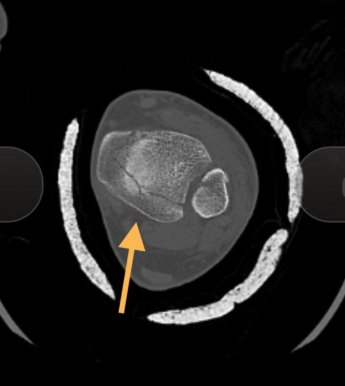
[3/9] In my eyes this is the time for CT, not because I think posterior mal fragment is big, but to determine apex of medial sided fracture (medial as suspected), and also to see if medial fracture and posterior malleolus is one fragment or 2, which determines surgical approach.
[4/9] Here we can see that the medial malleollus and posterior malleolus are a single fragment (or even if small crack, acting as one). This is Haraguchi type II, which has been shown to have worse outcomes. To learn more about the classification: pubmed.ncbi.nlm.nih.gov/31176480/?from…
[5/9] A 3D CT confirms what we should already know: the main fracture apex is medial/posteromedial (yellow), and another spike further posterior (green). So if you do your standard approach to medial mal, you will not reach here. Nor will the usual posterolateral approach.
[6/9] Unlike my usual approach to do medial side first, here I fixed the lateral side, hopeful that anatomic fixation would help me obtain length of the posterior portion of the medial mal fracture via the PITFL (posterior portion of syndesmosis). It probably didn’t matter much.
[7/9] Here, you go where money is—posteromedial. As long as you understand anatomy and are careful, you can avoid or retract neurovascular bundle. This spike was behind tib posterior; I used K wire to confirm before I cut periosteum. Remember: “Tom Dick (And Very Nervous) Harry!”
[8/9] I was able to see both spikes and confirm they were part of same fragment. I reduced main one and held it with screws closer to joint (cancellous lag in metaphyseal bone since can’t be bicortical in this direction), then 2.4 buttress plate to counteract upward shear forces.
[9/9] Final images. It doesn’t take huge or expensive implants to get what you want— 1/3 tubular for fibula and tiny cute plate as the buttress (with no distal screws because it doesn’t need them to counter shear force).
Missing some Tweet in this thread? You can try to force a refresh.

Enjoying this thread?

Keep Current with Yelena Bogdan, MD, FACS, FAAOS

Profile picture

Stay in touch and get notified when new unrolls are available from this author!

Read all threads

This Thread may be Removed Anytime!

Twitter may remove this content at anytime, convert it as a PDF, save and print for later use!

Try unrolling a thread yourself!

how to unroll video

1) Follow Thread Reader App on Twitter so you can easily mention us!

2) Go to a Twitter thread (series of Tweets by the same owner) and mention us with a keyword "unroll" @threadreaderapp unroll

You can practice here first or read more on our help page!

Follow Us on Twitter!

Did Thread Reader help you today?

Support us! We are indie developers!


This site is made by just two indie developers on a laptop doing marketing, support and development! Read more about the story.

Become a Premium Member ($3.00/month or $30.00/year) and get exclusive features!

Become Premium

Too expensive? Make a small donation by buying us coffee ($5) or help with server cost ($10)

Donate via Paypal Become our Patreon

Thank you for your support!