Learning Diastolic Dysfunction can seem Overwhelming! But what if ANYONE could Learn/Understand #Diastology using a Simple "PUSH and PULL" Analogy?
New Blog Post! Step By Step #POCUS Guide, PDF Pocket Card, & Calculator.
👉🔗pocus101.com/diastology
#medtweetorial👇(1/18)
New Blog Post! Step By Step #POCUS Guide, PDF Pocket Card, & Calculator.
👉🔗pocus101.com/diastology
#medtweetorial👇(1/18)
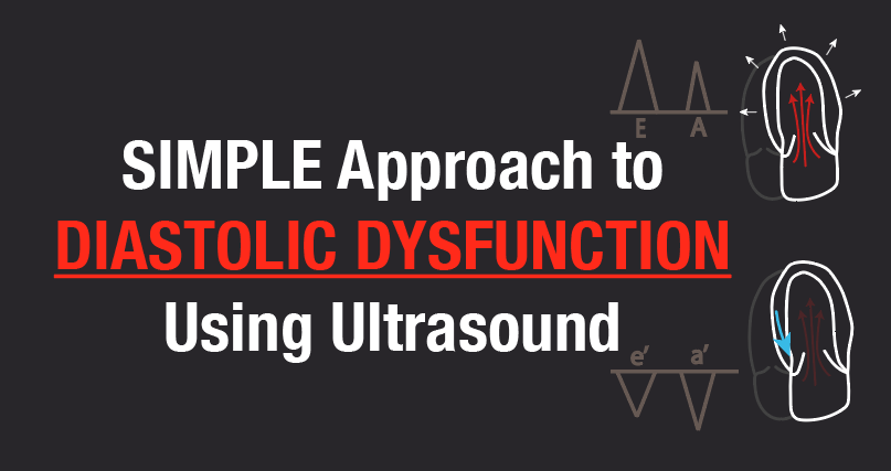

(2/18) Learning diastolic dysfunction doesn’t have to be hard. In this Post, we will go over the TWO main findings: Mitral Inflow and Tissue Doppler Measurements.
Download the summary Pocket Card PDF:
👉🔗pocus101.com/diastology
Download the summary Pocket Card PDF:
👉🔗pocus101.com/diastology

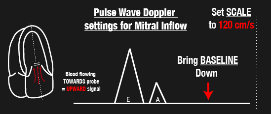
(3/18) Mitral Inflow measures BLOOD coming into the LV during diastolic filling (E and A waves).
Here are some Pulse Wave Doppler tips:
1. Place the sample gate at mitral valve tips
2. Bring the baseline down
3. Set Scale to about 120cm/s
👉🔗pocus101.com/diastology
Here are some Pulse Wave Doppler tips:
1. Place the sample gate at mitral valve tips
2. Bring the baseline down
3. Set Scale to about 120cm/s
👉🔗pocus101.com/diastology
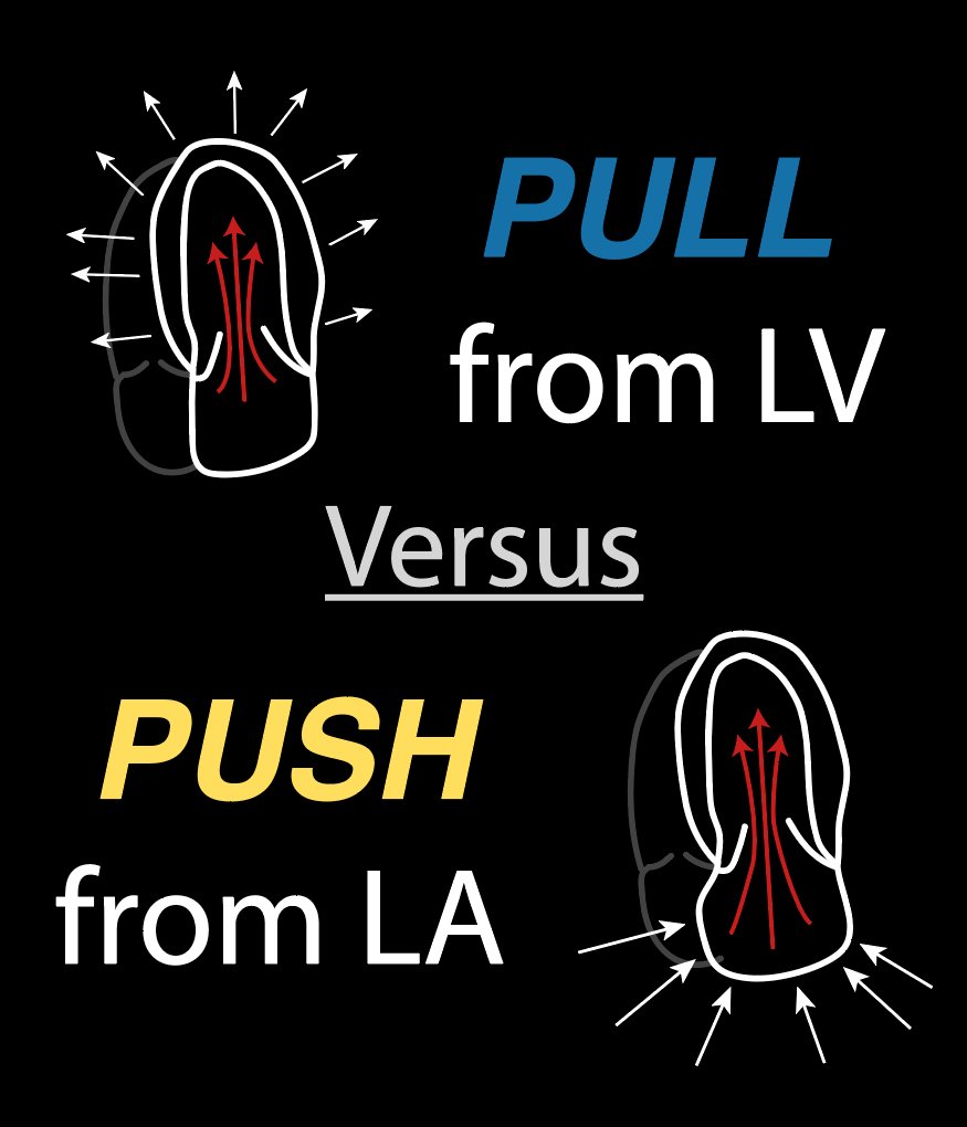
(4/18) Mitral Inflow waveforms are easy to understand if you just ask yourself: is it primarily the LV “PULLING” in blood or the LA “PUSHING” in blood during early diastole (E wave)?
👉🔗pocus101.com/diastology
👉🔗pocus101.com/diastology

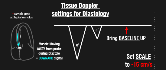
(5/18) Tissue Doppler measures the speed of LV muscle relaxation during diastole.
Here are some Tissue Doppler tips:
1. Place sample gate at septal (or lateral) annulus
2. Bring the baseline up
3. Set scale to about -15cm/s
👉🔗pocus101.com/diastology
Here are some Tissue Doppler tips:
1. Place sample gate at septal (or lateral) annulus
2. Bring the baseline up
3. Set scale to about -15cm/s
👉🔗pocus101.com/diastology
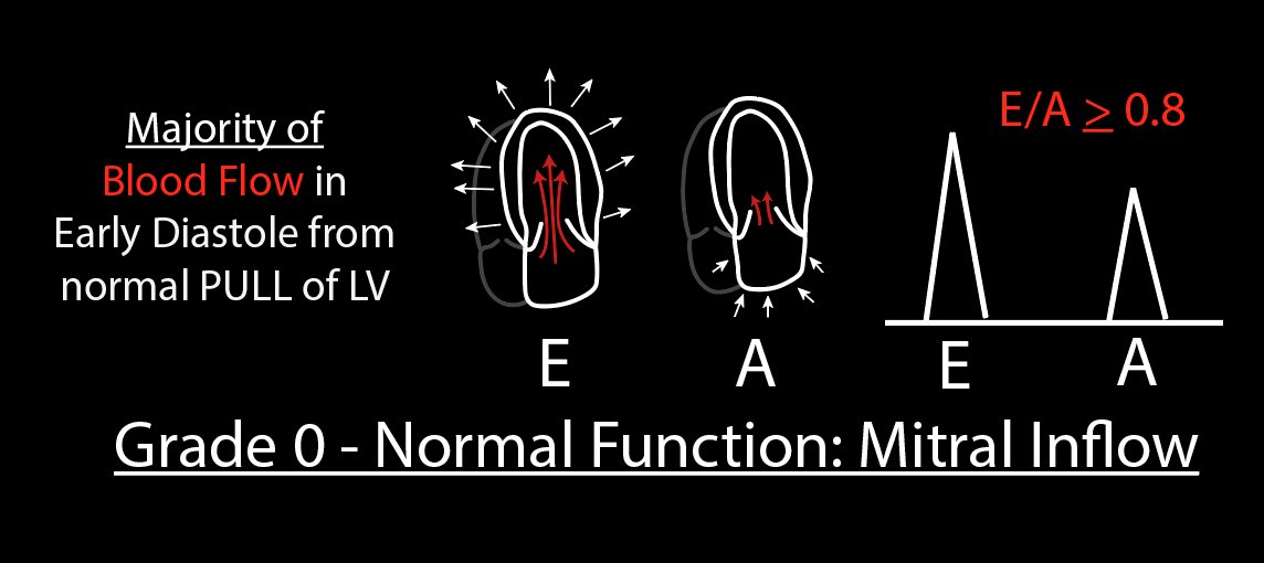
(6/18) Mitral Inflow Pattern for Grade 0 Diastolic Function
• E wave: majority of blood flow resulting from Passive PULL of left ventricle relaxation (large E wave)
• A wave: Atrial kick with small amount of Blood Flow (small A wave)
• E/A >= 0.8
👉🔗pocus101.com/diastology
• E wave: majority of blood flow resulting from Passive PULL of left ventricle relaxation (large E wave)
• A wave: Atrial kick with small amount of Blood Flow (small A wave)
• E/A >= 0.8
👉🔗pocus101.com/diastology
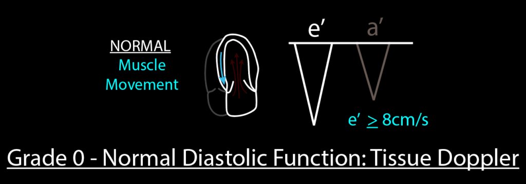
(7/18) Tissue Doppler Pattern for Grade 0 Diastolic Function - NORMAL
•Normal LV muscle relaxation
•e’ >= 8cm/s (septal annulus)
👉🔗pocus101.com/diastology
•Normal LV muscle relaxation
•e’ >= 8cm/s (septal annulus)
👉🔗pocus101.com/diastology
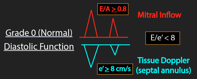
(8/18) Summary for Grade 0 Diastolic Function (Normal)
•Mitral Inflow: E/A >= 0.8
•Tissue Doppler: e’ >= 8cm/s
•E/e’ < 8
👉🔗pocus101.com/diastology
•Mitral Inflow: E/A >= 0.8
•Tissue Doppler: e’ >= 8cm/s
•E/e’ < 8
👉🔗pocus101.com/diastology
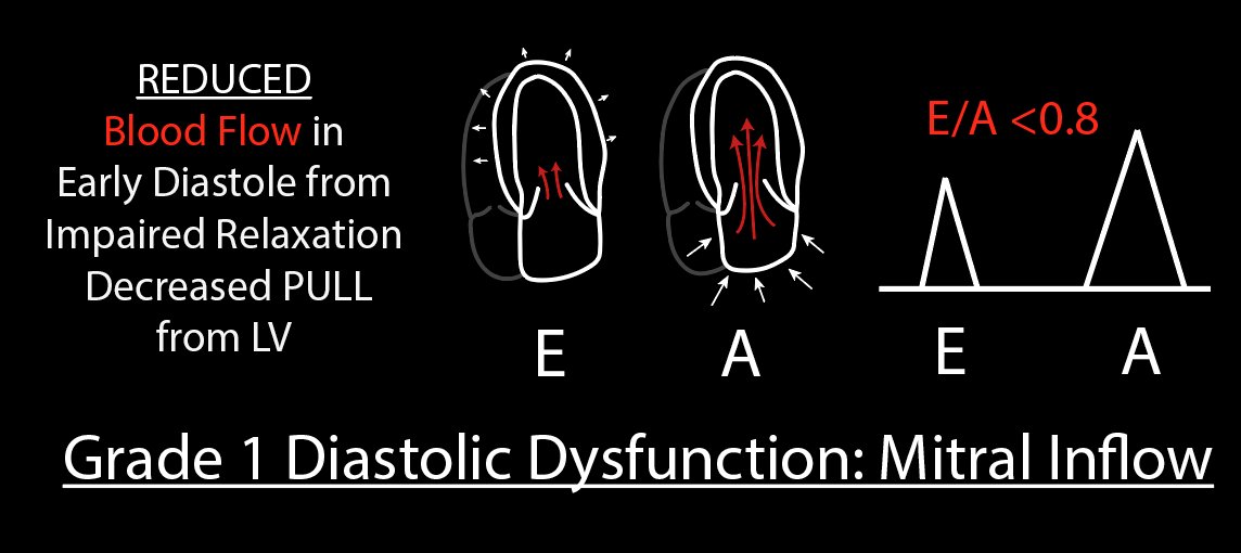
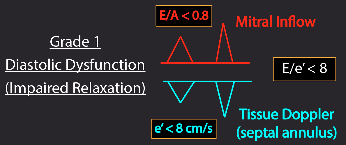
(9/18) Mitral Inflow Pattern for Grade 1 Diastolic Dysfunction
• E wave: Decreased “PULL” from LV due to impaired relaxation (small E wave)
• E/A < 0.8 (only Grade 1 has this mitral inflow pattern)
👉🔗pocus101.com/diastology
• E wave: Decreased “PULL” from LV due to impaired relaxation (small E wave)
• E/A < 0.8 (only Grade 1 has this mitral inflow pattern)
👉🔗pocus101.com/diastology
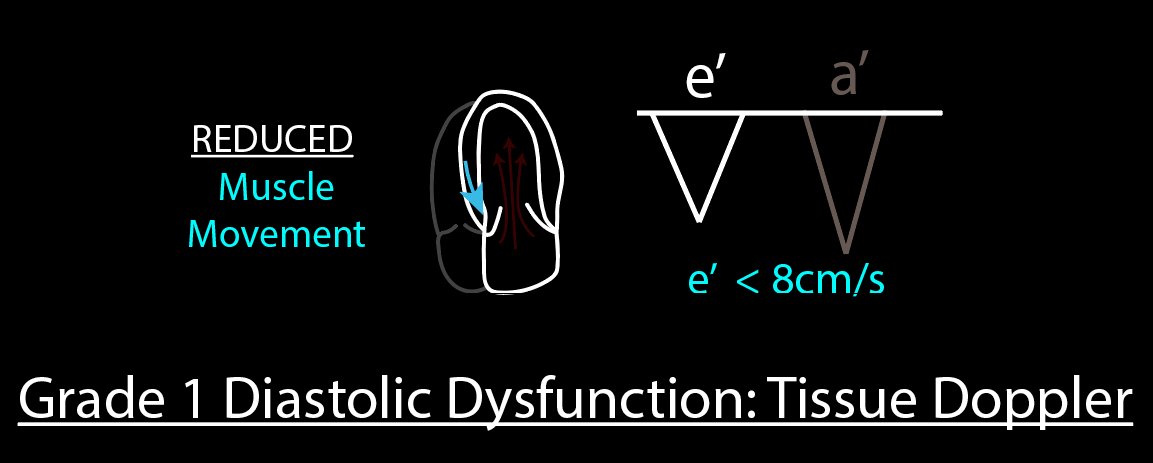
(10/18) Tissue Doppler Pattern for Grade 1 Diastolic Dysfunction
•Impaired left ventricular muscle relaxation
•e’ < 8cm/s
👉🔗pocus101.com/diastology
•Impaired left ventricular muscle relaxation
•e’ < 8cm/s
👉🔗pocus101.com/diastology
(11/18) Summary for Grade 1 Diastolic Dysfunction – IMPAIRED RELAXATION
•Mitral Inflow: E/A < 0.8 (Most Important Factor)
•Tissue Doppler: e’ < 8cm/s
•E/e’ < 8
👉🔗pocus101.com/diastology
•Mitral Inflow: E/A < 0.8 (Most Important Factor)
•Tissue Doppler: e’ < 8cm/s
•E/e’ < 8
👉🔗pocus101.com/diastology
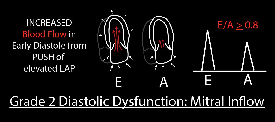
(12/18) Mitral Inflow Pattern for Grade 2 Diastolic Dysfunction
• E wave: Increased “PUSH” from left atrium due to increased Left Atrial Pressure (large E wave)
• E/A >= 0.8 ("Pseudonormal" since it looks like Grade 0)
👉🔗pocus101.com/diastology
• E wave: Increased “PUSH” from left atrium due to increased Left Atrial Pressure (large E wave)
• E/A >= 0.8 ("Pseudonormal" since it looks like Grade 0)
👉🔗pocus101.com/diastology
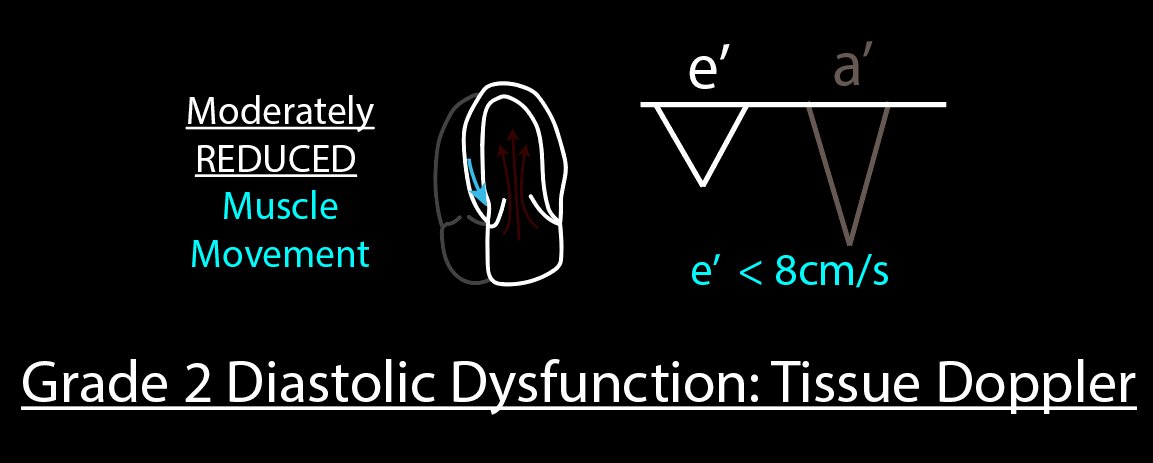
(13/18) Tissue Doppler Pattern for Grade 2 Diastolic Dysfunction
You will need to use tissue doppler to differentiate between Grade 0 and Grade 2 since the E/A ratio will be similar
•Moderately Reduced LV muscle relaxation
•e’ < 8cm/s
👉🔗pocus101.com/diastology
You will need to use tissue doppler to differentiate between Grade 0 and Grade 2 since the E/A ratio will be similar
•Moderately Reduced LV muscle relaxation
•e’ < 8cm/s
👉🔗pocus101.com/diastology
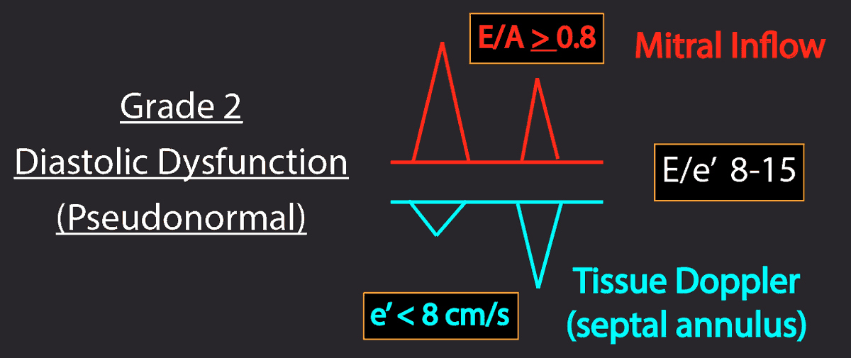
(14/18) Summary for Grade 2 Diastolic Dysfunction - PSEUDONORMAL
•Mitral Inflow: E/A >= 8
•Tissue Doppler: e’ < 8cm/s
•E/e’: 8-15
👉🔗pocus101.com/diastology
•Mitral Inflow: E/A >= 8
•Tissue Doppler: e’ < 8cm/s
•E/e’: 8-15
👉🔗pocus101.com/diastology
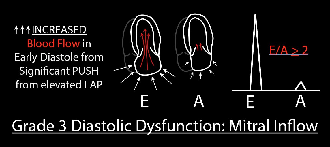
(15/18) Mitral Inflow Pattern for Grade 3 Diastolic Dysfunction
•E wave: Significant Increased “PUSH” from left atrial due to Severely increased Left Atrial Pressure (large E wave)
•A wave: small A wave
•E/A > 2
👉🔗pocus101.com/diastology
•E wave: Significant Increased “PUSH” from left atrial due to Severely increased Left Atrial Pressure (large E wave)
•A wave: small A wave
•E/A > 2
👉🔗pocus101.com/diastology
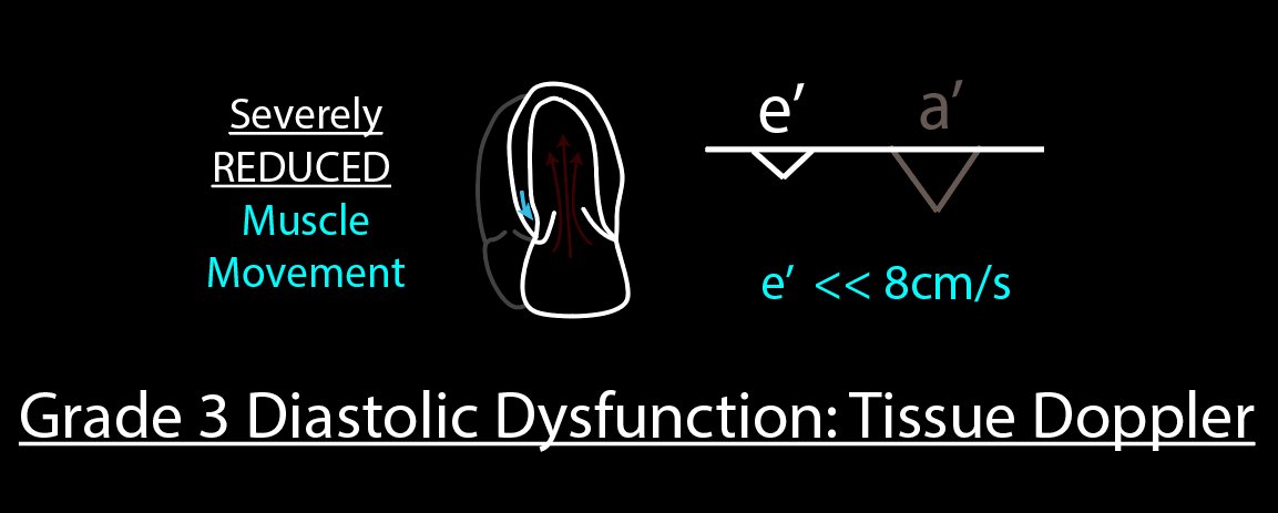
(16/18) Tissue Doppler Pattern for Grade 3 Diastolic Dysfunction
•Severely reduced LV muscle relaxation
•e’ << 8cm/s
👉🔗pocus101.com/diastology
•Severely reduced LV muscle relaxation
•e’ << 8cm/s
👉🔗pocus101.com/diastology
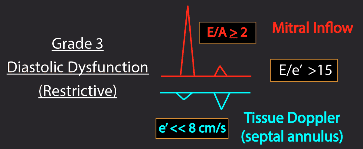
(17/18) Summary for Grade 3 Diastolic Dysfunction - RESTRICTIVE
•Mitral Inflow: E/A >= 2 (very high E wave)
•Left Atrial Enlargement
•Tissue Doppler: e’ << 8cm/s
•E/e’ > 15
👉🔗pocus101.com/diastology
•Mitral Inflow: E/A >= 2 (very high E wave)
•Left Atrial Enlargement
•Tissue Doppler: e’ << 8cm/s
•E/e’ > 15
👉🔗pocus101.com/diastology
(18/18) Now that you know how to assess diastolic function in your patients try it in the following scenarios:
• Preload evaluation prior to IVFs
• Diuresis Management of CHF patients
• Diagnosing ARDS (should have normal diastolic function)
👉🔗pocus101.com/diastology
• Preload evaluation prior to IVFs
• Diuresis Management of CHF patients
• Diagnosing ARDS (should have normal diastolic function)
👉🔗pocus101.com/diastology