Fundoscopy 👁🗨 got you down? See clearly into the 👀 with #POCUS!
1⃣Learn How to Perform Ocular Ultrasound
2⃣Recognize Retinal Detachment, PVD, ⬆️ ICP, + MORE!
3⃣Free Ocular Ultrasound PDF Pocket Card!
✅New Blog Post! 👉🔗pocus101.com/Ocular
#medtweetorial👇(1/21)
1⃣Learn How to Perform Ocular Ultrasound
2⃣Recognize Retinal Detachment, PVD, ⬆️ ICP, + MORE!
3⃣Free Ocular Ultrasound PDF Pocket Card!
✅New Blog Post! 👉🔗pocus101.com/Ocular
#medtweetorial👇(1/21)


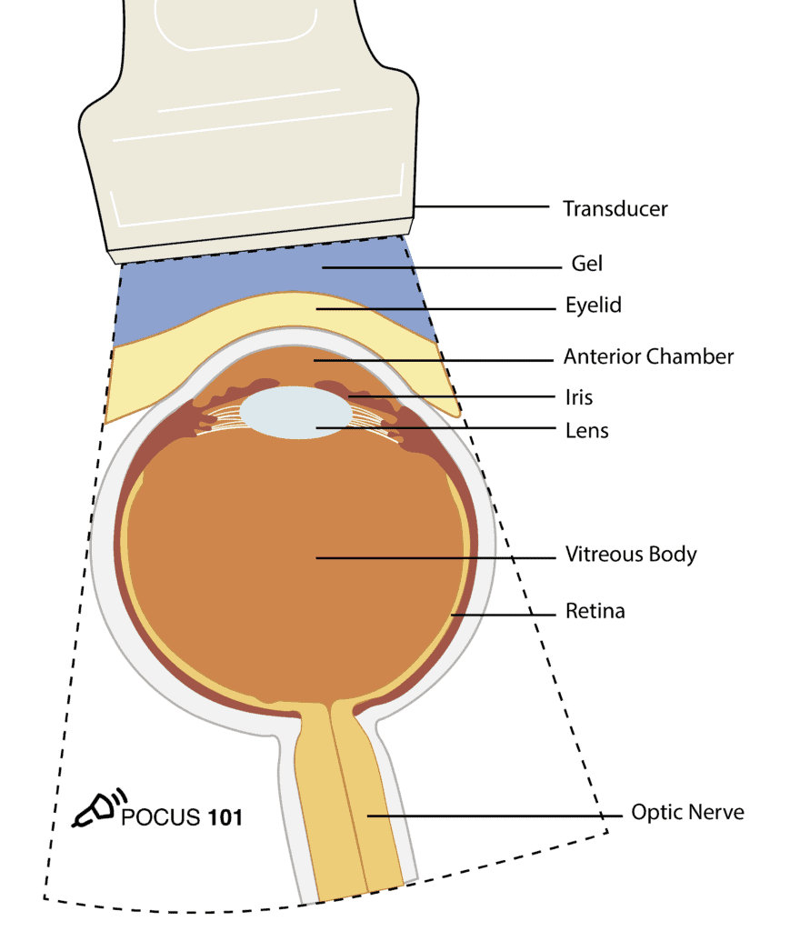
(3) Obtain the Transverse View:
Place the probe lightly on the gel covering the patient’s eye with the probe indicator pointed towards the patient’s right to obtain a transverse view.
👉🔗pocus101.com/Ocular
Place the probe lightly on the gel covering the patient’s eye with the probe indicator pointed towards the patient’s right to obtain a transverse view.
👉🔗pocus101.com/Ocular
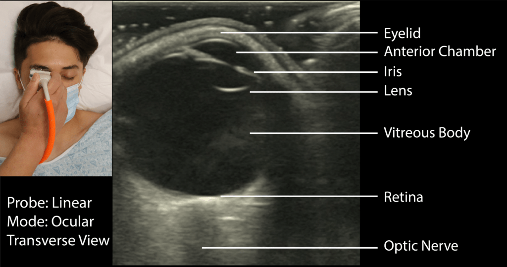
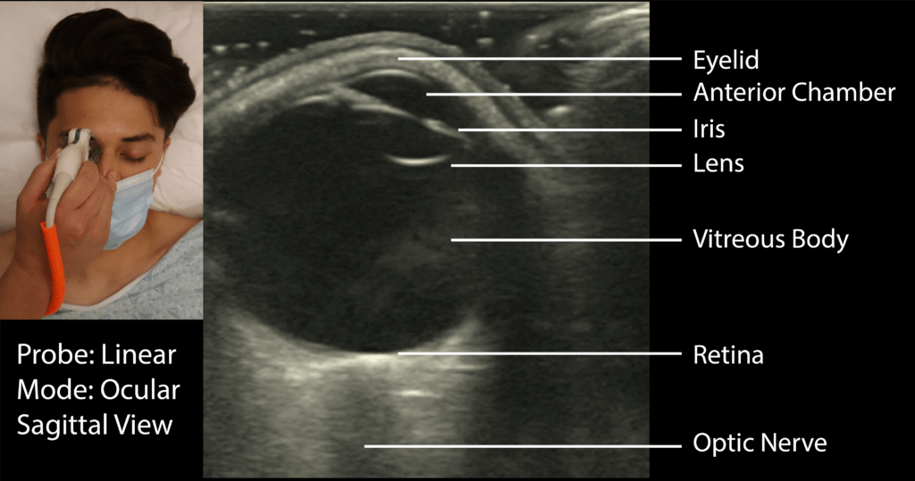
(4) Identify the Following Structures
•Eyelid
•Anterior Chamber
•Lens
•Iris
•Vitreous Body
•Retina
•Optic Nerve
•*Make sure to tilt/fan through the entire eye
👉🔗pocus101.com/Ocular
•Eyelid
•Anterior Chamber
•Lens
•Iris
•Vitreous Body
•Retina
•Optic Nerve
•*Make sure to tilt/fan through the entire eye
👉🔗pocus101.com/Ocular
(5) Obtain TheSagittal View
Next, turn the probe 90° clockwise so the indicator points superiorly towards the patient’s head to obtain a sagittal view. Identify the same structures you found in the transverse view.
👉🔗pocus101.com/Ocular
Next, turn the probe 90° clockwise so the indicator points superiorly towards the patient’s head to obtain a sagittal view. Identify the same structures you found in the transverse view.
👉🔗pocus101.com/Ocular
(6) Assess extraocular Movements
Evaluate for extraocular movements. This is important when patients have severe periorbital edema from facial trauma.
👉🔗pocus101.com/Ocular
Evaluate for extraocular movements. This is important when patients have severe periorbital edema from facial trauma.
👉🔗pocus101.com/Ocular
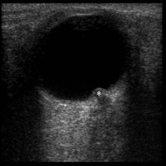
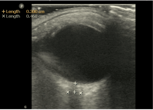
(7) Obtain Optic Nerve Sheath Diameter (ONSD)
Measure 3 mm (.3cm) posterior to where the optic nerve sheath attaches to the retina. This is the location where you will use to measure your optic nerve sheath diameter.
👉🔗pocus101.com/Ocular
Measure 3 mm (.3cm) posterior to where the optic nerve sheath attaches to the retina. This is the location where you will use to measure your optic nerve sheath diameter.
👉🔗pocus101.com/Ocular
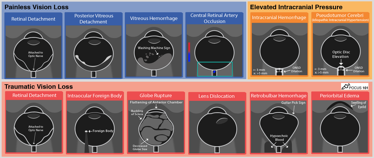
(8) Let's look at the different types of Ocular Ultrasound Pathology:
Download the Ocular Ultrasound Pathology PDF Pocket Card here for Reference.
👉🔗pocus101.com/Ocular
Download the Ocular Ultrasound Pathology PDF Pocket Card here for Reference.
👉🔗pocus101.com/Ocular
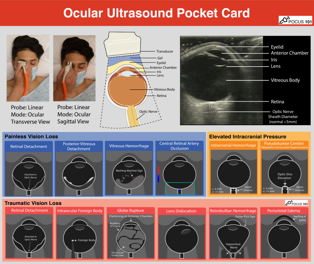
(9) There are three types of Retinal Detachments:
Rhegmatogenous, Traction, and Exudative.
👉🔗pocus101.com/Ocular
Rhegmatogenous, Traction, and Exudative.
👉🔗pocus101.com/Ocular
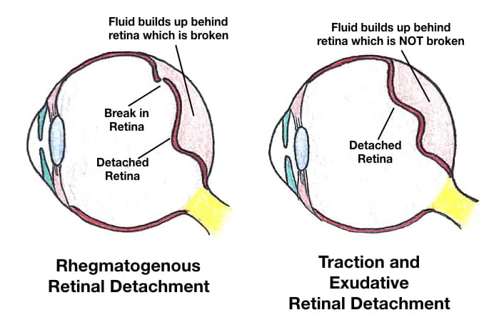
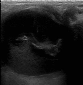
(10) Retinal Detachment: The defining characteristic is that a retinal detachment is tethered to the optic nerve and moves with the optic nerve.
👉🔗pocus101.com/Ocular
👉🔗pocus101.com/Ocular
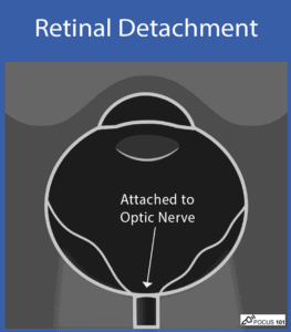
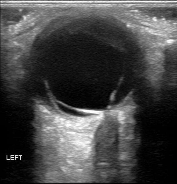
Retinal Detachment Video (notice attachment to optic nerve)
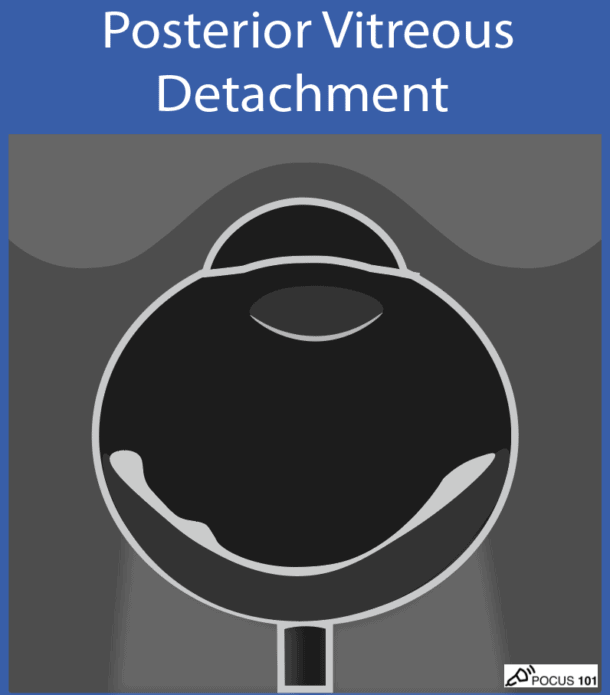
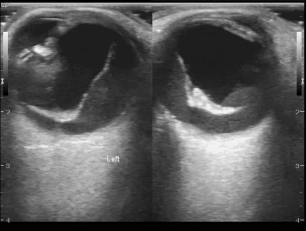
(11) Posterior Vitreous Detachment looks like a thin, hyperechoic membrane lifted off the posterior surface of the globe that is NOT tethered to the optic nerve.
👉🔗pocus101.com/Ocular
👉🔗pocus101.com/Ocular
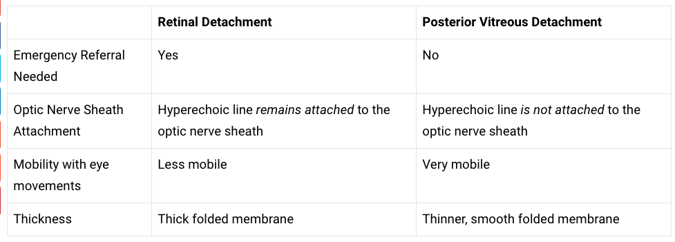
(12) Here is a table to help differentiate Retinal Detachment from Posterior Vitreous Detachment
👉🔗pocus101.com/Ocular
👉🔗pocus101.com/Ocular
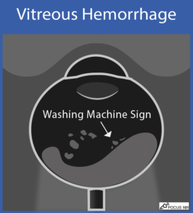
(13) Vitreous Hemorrhage: looks like echogenic material in the posterior chamber. If you ask the patient to move their eye side-to-side, you may see the washing machine sign
👉🔗pocus101.com/Ocular
👉🔗pocus101.com/Ocular
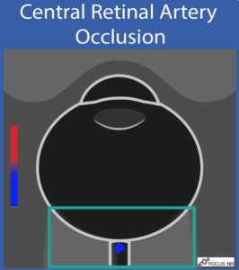
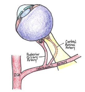
(14) Central Retinal Artery Occlusion: is a rare finding and color Doppler mode can be used to diagnose it with ocular ultrasound. You may find diminished or absent flow of the central retinal artery.
👉🔗pocus101.com/Ocular
👉🔗pocus101.com/Ocular
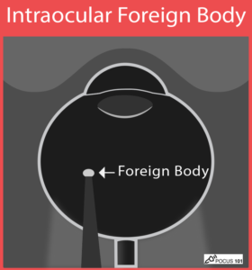
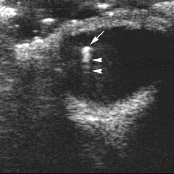
(15) Ocular Foreign Body: you can find a bright, hyperechoic object with an associated reverberation artifact (if the object is metallic).
👉🔗pocus101.com/Ocular
👉🔗pocus101.com/Ocular
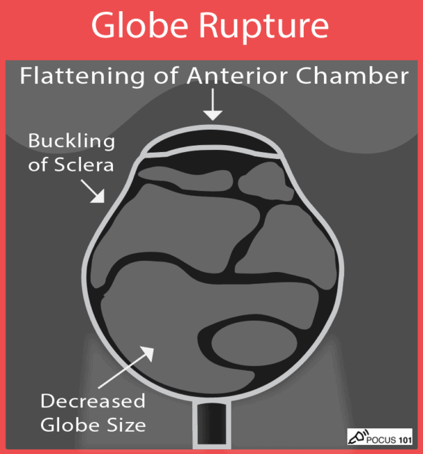
(16) Globe Rupture: you may find buckling of the sclera, asymmetric loss of the normal spherical shape of the globe, decreased size of globe, flattening or compression of the anterior chamber, or vitreous hemorrhage
👉🔗pocus101.com/Ocular
👉🔗pocus101.com/Ocular
Globe Rupture Video:
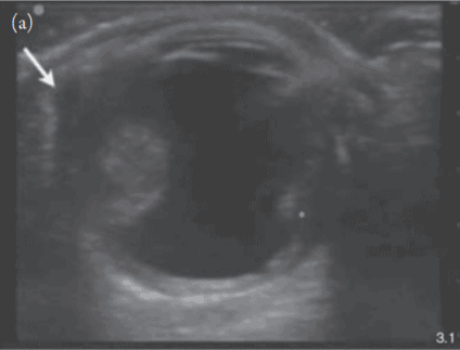
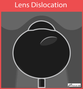
(17) Lens Dislocation: you will see the lens as a bi-convex structure with hyperechoic borders floating posteriorly in the vitreous body.
👉🔗pocus101.com/Ocular
👉🔗pocus101.com/Ocular
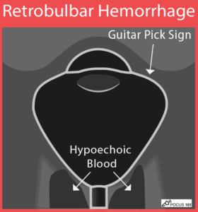
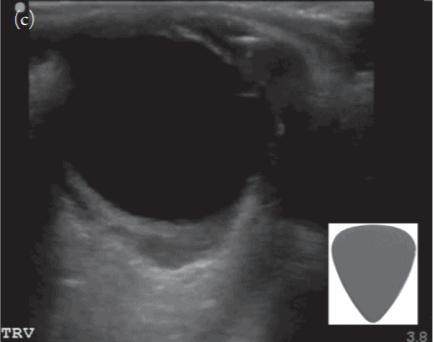
(18) Retrobulbar Hemorrhage: you may see a “Guitar Pick Sign” where the increased pressure from the retrobulbar hematoma distorts the spherical globe into a conical shape
👉🔗pocus101.com/Ocular
👉🔗pocus101.com/Ocular
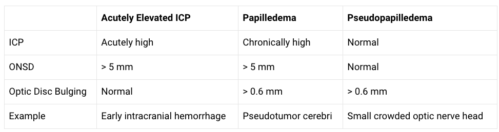
(19) Learn differences between Elevated ICP, Papilledema, and Pseudopapilledema
👉🔗pocus101.com/Ocular
👉🔗pocus101.com/Ocular
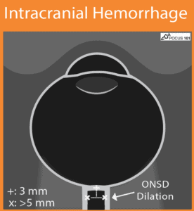
(20) Intracranial Hemorrhage with Increased ICP: you will find elevated ONSD measurements > 5 mm.
👉🔗pocus101.com/Ocular
👉🔗pocus101.com/Ocular
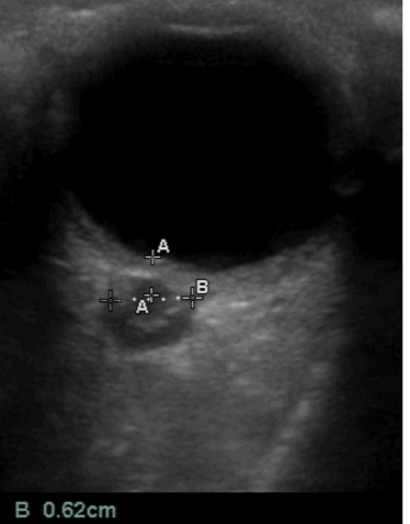
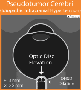
(21) Pseudotumor Cerebri (Idiopathic Intracranial Hypertension): you will find dilated ONSD measurements > 5 mm (increased ICP) and signs of Papilledema (optic disc bulging/elevation).
👉🔗pocus101.com/Ocular
👉🔗pocus101.com/Ocular