#POCUS is the FASTEST way to assess your patient's Bladder!
1⃣Learn How to Perform Bladder Ultrasound
2⃣Measure Bladder Volume Correctly
3⃣Recognize Bladder Pathology
4⃣FREE Bladder Volume Calculator!
✅New Blog Post! 👉🔗pocus101.com/bladder
#medtweetorial (1/20)
1⃣Learn How to Perform Bladder Ultrasound
2⃣Measure Bladder Volume Correctly
3⃣Recognize Bladder Pathology
4⃣FREE Bladder Volume Calculator!
✅New Blog Post! 👉🔗pocus101.com/bladder
#medtweetorial (1/20)
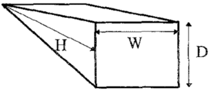
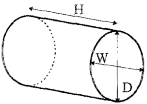
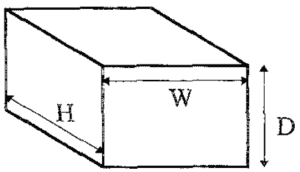
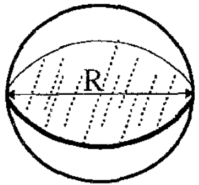
1 Although it is easy to think of the bladder as a sphere it actually takes on other shapes in the body: Triangular Prism, Cylinder (Ellipsoid), or Cuboid
👉🔗pocus101.com/bladder
👉🔗pocus101.com/bladder
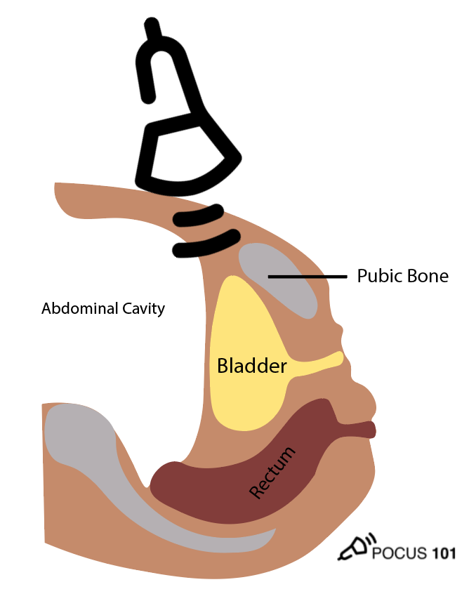
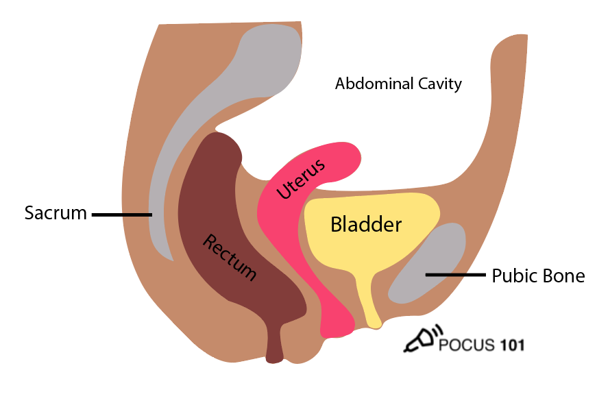
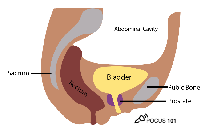
2 In addition to the bladder itself, it is also important to understand the surrounding structures including the pubic bone, abdominal cavity and rectum. The uterus and prostate are also important landmarks for females and males respectively.
👉🔗pocus101.com/bladder
👉🔗pocus101.com/bladder
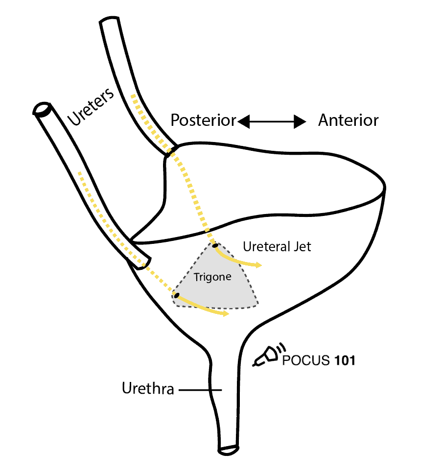
3 The ureters enter the top of the bladder but do not release urine until they reach the trigone of the bladder located inferiorly. This is important to know when you are evaluating “ureteral jets” on ultrasound.
👉🔗pocus101.com/bladder
👉🔗pocus101.com/bladder
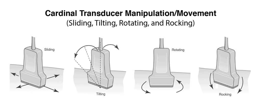
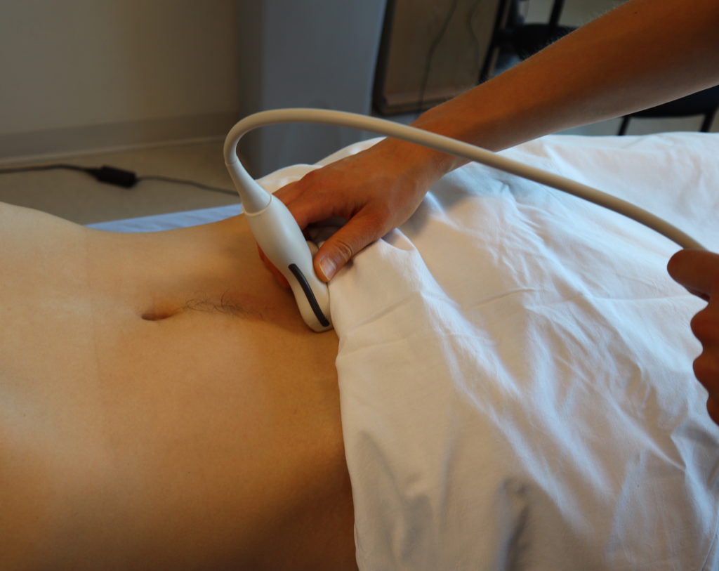
4 Obtain the Longitudinal/Sagittal view: Place the transducer with the indicator pointing towards the patient’s head in the patient’s midline, right above the pubic symphysis. Rock the probe so that it points down towards the pelvic cavity.
👉🔗pocus101.com/bladder
👉🔗pocus101.com/bladder
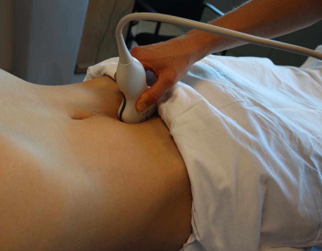
5 One of the most important things to remember when performing bladder ultrasound is that the bladder is directly posterior to the pubic symphysis. If you are unable to get proper images, most likely your ultrasound probe is placed too superiorly.
👉🔗pocus101.com/bladder
👉🔗pocus101.com/bladder

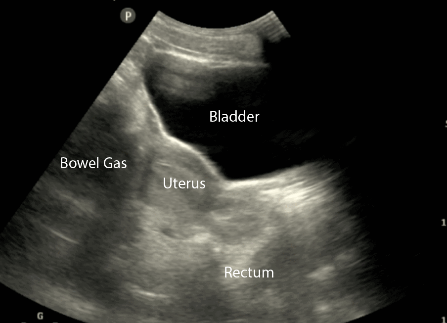
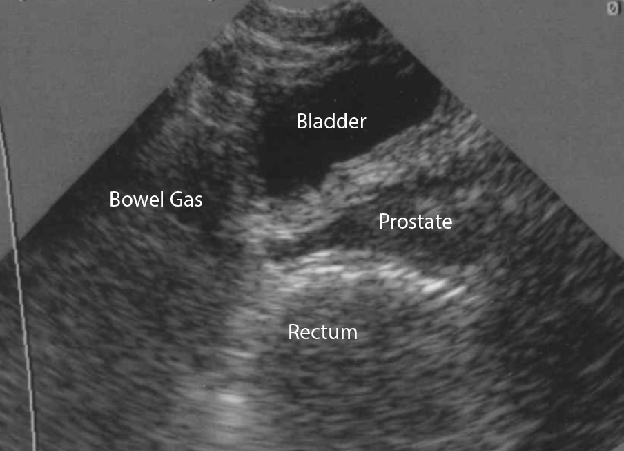
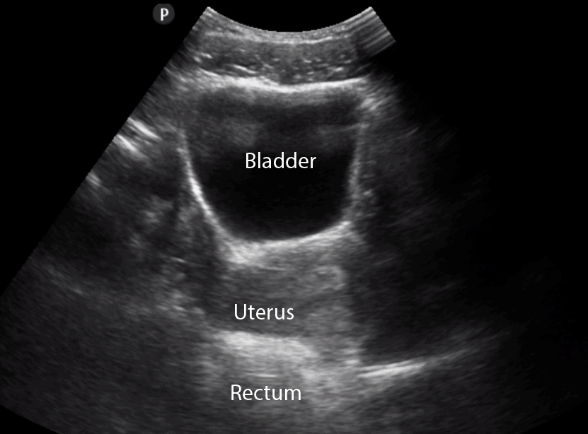
6 In the longitudinal (sagittal) view, identify the Bladder, Bowel Gas, Uterus (females), Prostate (males), and Rectum. Make sure to fan through the entire bladder.
👉🔗pocus101.com/bladder
👉🔗pocus101.com/bladder
7 Next, center the bladder and then rotate the transducer 90 degrees counterclockwise to get the transverse view. The indicator should now point to the patient’s left side. Make sure to tilt the ultrasound probe so it scans into the pelvic cavity.
👉🔗pocus101.com/bladder
👉🔗pocus101.com/bladder
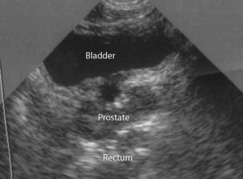
8 In the transverse view, identify the Bladder, Uterus (females), Prostate (males), and Rectum. Make sure to fan through the entire bladder.
👉🔗pocus101.com/bladder
👉🔗pocus101.com/bladder
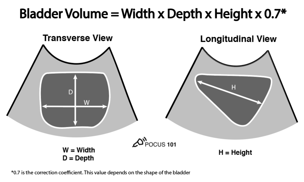
9 Calculate the Bladder Volume.
Ultrasound can be used to estimate bladder volume using a simple to remember formula: Height x Width x Length x 0.7.
👉🔗pocus101.com/bladder
Ultrasound can be used to estimate bladder volume using a simple to remember formula: Height x Width x Length x 0.7.
👉🔗pocus101.com/bladder

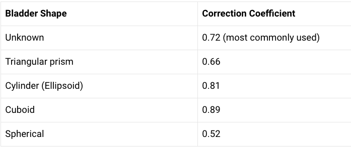
10 The actual formula used to calculate bladder volume is Width x Depth x Height x Correction Coefficient. For more accurate bladder volume measurements, use the correction coefficient that most closely corresponds to the patient’s bladder shape.
👉🔗pocus101.com/bladder
👉🔗pocus101.com/bladder
11 Don't want to remember all these numbers? Check out our Bladder Volume Calculator that will output volumes for ALL of the shapes: pocus101.com/bladder-volume…
Normal bladder volume should be less than 300-400 mL and Post Void Residual (PVR) should be less than 50-100mL
Normal bladder volume should be less than 300-400 mL and Post Void Residual (PVR) should be less than 50-100mL
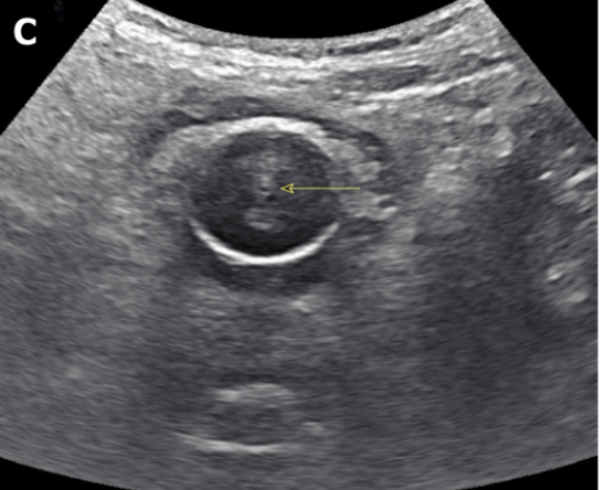
13 Foley Catheter malfunctions are a pain to troubleshoot without ultrasound. Here is what an ultrasound should look like for a normally functioning Foley Catheter. This image from THE @NephroP article: wjgnet.com/2220-6124/full…
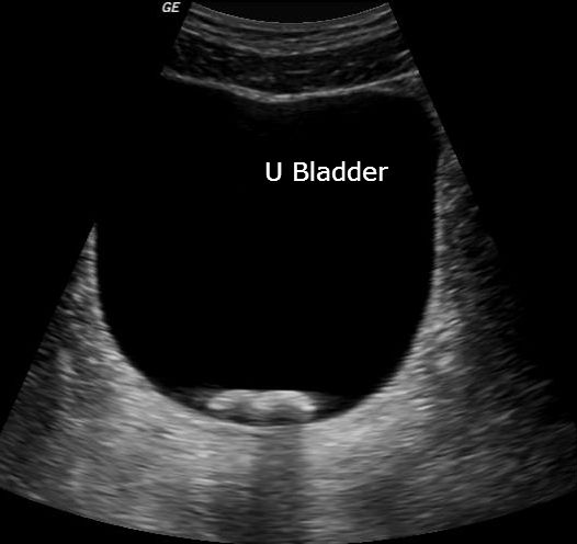
14 Here is an example of a foley catheter that is NOT draining. Notice the distended bladder.
Also if you look closely 🔍 you will see the free abdominal fluid posterior to the bladder
👉🔗pocus101.com/bladder
Also if you look closely 🔍 you will see the free abdominal fluid posterior to the bladder
👉🔗pocus101.com/bladder
15 Ureteral jets are a normal and periodic efflux of urine from the ureter into the bladder. Visualization of bilateral ureteral jets rules out complete obstruction of a specific ureter with high specificity.
👉🔗pocus101.com/bladder
👉🔗pocus101.com/bladder
16 Bladder stones are often seen after renal stones travel from the ureters into the bladder or from bladder stasis in patients with chronic urinary retention. They appear hyperechoic and mobile with acoustic shadowing.
👉🔗pocus101.com/bladder
👉🔗pocus101.com/bladder
17 Bladder masses are generally echogenic, irregularly shaped, and are found either mounted on the bladder wall or in areas of irregularly increased bladder wall thickness.
👉🔗pocus101.com/bladder
👉🔗pocus101.com/bladder
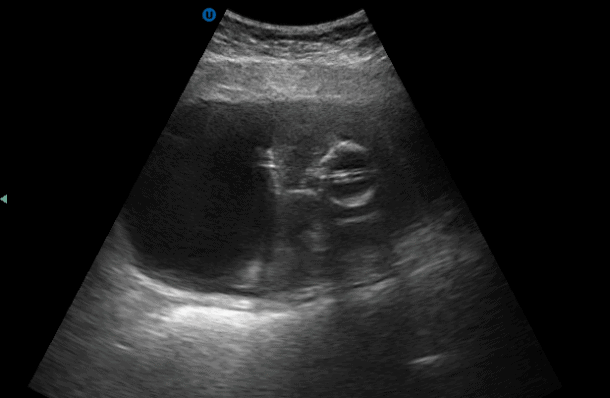
19 Blood clots can also appear to look like bladder masses. In this image, the clot has clogged the foley and requires irrigation.
👉🔗pocus101.com/bladder
👉🔗pocus101.com/bladder
20 Don't forget to check out the bladder volume calculator! pocus101.com/bladder-volume…
Sorry this should be towards the RIGHT side, NOT left. Typo and the blog post has been edited. Thanks @DrZebra1 for catching that!!